복부 둔상 후 발견된 복강동맥 박리 1례
서울대학교 의과대학 외과학교실, 서울대학교 의과대학 응급의학교실*, 분당 서울대학교병원 외과**
서윤석∙김성춘*∙라환도**∙한호성**
─ Abstract ─
Celiac Artery Dissection after Abdominal Blunt Trauma
Yun Suhk Suh, M.D., Seong Chun Kim, M.D.*, Hwan Do Ra, M.D.**, Ho-Seong Han, M.D.**
Department of Surgery, Seoul National University, College of Medicine, Seoul, Korea, Department of Emergency Medicine, Seoul National University, College of Medicine, Seoul, Korea*,
Department of Surgery, Seoul National University Bundang Hospital, Gyeonggi, Korea**
We report a case of celiac artery dissection after abdominal blunt trauma. A 29-year-old man visited the emergency room for acute left periumbilical pain after abdominal blunt trauma from his child. Computed tomography showed a wedge-shaped splenic infarction with splenic artery thrombus. He was hospitalized for careful observation, and after two days, follow-up computed tomographic angiography showed a progressed celiac artery dissection that involved common hepatic artery and an increased extent of splenic infarction. He underwent conventional angiography, and a self-expandable stent was placed between the celiac axis and the common hepatic artery. After two days, follow-up computed tomographic angiography showed good hepatic arterial blood flow via the stent and no progression of splenic infarction. After ten days, he was discharged without complications. (J Korean Soc Traumatol 2006;19:196-200)
Key Words: Celiac artery dissection, Abdominal blunt trauma, Stent
� Address for Correspondence : Ho-Seong Han, M.D.
Department of Surgery, Seoul National University Bundang Hospital 300 Gumi-dong, Bundang-gu, Seongnam-si, Gyeonggi-do 463-707, Korea Tel : 82-31-787-7099, Fax : 82-31-787-4055, E-mail : hanhs@snubh.org
접수일: 2006년 10월 30일, 심사일: 2006년 10월 30일, 수정일: 2006년 11월 20일, 승인일: 2006년 12월 5일
Ⅰ. 서 론
복부 둔상에 의한 복강 내 장기 손상은 비교적 흔하게 볼 수 있는 외상성 질환이나, 이러한 장기 손상 중에서 복 부 혈관 손상은 그 예를 쉽게 찾아 보기 힘들며, 특히 복 강동맥의 박리는 매우 드문 질환으로 높은 이환율과 사망 률을 보이는 질환이다.(1,2)
또한 대개의 복부 둔상 환자들이 응급실로 내원한 상황 에서, 명백한 다른 장기의 손상을 동반하거나 비 특이적인 증상들을 호소하는 경우가 많아 복부 혈관 손상에 대해 먼 저 의심하고 빠른 진단을 내리기 수월하지 않은 경우가 많 으며, 그 표준 치료 방법 역시 아직까지 명확히 제시되고 있지는 않다.(3,4)
이에 저자들은 복부 둔상 이후 발견된 복강동맥 박리를
1예 경험하였기에 문헌 고찰과 함께 보고하는 바이다.
Ⅱ. 증 례
환자는 29세 남자 환자로 내원 20분전부터 발생한 급성 복통을 주소로 응급실로 내원하였다.
병력 청취상 내원 당일 환자의 2세 아이가 환자의 배 위 에서 뛰어 놀면서 발생한 가벼운 복부 둔상 외에는 특이 사항은 없었다.
복부 시진 상 특이 소견 없었으며, 복부 통증의 양상은 배꼽 주위에서 좌상복부 사이의 압통을 동반한 지속적으로 느껴지는 산통이었으며 반발통이나 방사통은 없었다.
내원 당시 혈압은 수축기 혈압은 156 mmHg, 이완기 혈압은 89 mmHg, 맥박수는 분당 88회, 호흡수는 분당 20회, 체온은 36.4�C 소견을 보였다.
과거력 상 5년전 기흉으로 보존적 치료 받은 병력 이외 에는 특이 사항은 없었다.
가족력상 특이사항은 없었다.
내원 당시 시행한 임상병리학적 소견상 말초 혈액 검사 에서 WBC 9910/mm3, Hb 15.9 g/dL, PLT 289,000 /mm3, AST 44 IU/L, ALT 101 IU/L, PT 1.02 INR, aPTT 32.8 sec, 의 소견을 보였다. 항스트렙톨리 신-O검사(Anti-streptolysin O, ASO), 류머티즘 인자 (Rheumatoid factor, RF), 형광항핵항체(Fluorescent anti-nuclear anbibody, FANA) 모두 음성이었으며, 루프스항응고(Lupus anticoagulant, LA)는 1.36으로 약양성(weakly positive) 소견을 보였고, 단백질 C 101%, 단백질 S 68%, 항DNA항체(anti-DNA anti- body) 6.8 IU/ml 의 정상 소견을 보였다.
소변 검사 결과 RBC 1-4/HPF였다.
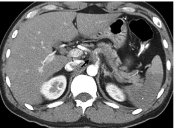
내원 당시 시행한 복부전산화단층촬영 소견상 비장동맥 내의 미만성 혈전을 동반한 비장 경색 소견을 보여, 입원 후 경과 관찰을 시행하였다(Fig. 1).
내원 1일째 시행한 임상병리학적 소견상 말초혈액검사결 과는 AST 39 IU/L, ALT 50 IU/L 외에 특이 사항은 없었다.
내원 2일째 추적관찰을 위해 시행한 전산화단층촬영 혈 관조영술(computed tomographic angiography, CT angiography)에서 복강동맥의 박리 소견이 관찰되었으 며, 총간동맥을 침범하는 양상이었고, 비장 경색이 더 진 행하는 양상을 보였다(Fig. 2). 전산화단층촬영 결과를 후 향적으로 비교하였을 때, 내원 당시에도 비장 경색 외에 이미 복강동맥의 박리 소견이 있었으며, 총간동맥과 비장
Fig. 2. CT angiography at second hospital day.
There was a celiac axis dissection (arrow) which involved common hepatic artery and progressed splenic infarction was identified.
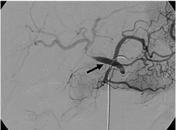
Fig. 3. Conventional angiography.
There was a focal stenosis (arrow) between celiac artery and common hepatic artery due to the dissection at celiac axis.
Fig. 1. Abdomen computed tomography.
Retrospectively suspicious celiac artery dissection (dot- ted arrow) and splenic infarction with splenic artery thrombosis (solid arrow) were showed.
동맥이 기시하는 부위에 미세 혈전 소견이 같이 있었음을 알 수 있었다(Fig. 1).
환자는 내원 2일째 시행한 재래식 혈관조영술(conven- tional angiography)에서, 복강동맥의 박리로 인해 복강 동맥의 협착 소견이 있었으며, 좌위동맥이 기시하는 부위의 총간동맥부위까지 협착 소견이 발견되었다(Fig. 3). 상장 간막동맥 조영상, 간으로의 혈류는 췌십이지장동맥에서의 우회 혈류로 유지되는 양상이었다. 환자는 내경 8 mm, 길 이 6 cm의 자가 확장형 스텐트(self expandable stent) 를 복강동맥에서 총간동맥까지 삽입 받았다(Fig. 4).
내원 4일째 시행한 추적 관찰 전산화단층촬영 혈관조영 술 상에서 간동맥으로의 혈류는 대부분 회복되었으며, 비 장동맥으로의 혈류는 일부분 회복되었으며 비장 경색은 더 이상 진행하지 않는 소견을 보였다(Fig. 5).
환자는 이후 항혈전제를 복용하면서, 내원 14일째 특별 한 합병증 없이 퇴원하였다.
Ⅲ. 고 찰
1947년 Baurersfeld(5)가 처음으로 복부 혈관의 자연 박리를 보고한 이후, 복강 동맥에 국한된 혈관 박리는 매 우 드물며, 그 원인으로는 대개 의인성이거나, 죽상경화 증, 외상, 임신, 섬유근육병, 염증성 질환, 혈관벽의 선천 성 질환에 의한 2차성 질환 등으로 보고된 바 있다.(2)
이러한 복부 혈관 손상의 가장 흔한 원인으로는 일반적 으로 자상이나 총상 등의 관통상에 의한 경우가 90~95%
로 대부분이며, 본 증례에서와 같이 복부 둔상에 의한 혈 관 손상에 의한 경우는 비교적 그 예가 드물어 대개 5~10
%로 보고 되고 있다.(1)
그 중에서 복강동맥의 손상은 더욱 드물며, 본 증례에서
와 같이 다른 복부 혈관의 손상이 같이 동반되고 있는 경 우가 많으며, 사망률 또한 대개 38~75%로 보고되고 있 다.(1,2,6)
본 증례에서와 같이 복부 둔상에 의한 것으로 의심되는 복강동맥에 국한된 혈관 박리는 매우 드문 예라 할 수 있 으며, 자연적인 복강동맥의 동맥 박리는 더욱 그 예가 드 물어 2004년까지 문헌상 단 69예가 보고되어있는 실정이 다.(2,3)
Yasushi 등(3)은 이와 같은 복강동맥 박리의 병리 기전 에 관하여 갑작스러운 복압의 상승에 의한 복부 충격, 호 기 시 횡격막의 급격한 강제 상승에 의한 정중활꼴인대 (median arcuate ligament of the diaphargm)와 복 강 신경총의 복강 동맥 압박, 일시적으로 복강동맥 기시부 와 가까운 위치에 복부 충격이 가해짐으로 인해 발생하는 복강동맥 내막의 손상 등을 그 원인에 대한 가설로 보고한 바 있다. 자연적인 복강동맥의 혈관 박리의 경우 증례 보 고가 점차 늘어나고 있으나 이러한 경우 복강 동맥에 국한 된 혈관 박리 증상의 병리 기전은 아직 자세히 밝혀지지 않았다.(2,3)
본 증례에서와 같이 이학적 검진 소견상 가벼운 복부 둔 상이 유일한 병력이라면, 이러한 경미한 복부 둔상이 복강 동맥의 혈관 박리를 일으킨 원인으로 가장 가능성이 높을 것으로 추정되지만, 앞서 언급한 바와 같이 혈관염이나 결 체조직 질환 등의 기저 질환, 죽상경화증 등 아직 확실히 밝혀지지 않은 다른 여러 기전들의 복합적인 작용에 대해 서도 간과하지 말아야 할 것으로 보인다.(2,7)
복강동맥 박리의 증상으로는 급성 또는 만성 등의 다양 한 형태의 복통, 폐쇄성 황달, 혈량 저하에 의한 쇼크, 사 지마비, 하지 경색, 요통 등의 비 특이적인 증상들이 있을 수 있다.(2,4,5,8) 상기 증례에서 보인 바와 같이 이러한
Fig. 4. Conventional angiography after a stent insertion
↗: A self expandable stent (8 mm diameter, 6 cm long segment) was placed between celiac axis and common hepatic artery.
Fig. 5. Follow up CT angiography at fourth hospital day Successful restoration of hepatic arterial blood flow was identified with a patent stent.
비 특이적인 증상들로 인하여 진단이 늦어지는 경우가 흔 히 있으며, 일부 보고에서는 진단 지연율을 높게는 34.3%
까지 보고한 바 있다.(3,4)
진단 방법으로는 과거 복부 대동맥 박리의 진단에 사용 되었던 재래식 혈관 조영술이 혈관 내로의 도관 삽입을 통 하여 혈관 내벽의 피판이나 이중 내벽(double lumen)과 같은 질병특유증상을 관찰 할 수 있어 절대표준 방법으로 알려져 있다.(9) 하지만 재래식 혈관 조영술은 침습적인 검사 방법으로서 시간과 비용이 많이 들고 검사 자체의 합 병증 등의 이유로 최근 들어 도플러 초음파나 전산화단층 촬영 혈관조영술 후 3차원적 재구성을 이용한 방법들이 많 이 이용되고 있다.(9)
현재 시도되고 있는 복강동맥 박리의 치료 방법에 대하 여, 아직 명확히 확립된 표준 치료 방법은 없지만 크게 나 누어 세가지 방법으로 전통적인 수술방법, 경피적 풍선 개 창술(percutaneous balloon fenestration), 경피적 스 텐트 삽입술 등이 시도되어 왔다.(7)
일반적으로 혈량 저하에 의한 쇼크나 하지 경색 등의 증 상을 보이는 환자에 있어서 대부분 개복에 의한 응급 수술 을 시행하여 왔으나, 자연적인 동맥 박리에 비하여 외상에 의한 동맥 박리는 후복막 내 정맥 손상, 외상 시 동반 가 능한 장 손상에 의한 세균의 복강 내 감염 등의 이유로 예 후가 안 좋은 것으로 알려져 있다.(4,7)
최근 들어 수술의 위험도가 높은 외상성 복부 동맥 박리 를 보이는 환자들에게 혈관 내 스텐트 삽입술을 이용한 중 재적 방사선 치료가 다각도로 시도되고 있으며 매우 좋은 성적들을 발표 하고 있다.(3,4,10-12) 이러한 시술의 장점 으로는 복부 대동맥의 교차 클램프(cross-clamping)가 필 요 없으며, 후복막의 박리가 필요없고, 수혈량이 줄어들 며, 수술시간의 단축, 낮은 감염 위험성 등을 들 수 있어, 혈역학적으로 안정된 환자들에게 있어서는 혈관 내 스텐트 삽입술을 첫 번째 치료 방법으로 시도하는 보고가 발표되 고 있다.(4,7,10-12) 최근 Rodney 등(13)에 의해 다기관 공동 연구로 발표된 외상에 의한 혈관 손상 시 막 부착성 스텐트(covered stent)를 이용한 치료 역시 시술 이후 1 년 이내 85%의 환자들이 수술이 필요 없이 호전되었던 매 우 좋은 결과를 보였다.
이외의 보존적 치료 방법으로는 시술 이후 발생할 수 있 는 혈전색전증의 예방을 위하여 항응고제의 투여가 권장되 기도 한다.(14)
복부 둔상에 의한 복강동맥의 박리는 드문 질환임과 동 시에 비 특이적인 증상들로 인하여 빠른 진단을 내리기가 쉽지 않은 질환이지만, 높은 사망률과 이환율을 가지고 있 어 응급실로 내원하는 외상 환자에서 깊은 주의를 요하는 질환이다. 본 증례에서는 주의 깊은 환자의 병력 청취와 반복적인 전산화단층촬영 혈관조영술의 시도로 복부 둔상
이후 발견된 비장 경색을 동반한 복강동맥의 박리를 진단 해낸 이후 최근 시도 되고 있는 스텐트 삽입술을 통하여 성공적으로 환자를 치료하였음을 보여주고 있다.
REFERENCES
01) Asensio JA, FornoW, Roldan G, Petrone P, Rojo E, Ceballos J, et al. Visceral vascular injurs. Surg Clin North Am 2002;82:1-20.
02) Fenoglio L, Allione A, Scalabrino E, Alberto G, Benedetti V, Pomero F, et al. Spontaneous dissec- tion of the celiac artery: A pitfall in the diagnosis of acute abdominal pin. Presentation of two cases.
Dig Dis Sci 2004;49:1223-27.
03) Yasushi L, Yoshihiko Y, Yasuo H, Hidenori K, Ken K, Toshiharu T, et al. A case of isolated celiac axis injury by blunt abdominal trauma. J Trauma 2006;61:451-3.
04) Berthet JP, Marty-Ane CH, Vecrapen R, Picard E, Mary H, Alric P. Dissection of the abdominal aorta in blunt trauma: endovascular or conventional sur- gical management? J Vasc Surg 2003;38:997-1004.
05) Bret PM, Patensky C, Bretagnolle M, Paliard P, Burke M. Obstructive jaundice by a dissecting aneusym of celiac axis and hepatic artery. Dig Dis Sci 1987;32:1431-4.
06) Brown DB, Singh H, Atnip RG, Cardella JF, Waybill PN. Blunt traumatic injury to the superior mesenteric artery and celiac axis. J Trauma 1998;
9:738-5.
07) Milagros M, Inmaculada P, Franziska B, Maria- Jose S, Gonzalo G. A case of acute abdominal aortic dissection caused by blunt trauma. Emerg Radiol 2006;12:182-5.
08) Peterson AH, Williams DM, Rodriguez JL, Francis IR. Percutaneous treatment of a traumatic aortic dissection by balloon fenestration and stent replace- ment. Am J Roentgenol 1995;164:1274-6.
09) Soudack M, Gaitini D, Ofer A. Celiac artery aneurysm: diagnosis by color Doppler sonography and three-dimensional CT angiography. J Clin Ultrasound 1999;27:49-51.
10) Duebener LF, Lorenzen P, Richardt G, Misfeld M, Notzold A, Hartmann F, et al. Emergency endovas- cular stent-grafting for life-threatening acute type B aortic dissections. Ann Thorac Surg 2004;78;1261-6.
11) Halkos ME, Nicholas J, Kong LS, Burke JR, Milner R. Endovascular management of blunt abd- nominal aortic injury. Vascular 2006;14:223-6.
12) Picard E, Marty-Ane CH, Vernhet H, Sessa C, Lesnik A, Senac JP, et al. Endovascular manage- ment of traumatic infrarenal abdominal aortic dis- section. Ann Vasc Surg 1998;12:515-21.
13) Rodney W, Zvonimir K, Matthew J, David W,
Michael B, Ellen O. Results of a multicenter trial for the treatment of traumatic vascular injury with a covered stent. J Trauma 2006;60:1189-96.
14) Schievink WI. Spontaneous dissection of the carotid and vertebral arteries. N Engl J Med 2001;344:898- 906.