Available at http://www.formulastudy.com
HFS
Original Article / 원저
알코올 유발 간 손상 마우스 모델에서 복합 추출물 MJY2018의 간 보호 및 항산화 효과
김광연
1#, 박광일
1․2․3#, 조원경
1, 양주혜
1, 마진열
1*1
한국한의학연구원 한의기술응용센터,
2경상대학교 수의과학대학,
3
경상대학교 동물의학연구소
Herbal formula MJY2018 protects against Alcohol-induced liver injury mice model
Kwang-Youn Kim
1#, Kwang-Il Park
1․2․3#, Won-Kyung Cho
1, Ju-Hye Yang
1, Jin-Yeul Ma
1*1
Korean Medicine (KM) Application Center, Korea Institute of Oriental Medicine (KIOM)
2
Department of Veterinary Physiology, College of Veterinary Medicine, Gyeongsang National University
3
Institutes of Animal Medicine, Gyeongsang National University
ABSTRACT
Objectives : This study investigated the liver-protective effects of MJY2018, a Herbal formula, against alcoholic fatty liver disease and anti-oxidative effects.
Methods : Its effects were investigated in an alcoholic fatty liver disease model in male C57BL/6 mice, which were fed Lieber−DeCarli liquid diet containing ethanol. MJY2018 (100 and 200 mg/kg bw/day) or silymarin (50 mg/kg bw/day) were orally administered daily in the alcoholic fatty liver disease mice for 16 days.
Results : The results indicate that MJY2018 promotes hepatoprotection by significantly reducing aspartate transaminase (AST) and alanine transaminase (ALT) levels as indicators of liver damage in the serum.
Furthermore, MJY2018 reduced accumulation of triglyceride and total cholesterol, increased levels of superoxide dismutase (SOD) and glutathione (GSH) in the livers of the alcoholic fatty liver disease mice model.
Additionally, it improved the serum alcohol dehydrogenase (ADH) activity.
ⓒ 2020 The Korean Medicine Society For The Herbal Formula Study
This paper is available at http://www.formulastudy.com which permits unrestricted non-commercial use, distribution, and reproduction in any medium, provided the original work is properly cited.
Conclusions : These results indicate that MJY2018 were effective in improving and protecting oxidative stress and alcoholic liver disease.
Key words : herbal formula, MJY2018, alcohol, liver damage, antioxidant.
Ⅰ. 서론
a)간에서 대부분의 알코올은 대사되나, 간이 분해하 는 능력을 초과하는 과량의 알코올은 알코올성 지방 간, 알코올성 간염 및 간경화증 등을 초래하게 된다
1,2). 장기간 알코올을 섭취하게 되면 단백질 및 지질 대사 장애를 유발하여 지방산 합성 증가 및 중성지방 의 분비장애를 유도하고, 생체내 활성산소종을 생성 하여 산화 스트레스를 유발함으로 알코올성 간 손상 의 주요 원인이 된다. 만성 알코올 섭취는 catalase (CAT), glutathione peroxidase (GPx), superoxide dismutase (SOD), glutathione reductase (GR) 등 의 항산화 효소와 glutathione(GSH) 등과 같은 비효 소적 항산화 물질들의 활성을 저해함으로써 산화 스 트레스를 촉진하여 간세포의 손상을 유도한다3,4). 우리나라의 경우 과음, 폭음 및 잦은 음주 형태의 과도한 음주습관 때문에 간질환의 발생이 증가하고 있으며, 대부분의 경우 숙취 제거를 위한 음료나 약 물에 대한 연구가 많이 수행되고 있다. 최근 많은 연 구에서 혈중 알코올 농도와 숙취 해소에 대한 한약재 에 대한 관심이 증가되고 있으며, 간질환 치료에 효 능을 나타내는 한약재에 대한 연구도 활발히 진행 중 에 있다5,6).
갈매나무과(Rhamnaceae)에 속하는 헛개나무의 열 매인 지구자(Hovenia dulcis Thunb.)는 한의학에서 간에 쌓인 독을 풀어줌으로 알코올성 간염, 알코올성 지방간, 간경화 및 황달 등의 질환에 효능이 있다고 알려져 있으며 , 항알레르기 효과, 간독성 보호 효과 도 보고된바 있다7). 오미자나무과에 속하는 오미자나 무(Schizandra chinensis Baillon)의 열매인 오미자 는 간염, 두통, 신경쇠약 등의 치료 및 항산화 작용, 간보호 및 간기능 복구 효과가 보고되어 있다8,9). 콩
과(Fabeceae)에 속하는 감초(Glycyrrhiza uralensis Fischer)는 한방에서 다른 약의 작용을 순하게 하는 효과가 있고, 혈당 및 복부지방을 감소 및 간보호, 항균 효능 등이 보고된바 있다10,11). 갈매나무과 (Rhamnaceae)에 속하는 낙엽활엽교목의 열매인 대 추는 식품과 한방재료로서 널리 사용되며 간 보호작 용과 항산화 활성도 우수한 것으로 나타낸다고 보고 된 바 있다12). 운향과의 귤(Citrus aurantium) 또는 동속 근연식물의 성숙한 열매의 껍질인 진피는 예로 부터 한방에서 약재로 널리 사용되어왔으며, 기관지 염 등으로 인한 기침과 가래 증세를 치료제로 사용되 었으며13), 항염증효과, 항알레르기 효과, 지방세포 감 소 등이 있다14). 생강과(Zingiber-aceae)에 속하는 아열대 또는 열대 원산의 다년생 초본 식물의 하나인 생강(Ginger, Zingiber officinale Rosc.)은 한방에 서는 소화불량, 구토, 설사에 효과가 있고, 혈액순환 을 촉진하며 항염증 및 진통에 효과가 알려져 있으 며, 항균작용15), 항염작용16), 혈청 콜레스테롤 저하효 과17), 항산화 작용18)을 나타낸다.
따라서 본 연구에서는 지구자, 진피, 오미자, 감초, 대추 및 생강을 포함한 복합 추출물이 알코올에 의해 간독성이 유발된 동물모델을 활용하여 확인하므로 알 코올성 간질환 치료의 후보 소재 개발을 위한 가능성 을 규명하고자 하였다.
Ⅱ. 재료 및 방법
1. 추출물 제조
지구자 500 g, 진피 315.5 g, 오미자 187.5 g, 감 초 125 g, 대조 62.5 g 및 생강 62.5 g을 영천약업 사(Yeongcheon, Korea)에서 구매하여 세척·건조한 후, 115℃에서 3시간동안 열수 추출하고 여과·동결
#These authors equally contributed to this work.
*Corresponding author : Jin-Yeul Ma. Korean Medicine (KM) Application Center, Korea Institute of Oriental Medicine (KIOM), 70, Cheomdan-ro, Dong-gu, Daegu, 41062, Republic of Korea.
Tel: +82-53-940-3812, Fax: +82-53-940-3899, E-mail: [email protected]
∙Received : April 24, 2020 / Revised : May 29, 2020 / Accepted : May 29, 2020
건조하여 MJY2018로 명명하고 사용하였다.
2. 실험동물 및 식이
체중 20±2 g인 6주령 C57BL/6 마우스를 ㈜ 샘타 코바이오코리아(Osan, Korea)로부터 분양받아, 온 도 25℃, 습도 50~55%에서 12시간씩 명암주기를 조 절하여 1주간 예비 사육하여 환경에 적응 시킨 후, 10마리씩 7개의 그룹으로 나누어 실험을 진행하였다.
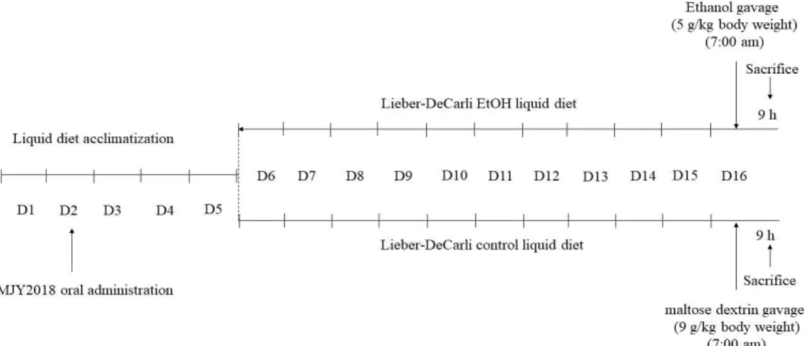
본 연구는 한국한의학연구원 동물실험윤리위원회의 승인을 받아 하에 수행하였다(IACUC-KIOM-D-18- 013). 실험방법은 NIAAA 모델에 따랐으며, 실험 설 계의 개요는 Figure 1에 표시하였다19). 1-5일에, 모 든 마우스가 액체식이에 적응하기 위해 대조군 Lieber-DeCarli 액체식이(control diet, Dyets, Inc., Bethlehem, PA)를 임의로 공급 하였다. 6일부 터 에탄올 단독 그룹, MJY2018 추출물-처리 그룹 (100, 200 mg/kg/body weight) 및 양성 대조군 (silymarin, 50 mg/kg/body weight)에 10일 동안 5% (v/v) 에탄올(36% ethanol-derived calories)을 함유 한 Lieber-DeCarli 에탄올 액체식이(EtOH diet, Dyets, Inc., Bethlehem, PA)를 급이하고, 대
조군, MJY2018 추출물 단독 대조군 (200 mg/kg/
body weight) 및 Silymarin 단독 대조군(50 mg/kg/
body weight)은 Lieber-DeCarli control 식이를 공 급 하였다. 16일에, 에탄올 및 대조군 급이한 마우스 를 각각 단일 투여량의 에탄올(5 g/kg/body weight) 또는 isocaloric maltose dextrin(9 g/kg/body weight)으로 이른 아침에 경구 투여하고, 9시간 후에 마우스로부터 혈액 및 간 조직을 수집하였다. 혈액은 37℃에서 30분 동안 방치한 후, 5000×g에서 20분간 원심분리 하여 혈청으로 분리하고, 간 조직은 액체 질소에서 급속하게 동결 동결시키고, 80℃에서 보관 하였다. 실험 전 기간 동안 MJY2018 추출물 및 silymarin은 매일 동일한 시간에 경구 투여 하였고, 식이섭취량 및 체중 또한 매일 일정 시간에 측정하였 다.
3. 조직학적 분석
간 조직의 일부를 10% para formaldehyde (Sigma Aldrich, St. Louis, MO, USA)에 고정시키고, 파라 핀에 매립하고, 단면으로 절단하여 hematoxylin- eosin (H&E)으로 염색하고 광학현미경으로 관찰하였다.
Figure 1. The basic overview of the model procedures (NIAAA model)
4. 생화학적 분석
혈액으로부터 분리한 혈청을 사용하여, aspartate aminotransferase (AST), alanine aminotransferase (ALT) 및 Alcohol Dehydrogenase (ADH) Assay Kit (Abcam, Cambridge, UK)를 사용하여 측정하였 다. 또한, triglyceride, total cholesterol, superoxide
dismutase (SOD) 및 Glutathione (GSH) assay kit (Cayman, Michigan, USA)를 사용하여 측정 하였다.
5. 통계처리
모든 실험값은 3회 이상 반복 실험한 결과를 기준 으로 하였으며 대조군과 각 실험군과의 평균 차이는
ANOVA로 분석을 실시하여 검정하였다. P-value<0.05 를 유의 수준으로 간주하였으며, mean±S.E. 값으로 표기 하였다.
Ⅲ. 결과
1. 알코올로 인한 식이량, 체중 변화에 대한 MJY2018 투 여 효과
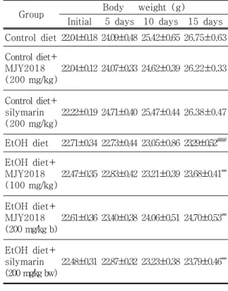
실험동물에 알코올과 MJY2018을 16일간 급이 한 후, 마우스의 식이효율 및 체중 증가율을 측정하였 다. 식이섭취량은 정상식이만 급여한 마우스에서 13.5±1.7 ml/day으로 정상식이에 MJY2018을 투여 한 마우스(14.3±2.0 ml/day) 간에 유의차가 적었으 며, 알코올 식이로 급이 시킨 마우스(12.4±1.4 ml/day)는 유의적으로 감소되었다(Table 2). 알코올 식이에 MJY2018을 투여한 마우스는 알코올 식이만 급이한 마우스보다 식이섭취량이 유의성 있게 증가하 는 것을 확인하였다(Table 1). 체중 증가율은 정상식 이만 급여한 마우스에서 26.75±0.63 g으로 정상식 이에 MJY2018을 투여한 마우스(26.22±0.33 g) 간 에 유의차가 적었으며, 알코올 식이로 급이 시킨 마 우스(23.29±0.52 g)는 유의적으로 감소되었다(Table 2). 알코올 식이에 MJY2018을 투여한 마우스는 알코 올 식이만 급이 한 마우스보다 체중증가율이 유의성 있게 증가하는 것을 확인하였다(Table 2).
Table 1. Dietary Intake in Mice Fed either the Control or Ethanol-Containing Lieber
−DeCarli Diet
Group Dietary intake (mL) 5 days 10 days 15 days Control diet 14.1±2.1 13.8±0.5 13.5±1.7 Control diet + MJY2018
(200 mg/kg bw) 14.4±1.9 14.0±1.3 14.3±2.0 Control diet + silymarin
(200 mg/kg bw) 14.2±1.8 13.9±1.4 14.8±1.8 EtOH diet 14.7±2.1 13.0±1.4 12.4±1.4###
EtOH diet + MJY2018
(100 mg/kg bw) 13.7±2.4 13.2±1.6 13.9±2.1***
EtOH diet + MJY2018
(200 mg/kg bw) 13.2±2.8 12.1±1.7 12.8±1.3***
EtOH diet + silymarin
(200 mg/kg bw) 14.2±1.8 12.1±1.7 13.8±1.3***
Data are shown as the mean±standard error of the mean (SEM, n=10). Statistical analyses were different values according to the one-way ANOVA with Tukey’s range test (Control diet vs EtOH diet ###p < 0.001, EtOH diet vs EtOH diet+MJY2018, vs EtOH diet+silymarin ***p <
0.001).
Table 2. Body Weight of Control and Experimental Mouse Groups
Group Body weight (g)
Initial 5 days 10 days 15 days Control diet 22.04±0.18 24.09±0.48 25.42±0.65 26.75±0.63 Control diet+
MJY2018 (200 mg/kg)
22.04±0.12 24.07±0.33 24.62±0.39 26.22±0.33
Control diet+
silymarin (200 mg/kg)
22.22±0.19 24.71±0.40 25.47±0.44 26.38±0.47
EtOH diet 22.71±0.34 22.73±0.44 23.05±0.86 23.29±0.52###
EtOH diet+
MJY2018 (100 mg/kg)
22.47±0.35 22.83±0.42 23.21±0.39 23.68±0.41***
EtOH diet+
MJY2018 (200 mg/kg b)
22.61±0.36 23.40±0.38 24.06±0.51 24.70±0.53***
EtOH diet+
silymarin (200 mg/kg bw)
22.48±0.31 22.87±0.32 23.23±0.38 23.79±0.46***
Data are shown as the mean±standard error of the mean (SEM, n=10). Statistical analyses were different values according to the one-way ANOVA with Tukey’s range test (Control diet vs EtOH diet ###p < 0.001, EtOH diet vs EtOH diet vs EtOH diet+MJY2018, vs EtOH diet+
silymarin ***p < 0.001).
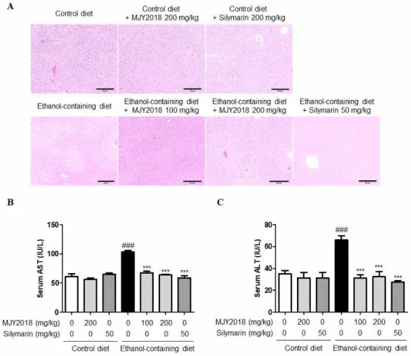
2. 알코올로 인한 간독성에 대한 MJY2018 투여 효과 MJY2018 투여가 알코올에 의해 유도된 간 손상에 대한 간 보호에 미치는 영향을 H&E 염색 및 혈청 내 aspartate aminotransferase (AST) 및 alanine aminotransferase (ALT)의 활성을 통해 확인하였다.
먼저, 간 보호 효과를 조직학적으로 확인하기 위해서 H&E 염색을 진행한 결과, 에탄올 식이를 급이 한 그 룹의 간 조직내에서 염증 및 괴사 등의 병변을 확인 하였고 희미한 미세 소포성 지방증이 관찰하였다. 이 러한 알코올로 인한 간 조직내 병변은 MJY2018을 투여한 마우스에서 감소한 것을 관찰하였다(Figure
2A). 또한, 간 기능 및 손상 정도를 나타내는 혈청 AST 및 ALT 활성을 측정한 결과, 알코올 식이 투여 로 인해 증가하였던 혈청 ALT 및 AST 활성은 MJY2018 및 양성 약물인 silymarin을 투여한 그룹 에서 유의적으로 감소하였다(Figure 2B, 2C).
Figure 2. Effects of MJY2018 on ethanol-induced liver injury. (A) H&E-stained liver sections of EtOH-induced liver injury model, visualized at 100×, n=10. (B) Activities of serum aspartate aminotransferase (AST). (C) Activities of serum alanine aminotransferase (ALT). Data are shown as the mean±standard error of the mean (SEM, n=10). Statistical analyses were different values according to the one-way ANOVA with Tukey’s range test (Control diet vs EtOH diet
###p<0.001; EtOH diet vs EtOH diet vs EtOH diet+MJY2018, vs EtOH diet+silymarin ***p<0.001).
3. 알코올로 인한 Triglyceride 및 Total Cholesterol 축 적에 대한 MJY2018 투여 효과
혈청에서 알코올로 유도된 triglyceride 및 total cholesterol 함량을 측정한 결과, 알코올 식이로 인 해서 혈청 triglyceride 및 total cholesterol의 함량
이 유의하게 증가한 것을 확인하였으며, 이는 MJY2018 및 양성 약물인 silymarin의 투여로 인하여 명백하게 triglyceride 및 total cholesterol 증가를 억제하였 다(Figure 3A, 3B).
Figure 3. Effects of MJY2018 on alcoholic fatty liver disease. (A) Hepatic triacylglycerol levels. (B) Hepatic cholesterol levels. Data are shown as the mean±standard error of the mean (SEM, n=10). Statistical analyses were different values according to the one-way ANOVA with Tukey’s range test (Control diet vs EtOH diet ###p<0.001; EtOH diet vs EtOH diet vs EtOH diet+MJY2018, vs EtOH diet+silymarin ***p<0.001, **p<0.05).
4. 알코올로 인한 Superxide Dismutase (SOD) 및 Glutathione (GSH) 활성에 대한 MJY2018 투여 효과
알코올에 의해 유도 되는 항산화 효능에 대한 MJY2018의 효과를 평가하기 위해, 혈청내 SOD 및 GSH 농도의 수준을 확인하였다. 알코올에 의해서
SOD 및 GSH의 함량이 유의적으로 감소하는 것을 확 인하였으며, MJY2018 및 양성 약물인 silymarin을 처리한 결과, 알코올에 의해 유도 된 SOD 및 GSH 활성 감소를 유의하게 억제한 것을 확인하였다 (Figure 4A, 4B).
Figure 4. Effects of MJY2018 on liver antioxidant defense. (A) Activities of serum superoxide dismutase (SOD) (B) Activities of serum glutathione (GSH). Data are shown as the mean±standard error of the mean (SEM, n=10). Statistical analyses were different values according to the one-way ANOVA with Tukey’s range test (Control diet vs EtOH diet
##p<0.05, ##p<0.05; EtOH diet vs EtOH diet vs EtOH diet+MJY2018, vs EtOH diet+silymarin **p<0.05).
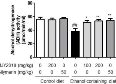
5. 알코올로 인한 Alcohol Dehydrogenase (ADH) 활성에 대한 MJY2018 투여 효과
MJY2018 투여가 알코올에 의한 alcohol dehydrogenase (ADH) 활성에 대한 효과를 확인한 결과, 알코올 식
이로 혈청 내 ADH의 활성은 유의적으로 감소 하였 으며, 알코올로 인해 감소한 ADH의 활성은 MJY2018 및 양성 약물인 silymarin에 의해 유의적으로 증가하는 것을 확인하였다(Figure 5).
Figure 5. Effects of MHY2018 on alcohol dehydrogenase activity (ADH). Data are shown as the mean±standard error of the mean (SEM, n=10). Statistical analyses were different values according to the one-way ANOVA with Tukey’s range test (Control diet vs EtOH diet
##p<0.05; EtOH diet vs EtOH diet vs EtOH diet+MJY2018, vs EtOH diet+silymarin
**p<0.05, *p<0.01).
Ⅳ. 고찰
알코올성 간질환의 초기 단계인 지방간은 광범위한 미세 및 거대 수포성 지방을 형성하는 lipid 및 triglyceride의 축적을 포함하며, 장기적인 알코올 섭 취는 미토콘드리아의 지질 과산화를 감소시키고 triglyceride의 합성을 증가시켜 지방 대사를 방해한
다20,21). 지속적인 알코올 섭취는 간에서의 산화 스트
레스 및 염증 반응을 증가로 인해 지방 간염, 간섬유 증 또는 간경변으로 이어진다22). 알코올성 간질환에 대해 간 보호 활성을 나타내는 식품 및 한약재에 대 한 여러 연구가 보고되어져 있다23). 더불어, 한약재 복합물 및 병용 처리는 주로 단일 한약재의 처리보다 약효의 상승작용에 대한 보고가 있으며, 항산화 효능 및 간보호 효능 등을 포함한 다양한 효과가 보고되어 져 있다24,25).
본 연구에서는 간 독성 보호, 항염증 및 항산화 효
과가 보고되어진 지구자, 오미자 감초, 대추, 진피 및 생강으로 이루어진 한약재 복합물인 MJY2018을 제조하여 알코올성 간질환 동물 모델에서 간 보호 및 항산화 활성을 알아보고자 하였다. 먼저, 알코올을 급이 한 마우스의 체중이 유의적으로 감소하는 것을 확인하였고, 간 기능 및 손상 정도를 나타내는 혈청 aspartate aminotransferase (AST) 및 alanine aminotransferase (ALT) 활성이 유의적으로 증가한 것을 확인하였다. 조직 병리학적인 관찰에서는 알코 올을 급이 한 마우스의 간에서 미세 지방소포의 증가 를 확인하고, 혈청내 total cholesterol 및 triglyceride 의 함량이 현저히 증가한 것을 확인하였다. 이는 한 약재 복합물인 MJY2018을 투여하였을 때, 양성약물 인 silymarin과 동일하게 농도 의존적으로 혈청내 AST, ALT, total cholesterol 및 triglyceride 수치 를 유의적으로 억제함으로써, 알코올성 간질환을 현 저하게 개선한 것을 확인하였다.
또한, 지속적인 알코올 섭취는 간에서 활성산소종
생산을 통해 산화 스트레스를 유도하고, 과도한 활성 산소종의 생성은 SOD와 GSH와 같은 항산화 방어체 계의 균형을 교란시켜 간 손상을 유발할 수 있다
26,27). 알코올 관련 독성으로 인한 항산화 효능 뿐만
아니라, 숙취 개선을 위한 식품 및 한약재를 활용한 소재 개발 연구도 활발히 진행 중이다28,29). 숙취 개 선에서 중요한 간에서의 알코올 대사는 ADH에 의해 acetaldehyde로 전환되고, acetaldehyde dehydrogenase (ALDH)를 통해 acetate로 대사된다. 이 때 대사장애 가 발생하면 acetaldehyde가 축적되어 알코올 분해 를 저해하게 되고 축적된 알코올로 인하여 간 손상을 초래한다30). 알코올을 급이 한 마우스의 혈청 내 SOD 및 GSH의 함량이 현저히 감소하고, 혈청내 ADH 활성 또한 저해되는 것을 확인하였다. 이는 MJY2018을 투여함으로 혈청내 SOD 및 GSH의 함량 이 증가되고, ADH 활성 또한 증가시킴으로, 간 보호 및 항산화 기능과 더불어 알코올에 의한 숙취를 효율 적으로 개선하는 것으로 사료된다.
Ⅴ. 결론
본 연구에서는 알코올에 의해 간손상이 유발된 마 우스의 in vivo 모델에서 MJY2018의 간보호 및 항 산화 효과를 평가하였다. MJY2018의 투여에 의해 알 코올에 의해 증가하였던 혈청 AST 및 ALT 활성을 감소시키고, 간조직 내 손상 및 염증을 억제하는 것 으로 확인되었다. 또한, MJY2018의 투여에 의해 항 산화 효소인 SOD 및 GSH의 활성을 증가시켰으며, ADH의 활성을 증가시키는 것을 확인되었다.
이러한 결과는 MJY2018의 처리가 in vivo 모델에 서 산화적 손상에 대해 간보호 효과가 있음을 입증한 것으로 항산화 효과와 더불어 숙취 개선을 할 수 있 는 유효한 약물 후보로 지속적인 연구가 필요할 것으 로 생각된다.
감사의 글
본 연구는 중소기업벤처부 과제 "동의고방 슈퍼푸 드 글로벌 명품화 사업"(Grant R0006219) 지원으로 수행되었으며, 이에 감사드립니다.
References
1. Chayanupatkul M, Liangpunsakul S, Alcoholic hepatitis: a comprehensive review of pathogenesis and treatment. World Journal of Gastroenterology, 2014;20(20):6279-86.
2. Yin H, Hu M, Liang X, Ajmo JM, Li X, Bataller R, et al., Deletion of SIRT1 from hepatocytes in mice disrupts lipin-1 signaling and aggravates alcoholic fatty liver.
Gastroenterology, 2014;146(3):801-11.
3. Wheeler MD, Nakagami M, Bradford BU, Uesugi T, Mason RP, Connor HD, et al., Overexpression of manganese superoxide dismutase prevents alcohol-induced liver injury in the rat.
Jouranl of Bioligical Chemistry, 2001;
276:36664-72.
4. Lu SC, Mato JM, S-Adenosylmethionine in cell growth, apoptosis and liver cancer. Journal of Gastroenterology and Hepatology, 2008;23:s73-s77.
5. LEE SJ, Kang MJ, Shin JH, Kim JG, Kang SK, Sung NJ, The Effect of Garlic and Medicinal Plants Extracts on the Liver Function and Lipid Metabolism of Rats Administered with Alcohol.
Journal of the Korean Society of Food Science and Nutrition, 2009;38(5);561-8.
6. Ko BS, Jang JS, Hong SM, Kim DW, Sung SR, Park HR, et al., Effect of new remedies mainly comprised of Hovenia dulcis Thunb on alcohol degradation and liver protection in Sprague Dawley male rats. Journal of the Korean Society of Food Science and Nutrition, 2006;35:828-34.
7. You Y, Jung KY, Lee YH, Jun W, Lee BY, Hepatoprotective Effects of Hovenia dulcis Fruit on Ethanol-Induced Liver Damage in vitro and in vivo. Journal of the Korean Society of Food Science and Nutrition, 2009;38(2):154-9.
8. Xiong J, Guo Y, Li LY, Hu H, Qu XL, Sun XZ, et al., A herbal composition of semen hoveniae, radix puerariae, and fructus schisandrae shows
potent protective effects on acute alcoholic intoxication in rodent models. Evidence- based Complementary and Alternative Medicine, 2012;2012:638197.
9. Cheng N, Ren N, Gao H, Lei X, Zheng J, Cao W, Antioxidant and hepatoprotective effects of Schisandra chinensis pollen extract on CCl4-induced acute liver damage in mice. Food and Chemical Toxicology, 2013;55:234-40.
10. Fiore C, Eisenhut M, Krausse R, Ragazzi E, Pellati D, Armanini D, et al., Antiviral effects of Glycyrrhiza species. Phytotherapy Research, 2008;22(2):141-8.
11. Jeon JH, Park D, Shin S, Kang DH, Moon SH, Kim YB, Effect of green tea and licorice extracts on cyclophosphamide teratogenicity.
Reproductive Toxicology, 2007;24(1):75.
12. Hong JY, Nam HS, Shin SR, Changes on the Antioxidant Activities of Extracts from the Ziziphus jujube Miller Fruits During Maturation.
Korean Journal of Food Preservation, 2010;
17(5):712-9.
13. Min SH, Park HO, Oh HS, A study on the properties of hot water extracts of Korean dried tangerine peel and development of beverage by using it. Korean Journal of Food and Cookery Science, 2002;18:51-6.
14. Kang SH, Lee YJ, Lee CH, Kim SJ, Lee DH, Lee YK, et al., Physiological Activities of Peel of Jeju-indigenous Citrus sunki Hort. Tanaka.
Korean Journal of Food Science and Technology, 2005;37(6):983-8.
15. Sheo HJ, The Antibacterial Action of Garlic, Onion, Ginger and Red Pepper Juice. Journal of the Korean Society of Food Science and Nutrition, 1999;28(1):94-9.
16. Thomson M, Al-Qattan KK, Al-Sawan SM, Alnaqeeb MA, Khan I, Ali M, The use of ginger (Zingiber officinale Rosc.) as a potential anti-inflammatory and antithrombotic agent.
Prostaglandins, Leukotrienes & Essential Fatty Acids, 2002;67(6):475-8.
17. Barbalata T, Deleanu M, Carnuta MG, Niculescu LS, Raileanu M, Sima AV, et al., Hyperlipidemia Determines Dysfunctional HDL Production and Impedes Cholesterol Efflux in the Small Intestine: Alleviation by Ginger Extract. Molecular Nutrition & Food Research, 2019;63(19):e1900029.
18. Shirpoor A, Rezaei F, Fard AA, Afshari AT, Gharalari FH, Rasmi Y, Ginger extract protects rat's kidneys against oxidative damage after chronic ethanol administration. Biomedicine &
Pharmacotherapy, 2016;84:698-704.
19. Bertola A, Mathews S, Ki SH, Wang H, Gao B, Mouse model of chronic and binge ethanol feeding (the NIAAA model). Nature Protocols, 2013;8(3):627-37.
20. Albano E, Free radical mechanisms in immune reactions associated with alcoholic liver disease. Free Radical Biology and Medicine, 2002;32:110−4.
21. Purohit, V, Gao B, Song BJ, Molecular mechanisms of alcoholic fatty liver. Alcoholism:
Clinical and Experimental Research, 2009;
33:191−205.
22. Albano E, Alcohol, oxidative stress and free radical damage. Proceedings of the Nutrition Society, 2006;65:278−90.
23. Cho HS, Lee SJ, Shin JH, Kang MJ, Cho HS, Lee HJ, et al., Antioxidative activity and nitrite scavenging effect of the composites containing medicinal plant extracts. Journal of Life Science, 2007;17: 1135-40.
24. Lee JM, Lee SH, Kim HM, Use of oriental herbs as medical food. Food Industry and Nutrition, 2000;5:50-6.
25. Lee EH, Baek SY, Kim KY, Lee SG, Kin SC, Lee HS, et al., Effect of Rheum undulatum Linne extract and Glycyrriza uralensis Fischer extract against arachidonic acid and iron- induced oxidative stress in HepG2 cell and CCl4-induced liver injury in mice. Herbal Formula Science, 2016;24(3):163-74.
26. Albano, E, Free radical mechanisms in immune reactions associated with alcoholic liver disease. Free Radical Biology and Medicine, 2002;32:110−4.
27. Lieber CS, Milestones in liver disease. Journal of Hepatology, 2004;40:198-202.
28. Lee EJ, Bae JH, Study on the Alleviation of an Alcohol Induced Hangover and the Antioxidant Activity by Mulberry Fruit. Korean Journal of Food And Nutrition, 2011;24(2):204-209.
29. Choi YJ, Jung KI, Anti-Diabetic, Alcohol- Metabolizing, and Hepatoprotective Activities of Moringa (Moringa oleifera Lam.) Leaf Extracts.
Journal of the Korean Society of Food Science and Nutrition, 2016;45(6): 819-27.
30. Rasineni K, Casey CA, Molecular mechanism of alcoholic fatty liver. Indian Journal of Pharmacology, 2012;44:299−303.