• 검색 결과가 없습니다.

γ-Aminobutyric Acid Metabolism in Plant under Environment Stressses

N/A
N/A
Protected

Academic year: 2021

Share "γ-Aminobutyric Acid Metabolism in Plant under Environment Stressses"

Copied!
7
0
0

로드 중.... (전체 텍스트 보기)

전체 글

(1)

γ-Aminobutyric Acid Metabolism in Plant under Environment Stressses

Tae-ho Ham, Sang-ho Chu, Sang Jun Han, and Su-Noh Ryu

Agricultural Science, Korea National Open Univerisity, Seoul 110-500, Republic of Korea

144

Corresponding author: (Phone) +82-2-3668-4631 (E-mail) ryusn@knou.ac.kr

<Received 7 March, 2012; Revised 25 May, 2012; Accepted 5 June, 2012>

ABSTRACT γ-Aminobutyric acid (GABA) is a non-protein amino acid that is widely distributed in plant and animal kingdom. GABA is found in tissues of the central nervous system (CNS) in animals. GABA functions as a the major inhibitory neurotransmitter in the CNS by acting through the GABA receptors. Clinical studies have revealed the relationship between an increased intake of GABA or analogues with several health benefits, including lowering of blood pressure in mildly hypertensive animals and humans. Furthermore, GABA would also has an inhibitory effect on cancer cell proliferation, stimulates cancer cell apoptosis and plays a role in alcohol-associated diseases and schizophrenia. In plants, interest in the GABA emerged mainly from experimental observations that GABA is largely and rapidly produced in large amounts in response to biotic and abiotic stresses. In this study, we speculated the properties and metabolism of GABA in plant and functions in relation to the responses to environmental stresses.

Keywords : GABA, GABA accumulation, GABA metabolism

GABA , a four-carbon non-protein amino acid, is a major component of the free amino acid pool and widely distribute in prokaryotic and eukaryotic organisms. GABA has an amino group on the γ-carbon rather than on the α-carbon, and exists in an unbound form. It is highly soluble in water; structurally it is a flexible molecule that can assume several conformations in solution, including a cyclic structure that is similar to proline.

GABA is zwitterionic(carries both a positive and negative charge) at physiological pH values(Deborah, et al., 2004). In plant, GABA has been treated merely as a metabolite for decades, but the recent evidence points towards a new possible role of GABA as a signal molecule in response to various stress. The interest in GABA shifted to animals when it was revealed that GABA occurs at high levels in the brain, playing a important role in neurotransmission. Clinical studies have related increased

intake of GABA or analogues to several health benefits, including regulation of blood pressure and heart rate, and alleviation of pain and anxiety(Mody et al., 1994). GABA is implicated in a number of neurological disorders including epilepsy, depression, anxiety, Alzheimer’s disease, Parkinson’s disease, schizophrenia and Huntington’s chorea(Deborah, et al., 2004). Furthermore, GABA would also have an inhibitory effect on cancer cell proliferation, stimulate cancer cell apoptosis(Oh, & Oh, 2004), and play a role in alcohol associated diseases and schizopherenia(Caputo, Vignoli, Maremmani, Bernardi, & Zoli, 2009; Oh & Oh 2003).

Recent studies show that GABA is also a strong secretagogue of insulin from the pancreas and effectively prevents diabetes (Adeghate & Ponery, 2002; Hagiwara et al.,2004).

Nowadays, there is a growing interest in natural, minimally processed, nutritional and healthy foods. A plant-based diet, focussing mainly on whole grains, has become one of the most important guidelines for lowering the risk of chronic human diseases(Lawrence & Machlin, 1995). Public interest in nutraceutical foods has led to investigations by biochemical technology to enhance nutritious compositions in cereals and fermentation food.

Although, enough amount of GABA is secreted naturally, there are various obstracles e.g. excess ingestion of estrogen, salicylate and food additives, low-protein diet, deficiency of zinc and vitamin B. Inspite of the supplement quantity of GABA is 500-3000 mg/day, the amount of GABA in cereals is 1-4 mg/100 g in rice, 4-8 mg/100 g in brown rice and 10-100 mg/g in germinated brown rice(BioWave, 2007). To increase amount of GABA in plant, various methods has been applied..

It was discovered in plants more than half a centrury ago (Steward, 1949) and the pathway, called GABA shunt, was first reported in potato(Solanum tuberosum) tuber(Dent et al., 1947). The pathway starts with the decarboxylation of glutamate by gultamate decarboxylase(GAD) to produce GABA and CO

2

in the cytosol. GABA is then presumably transported to the

(2)

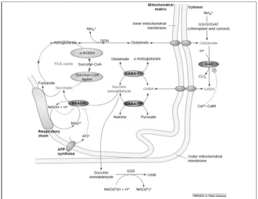
Fig. 1. The γ-aminobutyric aicd(GABA) shunt metabolic pathway and its regulation in plants. The glutamine-sysnthetase/

glutamate-synthase(GS/GOGAT) cycle is the principal nitrogen assimilation pathway into glutamate and amino acids in plants. The glutamate dehydrogenase(GDH) is thought to function primarily in glutamate catabolism but can also function in the opposite direction. The GABA shunt is composed of three enzymes (purple). Glutamate decarboxylase (GAD) is a cytosolic enzyme regulated(green) by the Ca

2+

/calmodulin(CaM) complex, which catalyses the irreversibl decarboxylation of glutamate to produce GABA. GABA is transported into the mitochondria, where it is converted into succinic semialdehyde by GABA transaminases using either α-ketoglutarate(by GABA-TK) or pyruvate(by GABA-TP) as amino acid acceptors.

Succinic semialdehyde is then reduced by succinic semialdehyde dehydrogenase(SSADH) to form succinate, which enters the tricarboxylic acid(TCA) cycle. Both ATP and NADH can inhibit the activity of the SSADH enzyme(green).

The succinyl-CoA ligase and the α-ketoglutarate dehydrogenase (α-KGDH) are two enzyme(pink) of the TCA cycle bypassed by the GABA shunt and sensitive to oxidative stress. Succinic semialdehyde can instead be reduced to γ-hydroxybutyric acid(GHB) via a succinic semialdehyde reductase(SSR) localized in the cytosol in animal cells and possibly in plants as well. In mammals, GHB is thought to be a neurotransmitter, whereas its role in plants in unkwon(Ref from Fig 1. Nicolas Bouche and Hillel Fromm, 2004).

mitochondria by an as yet unidentified GABA transporter, where it is converted to succinic semialdehyde(SSA). The presence of GAD activity and GABA in plants was first detected at least half a century ago(Satyanarayan and Nair, 1990; Bouche and Fromm, 2004). The pathway is composed of the cytosolic enzyme GAD and the mitochondrial enzymes GABA transaminase

(GABA-T) and succinic semialdehyde dehydrogenase(SSADH).

The regulation of this conserved metabolic pathway seems to have particular characteristics in plants.

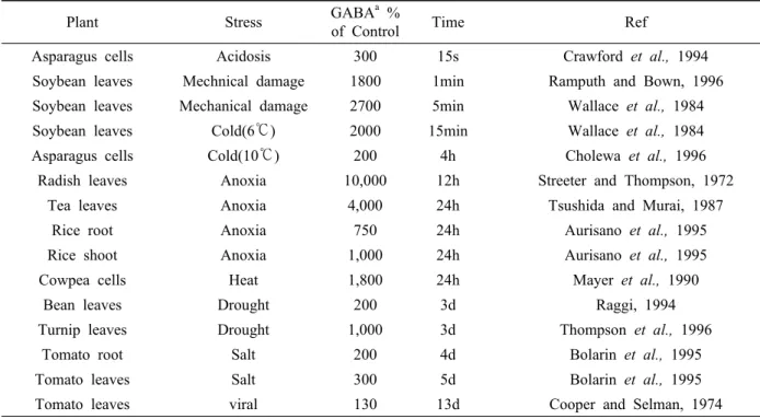
In response to environmental stresses, GABA production often increases so much that cellular levels of this non-protein amino acid exceed that of amino acid involved in protein synthesis(Shelp et al., 1999; Kinnersley, 2000). This situation also has been reported in drought-stressed cotton(Hanower and Brzozowsak, 1975), heat-stressed cowpea cells(Mayer et al., 1990), and soybeans subjected to cold stress and mechanical damage(Wallace et al., 1984).

In 1990s reviews have opinions that functions of GABA shunt associated with pH regulation, N storage, plant development, plant defense, and a role in carbon metabolism(Bown and Shelp, 1997; Satya Narayan and Nair, 1990). A couple of year later, it was suggested that GABA could function as an osmolyte and mitigate water stress(Shelp et al., 1999)

Metabolism of GABA

As mentioned above, GABA metabolism can described by GABA shunt, simply. The first step of this shunt is the direct and irreversible α-decarboxylation of glutamate by glutamate decarboxylas(GAD). GAD is specific for L-glutamate, pyridoxal 5’-phosphate-dependent, inhibited by reagents known to react with sulfhydryl groups, possesses a calmodulin-binding domain.

In E. coli GAD activity is stimulated by low pH with maximal activity at 3.8. A function of GAD in E. coli appears to be the control of cellular pH. For nearly 20 years, plant GADs were thought to function by reducing cellular acidosis in a manner similar to bacterial GADs(Reggiani et al., 1988: Satya Narayan and Nair, 1990: Snedden et al., 1992). This hypothesis was based on observations that: (1) all plant GADs have maximal activity in the acidic range, at approximately pH 5.8; (2) elevated GAD activity has been identified in tissues with low cytoplasmic pH; and (3) the synthesis of GABA consumes a proton and raises pH. exhibits a sharp acidic pH optimum of~5.8, and 12% of maximal activity at pH 7.0.

There are two independent investigation to support the proposal that stress-induced GAD synthesis can contribute to pH regulation(Tsushida and Murai, 1987; Reggiani et al., 1988;

Snedden et al., 1992). That investigations monitored changes

in cytosolic pH and rates of GABA accumulation. A fluorescent

pH probe and an enzymatic assay for GABA were employed

(3)

Table 1. Stress-Related Kinetics of GABA Accumulation in Plants

Plant Stress GABA

a

%

of Control Time Ref

Asparagus cells Acidosis 300 15s Crawford et al., 1994

Soybean leaves Mechnical damage 1800 1min Ramputh and Bown, 1996

Soybean leaves Mechanical damage 2700 5min Wallace et al., 1984

Soybean leaves Cold(6℃) 2000 15min Wallace et al., 1984

Asparagus cells Cold(10℃) 200 4h Cholewa et al., 1996

Radish leaves Anoxia 10,000 12h Streeter and Thompson, 1972

Tea leaves Anoxia 4,000 24h Tsushida and Murai, 1987

Rice root Anoxia 750 24h Aurisano et al., 1995

Rice shoot Anoxia 1,000 24h Aurisano et al., 1995

Cowpea cells Heat 1,800 24h Mayer et al., 1990

Bean leaves Drought 200 3d Raggi, 1994

Turnip leaves Drought 1,000 3d Thompson et al., 1996

Tomato root Salt 200 4d Bolarin et al., 1995

Tomato leaves Salt 300 5d Bolarin et al., 1995

Tomato leaves viral 130 13d Cooper and Selman, 1974

a

for each stress the time to reach the greatest reported GABA accumulation relative to unstressed controls Ref. from Table 1. Alan M. Kinnersley & Frank J. Turano, 2000

to measure cytosolic pH changes and GABA accumulation in photosynthetic asparagus cells exposed to permeant weak acids. A cytosolic pH decrease of 0.6 occurred with a half-time of 2 sec, and 200-300% increase in GABA levels was observed within 15 sec.

The second enzyme involve in the GABA shunt, GABA transaminase(GABA-T), catalyses the reversible conversion of GABA to succinic semialdehyde using either pyruvate of α -ketoglutarate as amino acceptors. Use of the former leads to alanine production, whereas the latter leads to Glu formation and thus would potentially set up a futile cycle, since at least part of the Glu recycled by the transamination of GABA would eventually feed back into the GABA shunt.

The last step of the GABA shunt is catalysed by succinic semialdehyde dehydrogenase(SSADH), irreversibly oxidizing succinic semialdehyde to succinate. The partially purified plant enzyme has and alkaline pH optimum of~9;

Control of GABA accumulation is mediated primarily via GAD, decreased catabolism by GABA-T and SSADH cannot be ruled out. In vitro activity ratios of GAD:GABA-T are 15-20: 1, and GABA-T and SSADH have much higher in vitro

pH optima than GAD. This suggests that GABA-T restricts GABA metabolism in vivo, contributing to GABA-T accumulation.

Under conditions that influence the cells energy status, such as hypoxia, it is possible that a decrease in the NAD: NADH ratio might limit or cause competitive inhibition of SSADH activity(Shelp et al., 1995). The resultant succinic semialdehyde accumulation might, in turn, inhibit GABA-T activity. but, more information is needed on the role of GABA-transaminase and succinic semialdehyde dehydrogenase.

Mechanisms of GABA Accumulation

The first report of a dramatic increase in GABA accumulation in response to stress was made by Naylor and Tolbert(1956).

By feeding C

14

labeled glutamic acid to barley leaves, the effects of oxygen deprivation on glutamate metabolism were determined.

Under aerobic conditions 32.2% of the C

14

was recovered from

glutamine and 1.4% from GABA. Under anaerobic conditions,

only 0.6% of the C

14

was found in glutamine and 32.3% was

recovered from GABA. Table 1 have been selected to illustrate

the GABA accumulation induced by different stresses over

time. As showed in Table 1 the time period available to elicit

(4)

Fig. 2 . Proposed roles of GABA in plant stress responses. Hypothetical pathways by which GABA may function as a cellular barometer and transduce of environmental stress signals.

The nature and severity of stress in sensed through cellular changes in Ca

2+

and /or H+ that activate GAD(shaded) producing GABA. GABA binds to a GABA-like receptor, which releases Ca

2+

from an intracellular store (a). An increase in cytosolic Ca

2+

amplifiesthe Ca

2+

/CaM stress response signal and induces stress response genes(cold acclimation/anoxia). GABA-like receptors may also be involved in acquisition of minerals (b) that activate enzymes in stress-related metabolic pathways. GABA-mediated activation of genes for ethylene biosynthesis (c) induces physiological responses associated with “stress” ethylene. Non-signaling stress -related roles for GABA (d) include possible functions as an osmolyte, as and insect deterrent and within mitochon-dria GABA catabolism may provide carbon skeletons to replenish carboxylic acids depleted as a result of stress-related metabolism.(Ref from Figure 7., Alan and Frank, 2000)

effective stress responses against animal feeding, frost of flooding is likely to be shorter than that to protect plants from heat, drought, or salt stress. Differences in patterns of GABA accumulation may reflect differences in stress-related metabolism.

Calcium/Calmodulin

Glutamate decaboxylase(GAD) catalyzes the formation of GABA from glutamic acid and is activated by increases in the cytosolic concentration of H

+

or Ca

2+

(Ramputh and Bown, 1996).

Cytosolic levels of Ca

2+

are elevated in response to cold shock, heat shock, salinity, drought, touch, and osmotic stress(Sanders et al., 1999). Increased cytosolic Ca

2+

forms complexes with

calmodulin(CaM) and Ca

2+

/CaM activates GAD in the physiological pH range. Anoxia causes cytosolic acidosis(Aurisano et al., 1995), giving the greatest increases in GABA accumulation.

Mechanical damage that ruptures vacuolar membranes will release organic acids into the cytosol, increasing cytosolic acidification and the likelihood of GAD activation.

In plant, Ca

2+

released by response of various environmental stress, and make complex CaM. This Ca

2+

/CaM stimulate the activity of GAD, but not by Ca

2+

or CaM alone(Snedden et al., 1995; 1996; Turano and Fang, 1998; Yun and Oh, 1998). This found suggest that GAD and GABA is part of signal transduction pathway when plant exposed to stress. Differently from other kingdom, GADs of plant have 22~25 additional amino acid at C-terminal. These domains have been shown to be sufficient for the binding of CaM in the presence of Ca

2+

. The hypothesis that GABA functions as a signaling molecule provides a plausible explanation for the existence of a CaM-BD on the enzyme responsible for its biosynthesis. It is likely that the CaM-BD provides a mechanism for the control of GABA biosynthesis at physiological pH(Alan and Frank, 2000). Ca

2+

/CaM-dependent stimulation provides several levels of control for the activation of GAD activity and thus GABA accumulation. One level of control is stress-induced release of Ca

2+

into the cytosol that can be from various sources, e.g., apoplast, vacuole, and mitochondria.

Recent studies have shown that there may be stress-specific routes of Ca

2+

into the cytosol. For example, anoxia has been shown to release mitochondrial Ca

2+

into the cytosol(Subbaiah et al., 1998), while wind and cold shock produce Ca

2+

signaling pathways that are predominantly in the nucleus and cytoplasm, respectively(van der Luit et al., 1999).

GAD in acidosis

Various reports for nearly 20 years, stress-induced GABA synthesis results from cytosolic acidosis and the consequent activation of GAD, and GADs were thought to function by reducing cellular acidosis in a manner similar to bacterial GADs(Davies, 1980; Tsushida and Murai, 1987; Reggiani et al., 1988, Satya Narayan and Nair, 1990; Snedden et al., 1992;

Crawford et al., 1994).

The pH optimum for E. coli is approximately 3.8(Shukuya

and Schwert, 1960), in fungi it is 5.0(Hao and Schmidt, 1991)

for plants it is 5.8(Snedden et al., 1996), and mammal GADs

have pH optima of 7.0(Wu et al., 1974). These data suggest

(5)

Fig. 3. Biphasic regulation fo GAD activity. At physiological pH, stress-induced increases in cellular Ca

2+

complex with CaM and GAD is activated by Ca

2+

/CaM. Intracellular stores of Ca

2+

release Ca

2+

into the cytosol further amplifying the stress response. Below pH 6.8, acid pH activates GAD with a pH optima at 5.8 at which GAD activity is 3 to 9 times higher that at 7.3 in the presence of Ca

2+

/CaM. (Ref from Figure 4. Alan and Frank, 2000.)

two hypotheses (1) there was strong selective pressure in mammals against acidic pH stimulated activity and (2) there was strong selective pressures to maintain the acidic pH stimulation of GAD in plants and other organisms(Alan and Frank, 2000).

It’s difficult to prove above hypotheses, but there are data to support for latter one. First, in lower organisms, such as bacteria, GAD does play a role in protection against low pH(Castanie -Cornet et al, 1999), so there is a biological rationale for functional acidic pH-stimulated activity. Second, there are examples of decreases in cytosolic pH due to biotic stresses(Aurisano et al., 1995A, Roberts et al., 1984; Roos et al., 1999) and examples of acidic pH-dependent activation of various enzyme and protein systems in plants(Togniloli and Basso, 1987). Third, there are data to support acidic pH stimulation of plant GADs (Ramputh and Bown, 1996; Crawford et al., 1994), coupled with the occurrence of maximal GAD activity at pH 5.8, in purified preparations of the enzyme(Snedden et al., 1995;

1996) and in the absence of Ca

2+

/CaM. In fact, GAD activity at 5.8 is 3 to 9 times higher than activity at 7.3 in the presence of saturation Ca

2+

and CaM(Snedden et al., 1995; 1996).

BI-phasic Control

It’s clear that GAD activity has been linked to increases in

cytosolic Ca

2+

concentrations, and has been associated with decreases in pH. There are suggest that plants have bi-phasic control to the response of environmental stresses. From results of several investigations Alan and Frank(2000) divided the stimulation of GAD activity into two phases based on Ca

2+

/CaM -dependent(Phase I) and acidic pH-dependent(Phase II) activity.

Each phase may occur continuously or both phases may occur as distinct events in no predetermined order, depending on the factors discussed above. The stimulation of GAD by Ca

2+

/CaM in Phase I may serve as a rapid or initial response to stress and /or a response to a mild or transient stress. As cytosolic pH decreases due to the extended duration and/or severity of the stress, then GAD activity could be stimulated by acidic pH in a Ca

2+

/CaM-independent manner in Phase II. However, if the stress were transient and/or mild, Phase II may not be reached and the cells would revert to normal metabolism directly from Phase I. Alternatively, under severe conditions Phase II may be the only type of control(pH-dependent) of GAD activity. This is likely under conditions of severe anoxia where pH approaches 6.0, after exposure to a bacterial elicitor that decreases cytosolic pH, or where cellular damage results in disruption of vacuolar membranes releasing acids into the cytosol. but this is not clear at this time, and there is a need for continued research to determine the factors that may contribute to acidic pH-stimulated GAD activity and to the factors that interact with Ca

2+

/CaM -dependent stimulation of GAD activity.

Conclusion

GABA accumulation occurred in responds of biotic-abiotic stresses. In metabolism and mechanism, GABA accumulation related GAD activity, and GAD activity associated with signal molecules Ca

2+

complex and CaM, also with decreased pH(acidosis).

Although various results lead us step forward to understand the process, it is not yet clear how the factors contribute to both phases and these phases interrelate.

Reference

Adeghate, E. and A. S. Ponery. 2002. GABA in the endocrine pancreas : Cellular localization and function in normal and diabetic rats, Tissue Cell 34 : 1-6.

Aurisano, N., A. Bertani, and R. Reggiani. 1995. Anaerobic

accumulation of 4-aminobutyrate in rice seedlings ; causes and

significance. Phytochemistry 38 : 1147-1150.

(6)

Bouche, Nicolas and Fromm, Hillel. 2004. GABA in plants: just a metabolite?. Trends in Plant Science 9(3) : 110-115.

Bown, A. W. and B. J. Shelp. 1997. The metabolism and functions of γ -aminobutyric acid. Plant Physiology 115 : 1-5.

Hagiwara, H., T. Seki, and T. Ariga. 2004. The effect of pre -germinated brown rice intake on blood glucose and PAI-1 levels in streptozotocin-induced diabetic rats, Bioscience, Biotechnology, Biochemistry 68 : 444-447.

Caputo, F., T. Vignoli, I. Maremmani, M. Bernardi, G. And Zoli.

2009. Gamma hydroxybutyric acid(GHB) for the treatment of alcohol dependence : A review, International Journal of Environmental Research and Public Health 6(5) : 1917-1929.

Castanie-Cornet, M. P., T. A. Penfound, D. Smith, J. F. Elliott, and J. W. Foster. 1999. Control of acid resistance in Escherichia coli. Journal of Bacteriology 181 : 3525-3535.

Crawford, L. A., A. W. Bown, K. E. Breitkreuz, and F. C. Guinel.

1994. The synthesis of γ-aminobutyric acid in response to treatments reducing cytosolic pH. Plant Physiology 104 : 865-871.

Deborah, L., M. C. Crittenden, and J. T. Meredith Jordan. 2004.

Stabilization of Zwitterions in Solution: γ-Aminobutyric Acid (GABA), Journal of Physical Chemistry A 108 : 203-211.

Dent, C. E. 1947. Detection of the free amino acids of plant cells by partition chromatography. Nature 160 : 682-683.

Hanower, P. and J. Brzozowska. 1975. Influence d’un chocosmotique sur la composition des feuilles de cotonnier en acides amines libres. Phytochemistry 14 : 1691-1694.

Hao, R. and J. C. Schmit. 1991. Purification and Characterization of glutamate decarboxylase from Neurospora crassa conidia.

Journal of Biological Chemistry 266 : 5135-5139.

Kinnersley, A. M. and F. Lin. 2000. Receptor modifirs indicate that GABA is a potential modulator of ion transport in plants.

Plant Growth Regulation 32 : 65-76.

Kinnersley, A. M. and F. J. Turano. 2000. Gamma aminobutyric acid(GABA) and plant responses to stress, Critical Reviews of Plant Science 19 : 479-509.

Lawrence, J. and Ph, D. Machlin. 1995, Critical assessment of the epidemiological data concerning the impact of antioxidant mutrients on cancer and cardiovascular disease, Critical Reviews in Food Science and Nutrition 35 : 41-50.

Mayer, R. R., J. L. Cherry, and D. Rhodes. 1990. Effects of heat shock on amino acid metabolism of cowpea cells. Phytochemistry 94 : 796-810.

Mody, I., Y. Dekoninck, T. S. Otis, and I. Soltesz. 1994. Bringing the cleft at GABA synapses in the brain, Trends Neuroscienc.

17 : 517-525.

Naylor, A. W. and N. E. Tolbert. 1956. Glutamic acid metabolism in green and etiolated barley levaes. Physiologia Plantarum 9 : 2200-229.

Oh, C. H., S. H. Oh. 2004. Effects of germinated brown rice extracts with enhanced levels of GABA on cancer cell proliferation and apoptosis, Journal of Medicinal Food 7(1) :

19-23.

Oh, C. H., S. H. Oh. 2003. Brown rice extracts with enhanced levels of gamma-aminobutyric acid inhibit cancer cell proliferation, Faseb Journal 17(5) : A1157.

Ramputh, A. I. and A. W. Bown. 1996. Rapid γ-aminobutyric acid synthesis and the inhibition of the growth and development of oblique-banded leaf-roller larvae. Plant Physiology 111 : 1349-1352.

Roberts, J. K. M., J. Callis, D. Wemmer, V. Walbot, and O. Jardetzky.

1984. Mechanism of cytoplasmic pH regulation in hypoxic maize root tips and its role in survival under hypoxia. Proc.

Natl. Acad. Sci. U. S. A. 81 : 3379-3383.

Roos, W., S. Evers, M. Hiek, M. Tschope, and B. Schumann.

1999. Shifts of intracellular pH distribution as a part of the signal mechanism leading to the elicitation of benzophenanthridine alkaloids. Phytoalexin biosysnthesis in cultured cells of Eschscholtzia californica. Plant Physiology 118 : 349-364.

Reggiani, R., C. A. Cantu, I. Brambilla, and A. Bertain. 1988.

Accumulation and interconversion of amino acids in rice roots under anoxia. Plant Cell Physiology 29 : 981-987.

Satya Narayan, V. and P. M. Nair. 1990. Metabolism, enzymology and possible roles of 4-amninobutyrate in higher plants.

Phytochemistry 29(2) : 367-375.

Shelp, J. B., W. A. Bown, D. M. McLean. 1999. Metabolism and functions of gamma-aminobutyric acid, Trends in Plant Science 4(11) : 446-452.

Shelp. J. B., S. C. Walton, A. W. Snedden, G. L. Tuin, J. I. Oresnik, and B. D. Layzell. 1995. GABA shunt in developing soybean seeds in associated with hypoxia. Physiologia Plantarum 94 : 219-228.

Shukuya, R. and G. W. Schwert. 1960. Glutamic acid decarboxylase.

. Isolation procedures and properties of the enzyme. Journal of Biological Chemistry 235 : 1649-1652.

Snedden, W. A., I. Chung, R. H. Pauls, and A. W. Bown. 1992.

Proton/L-goutamate symport and the regulation of intracellular pH in siolated mesophyll cells. Plant Physiology 108 : 543-549.

Snedden, W. A., T. Arazi, H. Fromm, and B. J. Shelp. 1995.

Calcium/calmodulin activation of soybean glutamate decarboxylase.

Plant Physiology 108 : 543-549.

Snedden, W. A., N. Koutsia, G. Baum, and H. Fromm. 1996.

Activation of a petunia glutamate decarboxylase by calcium/

calmodulin or by a monoclonal anti-body which recognizes the calmodulin binding domain. Journal of Biological Chemistry 108 : 543-549.

Steward, F. C., J. F. Thompson, and C. E. Dent. 1949. γ-aminobutyric acid: a constituent of the potato tuber? Science 110 : 439-440.

Subbaiah, C. C., D. S. Bush, and M. M. Sacks. 1998. Mitochondrial contribution to the anoxic Ca2+ signal in maize suspension -cultured cells. Plant Physiology 118 : 759-771.

Tognioli, L. and L. Basso. 1987. The fusicoccin-stimulated

phosphorylation of a 33-kDa polypeptide in cells of Acer

pseudoplatanus as influenced by extarcellular and inetrcellular

(7)

pH. Plant Cell and Environment 10 : 233-239.

Tsushida, T. and T. Murai. 1987. Conversion of glutamic acid to γ-aminobutyric acid in tea leaves under anaerobic conditions.

Agricultural and Biological Chemistry 51 : 2865-2871.

Turano, F. J. and T. K. Fang. 1998. Characterization of two glutamate decarboxylase cDNA clones from Arabidopsis thaliana. Plant Physiology 117 : 1411-1421.

Van der Luit, A. H., C. Olivari, A. Haley, M. R. Knight, and A. J.

Terwavas. 1999. Distinct calcium signaling pathways regulate calmodulin gene expression in tobacco. Plant Physiology 121 : 705-714.

Wallace, W., J. Secor, and L. E. Schrader. 1984. Rapid accumulation of γ-aminobutyric acid and alanine in soybean leaves in response to an abrupt transfer to lower temperature, darkness, or mechanical manipulation. Plant Physiology 75 : 170-175.

Wu, J. Y., T. Matsuda, and E. Roberts. 1974. Purification and characterization of glutamate decarboxylase from mouse brain. Journal of Biological Chemistry 245 : 3029-3034.

Yun, S. J. and S. H. Oh. 1998. Cloning and characterization of

tobacco cDNA encoding calcium/calmodulin-dependent glutamate

decarboxylase. Molecules and Cells 8 : 125-129.

수치

Fig. 1. The γ-aminobutyric aicd(GABA) shunt metabolic pathway and its regulation in plants
Table 1. Stress-Related Kinetics of GABA Accumulation in Plants
Fig. 2 . Proposed roles of GABA in plant stress responses. Hypothetical pathways by which GABA may function as a cellular  barometer and transduce of environmental stress signals
Fig. 3. Biphasic regulation fo GAD activity. At physiological pH,  stress-induced increases in cellular Ca 2+  complex with  CaM and GAD is activated by Ca 2+ /CaM

참조

관련 문서

: Bone response to unloaded titanium implants with a sandblasted and acid-etched surface : A histomorphometric study in the canine mandible.. : Early

Taking account of these results, in this work, it looks that chlorogenic acid depresses the CA secretory effect revealed by cholinergic secretagogues as well as Ang II

Growth and rosmarinic acid accumulation in callus, cell suspension, and root cultures of wild Salvia fruticosa.. Plant Cell Tissue

Within the limitation of this study, it can be concluded that pretreatment with EDTA or phosphoric acid have negative effect in apical part on push out bond strength of fiber

 Manipulation of protein’s amino acid sequence to change its function or properties.

In this study very sensitive detection of TNT (2,4,6-trinitrotoluene), DNT (2,4-dinitrotoluene), NB (nitrobenzene), and PA (picric acid) has been achieved in

phenylalanine, carnosine, ornitine contents were significantly higher in carrot than in parsnip, while α-aminoadipic acid, leucine, tyrosine, γ-amino-n-butyric

이 스케일억 제제들로는 Acrylic acid-hydroxypropyl acrylate copolymer, Styrene sulfonic acid-maleic acid copolymer, Acrylic acid-sulfonic acid copolymer