이성 뇌농양의 임상양상과 치료
동아대학교 의과대학 이비인후과학교실
안중기·황찬호·배우용·이현직·강명구·김리석
Clinical Presentations and Managements of Otogenic Brain Abscess
Joong Ki Ahn, MD, Chan Ho Hwang, MD, Woo Yong Bae, MD Hyun Jik Lee, MD, Myung Koo Kang, MD and Lee Suk Kim, MD Departments of Otolaryngology and Head & Neck Surgery, College of Medicine,
Dong-A University, Busan, Korea -
--
- ABSTRACT ----
Background and objetives:Otogenic brain abscess is the second most common intracranial complication of middle ear infections next to meningitis. With advanced radiologic technique and early antibiotic treatment, the incidence has been reduced. It is, however, one of the most significant life-threatening complications of otologic disease. Its mortality rates have been reported as up to 10%. The purpose of this study was to review the clinical presentations and to investigate prognostic factors of otogenic brain abscess. Materials and method: The study group consisted of 7 patients whose otogenic brain abscesses were diagnosed and treated, between January 1994 and July 2004, retrospectively reviewed in Departments of Otolaryngology and Head & Neck Surgery, College of Medicine, Dong-A university. There were 6 males and 1 female, and their ages ranged from 12 to 66 years, mean 41.9±22.2 years. The diagnosis and postoperative follow-up were based on the computed tomography and magnetic resonance imaging. Results:The most common presenting symptom was headache with otorrhea. Other generalized symptoms and signs included otalgia, fever and symptoms of incre- ased intracranial pressure, 6 patients, even had altered mental status. Primary brain computed tomography could diagnose brain abscess in 6 patients, another 1 patient could be diagnosed by additional magnetic resonance imaging 5 days later. One of these was associated with acute otitis media, two with mastoidectomy, and four with chronic otitis media with cholesteatoma. There were eroded tegmens by cholesteatoma in all cases, but direct invasion of cholesteatoma into brain parenchyma was not found in any case. Aggressive medical or surgical management were performed in all cases. In addition to mastoidectomy, neurosurgical operation was performed in 6 patients. Six patients had no neurological sequelae, but one case, diagnosis was too late, had permanent hemiparesis. Conclusion:Early diagnosis was the most important prognostic factor of otogenic brain abscess. Magnetic resonance imaging should be more preferred than brain computed tomography for early dia- gnosis of brain abscess. (J Clinical Otolaryngol 2005;16:240-246)
KEY WORDS:Brain abscess·Otitis media·MRI.
논문접수일:2005년 8월 27일 심사완료일:2005년 9월 30일
교신저자:황찬호, 603-102 부산광역시 서구 동대신동 3가 1번지 동아대학교 의과대학 이비인후과학교실 전화:(051) 240-5428・전송:(051) 253-0712 E-mail:innerear@hanmail.net
서 론
이성 뇌농양(otogenic brain abscess)은 중이염에 의 한 두개 내 합병증 중에서 뇌막염 다음으로 흔한 질환이 다. 일반적으로 이성 뇌농양은 급성 중이염 환자의 0.3~
0.5%, 만성 중이염 환자의 3%에서 발생하며, 이성 뇌농 양의 80% 정도는 진주종성 중이염이 차지하는 것으로 알 려져 있다.1-3) 항생제 발달과 더불어 발생률과 사망률은 감소하는 추세이며, 전산화 단층촬영(computed tomo- graphy:CT) 및 자기공명영상(magnetic resonance imaging:MRI) 등의 진단적 영상기술의 발달로 사망 률이 현저히 감소하였다. 그러나 아직까지 10% 정도 의 높은 사망률이 보고 되고 있어 조기증상 및 임상소 견에 대한 정확한 이해와 관심이 요구되고 있다.4)5) 이 성 뇌농양의 치료는 전신적인 항생제 투여, 뇌 농양 병 소에 대한 신경외과적 처치, 중이염에 대한 수술적 치료 가 원칙이다. 하지만 수술적 처치로 원발 병소인 중이 염 수술과 뇌 농양의 배액을 동시에 시행할 것인지, 뇌 농양만을 먼저 제거할 것인지, 또는 중이염 수술만 시행 하고 뇌농양에 대해서는 항생제만을 사용할 것인지 등 수술방법의 선택에 대해서는 아직까지 논란의 여지가 남 아있다.5)6)
저자들은 이성 뇌농양으로 진단되어 치료받은 환자들 의 임상양상을 살펴보고 예후와 관련된 인자를 찾아보고 자 본 연구를 시행하였다.
대상 및 방법
1994년 1월부터 2004년 7월까지 동아대학교의료원 에서 이성 뇌농양으로 진단되어 치료받은 환자 7명의 의 무기록과 수술 기록지, 진단 방사선 검사를 후향적 방법 으로 조사하였다. 다른 원인에 의한 뇌농양이나 두개외 농양은 본 연구에서 제외하였다. 남자가 6명, 여자가 1 명이었으며, 연령은 12세에서 66세로 평균 41.9±22.2 세였다(Table 1). 뇌농양의 진단과 수술 후 농양의 잔 존 여부를 알기 위해 뇌 전산화 단층촬영 혹은 뇌 자기 공명영상을 이용하였다.
수술 시기는 4명의 환자(case 2, 3, 4, 7)에서 진단이 되는 즉시 응급으로 시행하였고, 뇌농양이 광범위하게 퍼 져 있던 환자(case 5)에서는 3주간의 정맥 항생제를 사 용하여 농양의 국소화와 피막의 형성을 유도한 후 수술 을 시행하였으며 수술 전 기존의 심장판막부전이 갑자 기 악화된 환자(case 6)는 정맥 항생제를 쓰면서 심장 기능을 안정화 시킨 후 10일 만에 수술을 시행하였다.
수술적 처치로는 신경외과적 두개절제술(혹은 개두술) 및 배농과 중이염에 대한 유양돌기절제술을 시행하였으 며 같은 시기에 동시에 시행한 경우가 4명이었고, 뇌농 양의 제거를 먼저하고 1주 후에 유양돌기절제술을 한 경 우가 1명이었다. 나머지 2명은 중이염 수술 후 합병증 으로 뇌농양이 생긴 경우로 1명은 두개절제술 및 농양 배농을 시행하였으나 다른 1명은 4주간 정맥 항생제만 을 사용하였다.
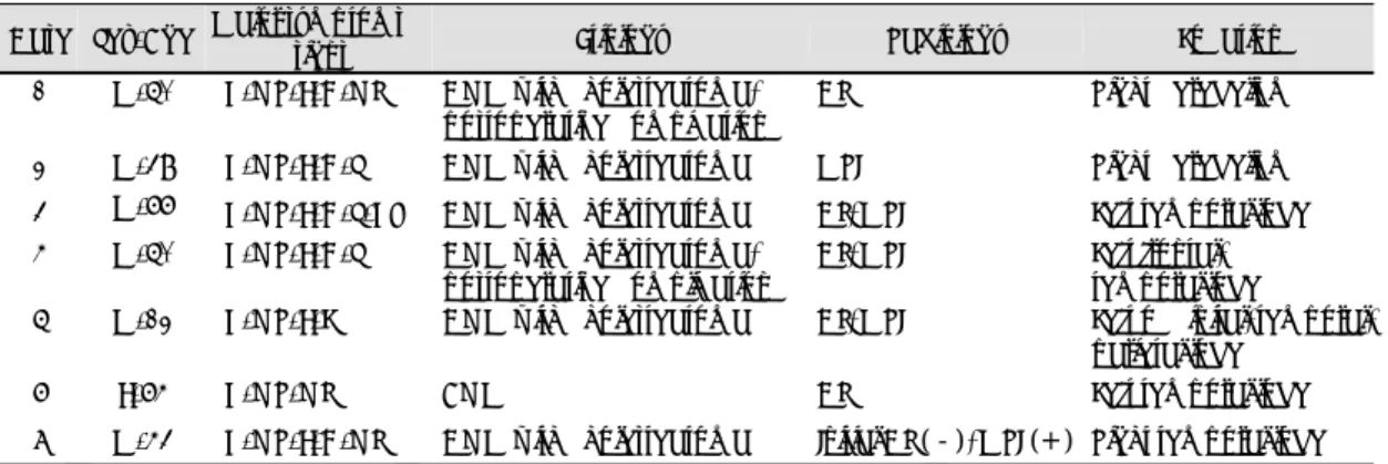
Table 1. Summary of 7 patients with otogenic brain abscess Case Sex/Age Major symptoms &
signs Etiology Radiology Location
1 M/50 H/OR/F/D/OT COM with cholesteatoma, postoperative compication
CT Right cerebellum
2 M/38 H/OR/F/D/S COM with cholesteatoma MRI Right cerebellum 3 M/66 H/OR/F/D/S/HP COM with cholesteatoma CT, MRI Left temporal lobe 4 M/50 H/OR/F/D/S COM with cholesteatoma,
postoperative complication
CT, MRI Left frontal, temporal lobe 5 M/12 H/OR/F/L COM with cholesteatoma CT, MRI Left occipital, temporal,
parietal lobe
6 F/64 H/OR/OT AOM CT Left temporal lobe
7 M/43 H/OR/F/D/OT COM with cholesteatoma Initial CT (-), MRI (+) Right temporal lobe AOM:acute otitis media, COM:chronic otitis media, H/OR/F/D/L/S/HP/OT:headache/otorrhea/fever/drowsy/
lethargy/seizure/hemiparesis/otalgia
내과적 처치로는 뇌부종을 감소시키기 위한 스테로이 드와 항경련제, 그리고 광범위 항생제를 같이 사용하였 다. 항생제는 세균배양 및 감수성 검사결과에 준하여 사 용하였으나 결과를 알기 전이나 결과에서 음성으로 나 온 경우는 여러 가지 항생제를 복합적으로 사용하였다 (Table 2). 수술 후 4~6주간 지속적으로 정맥 항생제 를 사용하였다.
결 과
환자들이 호소하는 가장 흔한 특이증상은 두통이었다.
6명의 환자에서 내원 전 7일 이상의 두통을 호소하였고, 환측의 측두골에서 시작된 깨질 듯한 통증이 시간이 지 날수록 악화되고 전체적으로 퍼지는 경향을 보였다. 그 리고 내원 당시에 모두 환측의 이루와 오심 구토를 동반 하였다. 또 다른 증상으로는 열이 6명, 의식 저하 소견 을 보인 환자가 6명, 이통이 3명, 경련이 3명, 그리고 좌 측 반신마비가 1명이었다(Table 3).
4명(57.1%)의 환자는 만성 진주종성 중이염으로 인 해 이성 뇌 농양이 발생하였고, 이들은 모두 유양돌기내 에 진주종의 침범이 심하였으나 진주종이 직접 뇌 실질 을 침범한 경우는 없었다. 1명(14.3%)은 외상성 고막 천공으로 생긴 급성 중이염이 원인이었고, 나머지 2명 (28.6%)은 만성 진주종성 중이염에 대한 유양돌기절제 술 이후 의인성으로 발생한 경우였다(Table 4).
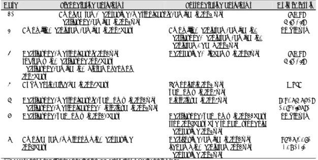
뇌 농양의 방향은 모두 이환된 중이염의 동측에서 발 생하였고, 위치는 측두엽과 측두엽을 포함한 주변 뇌로 Table 2. Pre and postoperative I.V antibiotics treatment of otogenic brain abscess
Case Preoperative antibiotics Postoperative antibiotics Culture result 1* Ampibactam, cefotaxime, metronidazole, amikacin for 2 wks
ceftriaxone, amikacin for 2wks
Proteus mirabilis 2* Ampicillin, cefolatam, amikacin for 4days Ampicillin, cefolatam, amikacin/
ceftriaxone, cefolatam, amikacin/
cefolatam/ each for 2wks
no growth
3* Ceftriaxone, metronidazole for 1wks tobramycin, ceftriaxone for 3days ceftriaxone, amikacin, chloramphenicol for 4days
Cefotaxime, clindamycin for 6wks Proteus mirabilis
4* Augmentin, arbekacin for 3days Teicoplanin for 2 wks vancomycin for 4wks
MRSA
5* Ceftriaxone, metronidazole, vancomycin for 1wks ceftriaxone, metronidazone, ciprofloxacin for 2wks
Ciprofloxacin for 4wks Pseudomonas aerugiinosa 6* Ceftriaxone, vancomycin for 10days Ceftriaxone, vancomycin for 11days
stop for 5days due to pancytopenia cefotaxime for 2wks
no growth
7* Ampibactam, micronomicin, cefotaxime for 5days
Ceftizoxime, amikacin for 1wks isepamicin, cefolatam for 1wks cefotaxime for 2wks
Burkholderia cepacia
*:He did not undergone operation, I.V antibiotics were only used Table 3. Symptoms and signs of the otogenic brain abs-
cess
Symptoms and signs Case (n)* (%)
Otorrhea 7 100
Headache 7 100
Nausea/Vomiting 7 100
Mental disturbance 6 085.7
Fever 6 086.7
Seizure 3 042.8
Otalgia 3 042.8
Left hemiparesis 1 014.3 Total (n) 7
*:Scores are not mutually exclusive
Table 4. Etiology of otogenic brain abscess
Etiology Case (n) (%) Chronic otitis media with
cholesteatoma 4 57.1
Tympanomastoid operation
(iatrogenic) 2 28.6
Acute otitis media 1 14.3 Total (n) 7
발생한 경우가 5명(71.4%)이고, 소뇌에만 국한되어 발 생한 예가 2명(28.6%)이었다(Table 5).
2명(28.6%)은 뇌 농양 외에 다른 문제가 없었고, 뇌 수막염이 같이 동반되어 발생한 예가 3명(42.8%), 경 막하 농흉을 가진 예가 2명(28.6%), 그리고 경막외 농 양이 1명(14.3%) 있었다(Table 6).
진단 방법으로는 5명에서 뇌 전산화 단층촬영과 뇌 자 기공명영상을 같이 사용하였고, 1명은 뇌 전산화 단층촬 영, 다른 1명은 뇌 자기공명영상만을 사용하였다. 그 중 1명에서는 초기의 뇌 전산화 단층촬영에는 뇌 농양이 발 견되지 않아 뇌수막염으로 진단 받았다가 5일 후 실시 한 뇌 자기공명영상에서 뇌농양이 발견되었다(Fig. 1).
5명에서 유양돌기절제술을 시행하였고, 추가적으로 경 미로 농양 절제술 1명, 개두술 2명, 그리고 두개절제술 을 2명에서 실시하였다. 유양돌기절제술 이후 생긴 뇌 농 Table 5. Location of otogenic brain abscess
Location Case (n)* (%) Temporal lobe only 3 42.8
Cerebellum 2 28.6
Temporal lobe+frontal lobe 1 14.3 Temporal lobe+parietal lobe
Temporal lobe+occipital lobe 1 14.3
Total (n) 7 100
*:Scores are not mutually exclusive
Table 6. Combined problems with otogenic brain abs- cess
Additional pathology Case (n)* (%)
Meningitis 3 42.8
Subdural empyema 2 28.6 Epidural abscess 1 14.3
None 2 28.6
Total (n) 7
*:Scores are not mutually exclusive
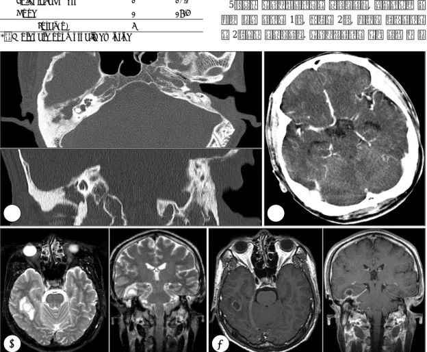
Fig. 1. Imaging study in a case of brain abscess (case 7). A:Temporal bone CT;Total haziness in right mastoid cavity and tegmen erosion are seen, B:Brain CT (enhance);There is no abnormal enhancement in brain parenchyma, C:Brain MRI, T2-weighted image;Irregular high signal lesion in right temporal lobe is seen, D:Brain MRI, T1-weighted contrast image;Low signal lesion with thin rim enhancement in right temporal lobe is seen.
A A A
A BBBB
CC
CC DD DD
양에서 1명은 두개절제술을, 다른 1명은 수술적 치료없 이 4주간 정맥 항생제만을 사용하였다(Table 7).
세균 배양검사 결과 Proteus 2명, Staphylococcus(MR- SA) 1명, Pseudomonas 1명, Burkholderia 1명, 그리고 배양되지 않은 경우가 2명이었다.
본원에서 진단되어 치료받은 환자들은 전례에서 완치 되었고, 1명을 제외하고는 후유증 없이 퇴원하였다. 후 유장애가 남은 환자는 두통이 시작되고 나서 뇌농양으로 진단되기까지 2주가 늦어졌던 경우로 지역병원에서 항 생제 치료를 받았으나 의식저하가 악화되고 반신마비가 나타나서 본원으로 전원되어 수술적 치료를 받은 예였 다. 뇌농양은 완치되었으나 반신마비는 회복되지 않았다.
고 찰
지난 50년간 항생제의 사용으로 이성 뇌 농양의 발생 빈도는 현저히 감소하였고, 사망률도 감소하였으나 아직 까지 1년에 1~2명 정도 계속해서 발생이 보고 되고 있 다.4) 동아대학교 병원에서 이번 연구기간 동안 만성 중 이염으로 유양돌기절제술을 시행 받은 환자는 4319명 이었고, 그 중에서 진주종성 중이염 환자는 857명으로 만성 중이염 환자의 이성 뇌농양 발생률은 0.16%, 진주 종에 의한 발생률은 0.78%이다. 항생제 사용 이전의 시 대에 뇌 농양으로 인한 사망률은 80% 정도였으나 최근 20년 동안 사망률은 10% 정도로 낮아졌다고 Sennaro-
glu 등이 보고하였다.4)7)8) 본 연구에서는 전례에서 완치 되어 사망한 예는 없었으나 신경학적 후유증을 남긴 경우 는 1예(14.3%)가 있었다.
이성 뇌 농양 환자의 증상은 농양 부위와 중이염의 병 리 소견에 따라 다를 수 있으나 가장 흔한 초기 증상은 두통으로 알려져 있다.9) 다른 증상으로는 이루, 이통, 경 련, 열, 그리고 뇌압 상승으로 인한 오심, 구토 등이 있을 수 있으며 심할 경우 의식 장애가 발생하기도 한다.5)9) 1예를 제외한 모든 예에서 7일 이상의 두통을 호소하였 으며 모두 오심과 구토를 동반하였고, 의식장애를 동반 한 경우도 6예(85.7%)나 있었다. 뇌 전산화 단층촬영 이 1970년대 후반부터 도입되어 이성 뇌 농양의 조기 진 단에 매우 유용하게 사용되었고, Sennaroglu 등4)은 사 망률이 10%정도로 낮아진 데는 뇌 전산화 단층촬영의 역할이 있었음을 강조하였다. 최근에는 뇌 자기공명영상 이 진단에 도움이 되고 있는데 급성 두개 내 감염을 평 가하는데 뇌 전산화 단층촬영보다 민감도가 더 높고, 만 성 출혈과도 감별이 가능하며, 농양에 인접한 뇌실질의 부종 또는 경색까지 확인이 가능하여 전산화 단층촬영 보다 조기에 뇌농양을 진단할 수 있게 되었다. 자기공명 영상에서 뇌농양은 T1 강조영상에서 뇌 실질보다는 저 음영으로 뇌척수 액 보다는 약간 고 음영으로 보이고, T2 강조영상에서는 뇌척수액과 뇌실질보다 고 음영으로 관
찰된다.10)11) 이 연구의 한 증례 중 두통, 고열을 주소로
응급실에 내원하여 뇌 전산화 단층촬영을 하였으나 특 Table 7. Summary of hospital management
Case Surgical management Medical management
1 none Antibiotics 4weeks
2 SM with T0
Abscess excision (translabyrinth approach)
Preoperative antibiotics 4 days postoperative antibiotics 6weeks 3 SM with T0
Craniectomy, left temporal 2day later, revision ICH removal
Preoperative antibiotics 2weeks postoperative antibiotics 6weeks 4 Craniectomy
right frontal, temporal, parietal
Preoperative antibiotics 3days postoperative antibiotics 6weeks 5 Craniectomy
left temporal, occipital 1week later, SM with T0
Preoperative antibiotics 3weeks postoperative antibiotics 4weeks 6 SM with T1
Craniotomy, left temporal
Preoperative antibiotics 10days postoperative antibiotics 4weeks 7 CWD with T0
Craniotomy, right temporal
Preoperative antibiotics 5days postoperative antibiotics 4weeks
SM:simple mastoidectomy, CWD:canal wall down mastoidectomy, T0:Tympanization, T1:tympanoplasty type 1, ICH:intracranial hemorrhage
이 소견은 없었고, 뇌척수 액 검사에서 세균성 뇌수막염 으로 진단되어 세균성 뇌수막염에 대한 정맥성 항생제 치 료를 하여 의식회복 등 증상의 호전이 있었으나 두통이 지속되고 이루가 심해져 촬영한 뇌 자기공명영상에서 우 측 측두엽에 농양이 발견되어 유양돌기절제술과 개두술 및 배농을 응급으로 시행한 예가 있었다. 이 환자는 수 술 36일 후 완치되어 합병증 없이 퇴원하였다. 이 예는 급・만성 중이염 환자에서 지속적인 화농성 이루와 두통 을 호소하는 경우 자기공명영상을 조기에 시행하여 두 개 내 합병증 유무를 반드시 확인해야 한다는 것을 환 기시켰다.
이성 뇌 농양의 발생 부위는 크게 측두엽 농양과 소뇌 농양으로 구분 할 수 있으며 Murthy 등12)은 소뇌가 측 두엽보다 4배 더 흔하게 발생한다고 보고하였으나 Sen- naroglu 등4)은 측두엽 농양이 54%, 소뇌 농양이 44%, 그리고 2%에서 양측성으로 발생하였다고 보고하였다.
또한 Kaplan13)에 의하면 이성 뇌 농양의 4~15%는 다 발성임을 주의해야 한다고 하였다. 본 연구에서는 3예는 측두엽 농양, 2예는 측두엽을 포함한 다발성 농양이었고, 2예만 소뇌에 국한되어 발생하여 측두엽에 더 흔하게 나 타났다.
이성 뇌 농양의 발생 기전은 진주종이나 골염에 의한 골 조직의 파괴, 기존의 해부학적인 통로, 혈전성 정맥염, 그 리고 혈행성 전파 등을 통해 생긴다고 알려져 있다.5)10) 본 증례들 중 만성 중이염에 의한 4예뿐 아니라 유양돌 기절제술 후 생긴 2예 모두 진주종성 중이염이었다. 수 술 소견상 진주종에 의한 골 파괴 소견은 있으나 진주종 이 직접적으로 뇌 경질막을 뚫고 침범한 소견은 관찰되 지 않았다. 따라서 진주종 자체가 이성 뇌농양의 직접적 인 원인이라기 보다 염증이 확산하는데 통로를 만들어 주는 역할을 하는 것으로 생각된다.
뇌 농양을 일으키는 균주는 혐기성을 포함하는 혼합 감염의 형태가 가장 흔하다.5) 호기성균으로 Group A streptococcus, Streptococcus pneumoniae가 흔하고, 혐 기성 균주로는 Bacterioides fragilis가 대표적이며 최근 Pseudomonas aeruginosa, Proteus mirabilis와 같은 그 람음성세균의 감염이 증가 추세에 있다고 보고되고 있 다.4)5) 그리고 Sennaroglu 등4)은 세균배양검사의 음성
률이 39%라고 보고하였는데, 이것은 항생제 사용의 증 가 때문이라고 하였다. 본 연구에서는 술 전에 실시한 세 균배양 검사에서 Proteus 2예, Staphylococcus(MRSA) 1예, Pseudomonas 1예, Burkholderia 1예 등 다양한 균 주가 나와서 이성 뇌농양과 특이 균주와의 관련성은 적 었다. 그리고 2예에서는 배양되지 않아서 28.6%의 음성 률을 보였다.
이성 뇌 농양의 조기 수술적 치료는 논란의 여지가 있 다. Kulali 등14)은 개두술과 농양 절제술을 선호하였다.
그러나 이 치료방법은 뇌 조직에 손상을 줄 수 있으며 특히 다발성 농양일 때 더 위험하다. Singh and Maha- raj15) and Mathews and Marus16)들에 의하면 신경외 과적으로 뇌 농양의 우선 처치 후 유양돌기절제술을 동 시에 시행하여 원발 병소를 치료하였고, Murthy 등12) 은 일차적으로 신경외과적 배농을 시행하고, 이후에 유 양돌기절제술을 하였다. 본 병원에서는 4예는 동시에 시 행하였고, 1예는 신경외과적 배농을 먼저 시행한 후 유 양돌기절제술을 시행하였다. 유양돌기절제술 후 생긴 2 예에서 1예는 개두술 및 배농을 시행하였고, 다른 1예 는 4주간 정맥 항생제만을 사용하였다. 수술 이후에도 전산화 단층촬영, 자기공명영상 소견에 따라 4~6 주간 정맥 항생제를 계속 사용하였다. 정맥 항생제 부작용이 있었던 예에서는 입원 치료 도중 전 혈구 감소증이 있었 으나 모든 정맥 항생제를 중단하고 5일이 지나자 다시 회 복되어 혈구감소 부작용이 없는 cefotaxime만 남은 기 간 사용하여 완치되었다. 진단이 늦었던 예로서 1예는 첫 내원 당시 우측 반신마비 증상을 보였는데, 두통이 13일 간 있다가 갑자기 의식의 변화가 생겨 지역병원 응급실 을 방문하여 시행한 뇌 전산화 단층촬영, 뇌자기공명영 상에서 이성 뇌농양으로 진단받고 2주간 정맥 항생제를 사용하였으나 추적 관찰 전산화 단층촬영에서 뇌농양이 진행하는 소견을 보이고 우측 반신마비 증상을 보여 본 원으로 전원된 예로써 환자는 수술 후 35일 만에 회복 되어 퇴원하였으나 영구적인 반신마비의 신경학적 장애 를 남겼다. 이 한 예를 제외한 전 예에서 모두 신경학적 합병증 없이 모두 회복되어 퇴원하였고, 외래 관찰 기간 중에도 새로운 증상은 없었으며 만성 중이염도 완치 되 었다.
결 론
이성 뇌농양의 예후에 영향을 미치는 가장 중요한 인 자는 조기진단이었으며 급・만성 중이염 환자에서 지속 적인 화농성 이루와 두통을 호소하는 경우 자기공명영상 을 조기에 시행하여 두개 내 합병증 유무를 반드시 확 인해야 하겠다.
조기진단과 적절한 수술적 처치, 적극적인 항생제 치 료를 한다면 뇌농양에 의한 합병증을 줄일 수 있고, 완 치율을 높일 수 있을 것이다.
중심 단어:뇌농양・중이염・자기공명영상.
REFERENCES
1) Bradley PJ, Mannino KP, Shaw MDM. Brain abscess se- condary to otitis media. J Laryngol Otol 1984;98:1185-91.
2) Stuart EA, O’Brien FH, McNally WJ. Some observations in brain abscesses. Arch Otolaryngol 1955;61:212-6.
3) Kangsanarak J, Navacharoen N, Fooanant S, Ruckphaopunt K. Intracranial complications of suppurative otitis media:
13 years’ experience. Am J Otol 1995;16:104-9.
4) Sennaroglu L, Sozeri B. Otogenic brain abscess: review of 41 cases. Otolaryngol Head & Neck Surg 2000;123:751-5.
5) Chung JW, SEO YI, HA SY, Yoon TH, LEE KS. Otogenic
brain abscess. Korean J Audio 1997;1:196-202.
6) Brand B, Caparosa RJ, Lubic LG. Otorhinological brain abscess therapy-past and present. Laryngoscope 1984;94:
483-7.
7) Ballantine HT Jr, White JC. Brain abscess; influence of the antibiotics on therapy and mortality. N Engl J Med 1953;248:14-9.
8) Proctor CA. Intracranial complication of otic origin. Lary- ngoscope 1996;78:288-308.
9) Kim CS, Kim YJ, Kim HK. Clinical study on the Otogenic Brain Abscess. Korean J Otolaryngol 1986;29:763-71.
10) Samuel J, Fernandes CM, Steinberg JL. Intracranial oto- genic complications: a persisting problem. Laryngoscope 1986;96:272-8.
11) Zimmerman RA, Bilaniuk LR, Sze G. Intracranial infe- ction. In: BrantZawadzki M, Norman D. Magnetic resonance imaging of the central nervous system. New York; Raven Press;1987. p.235-57.
12) Murthy PS, Sukumar R, Hazarika P, Rao AD, Mukulchand, Raja A. Otogenic brain abscess in childhood. Int J Pediatr Otorhinolaryngol 1991;22:9-17.
13) Kaplan RJ. Neurological complications of infections of head and neck. Otolaryngol Clin North Am 1976;9:729-49.
14) Kulali A. Ozatik N, Topcu I. Otogenic intracranial abs- cesses. Acta Neurochir(Wien);1990. p.140-6.
15) Singh B, Maharaj TJ. Radical mastoidectomy: its place in otitic intracranial complications. J Laryngol Otol 1993;
107:1113-8.
16) Mathews TJ, Marus G. Otogenic intradural complications (a review of 37 patients). J Laryngol Otol 1988;102:121-4.