Introduction
Implant restorations are commonly used for func- tional and esthetic rehabilitation in missing areas instead of conventional fixed bridges. Despite the high long term success rate of implant prosthesis, implant restorations in the esthetic zone could make some trouble concerning aesthetics and the surgical technique to restore the favorable esthetics is very dif- ficult.
Clinicians often meet a non-esthetic implant resto- ration in the esthetic zones, and need to be aware of and be capable of handling complications that can lead to the unaesthetic implant restorations. Com-
plications can range from prosthetic problems stem- ming from a misalignment of implants to biological problems such as failure of guided bone regeneration (GBR), deficiency of soft tissue and peri-implantitis.
1Lack of an attached gingiva can jeopardize the maintenance of periodontal health and the long term success of dental implants.
2Many procedures could be used to increase the width and thickness of the attached gingiva. These procedures include an inlay graft, onlay graft, vascularized interpositional perios- teal connective tissue graft (VIP-CTG), palatal roll and palatal rotating flap and so on.
3Peri-implantitis or a misplaced implant without considering the biologic width can cause a loss of
*Correspondence to: Ju-Youn Lee
Associate Professor, Department of Periodontology, School of Dentistry, Pusan National University, 20 Geumo-ro, Mulgeum-eup, Yangsan, 626-787, Republic of Korea
Tel: +82-55-360-5202, Fax: +82-55-360-5194, E-mail: [email protected] Received: March 10, 2015/Last Revision: May 7, 2015/Accepted: May 31, 2015
Multidisciplinary approach of the problem of unaesthetic implants in the maxillary anterior dentition
Ji-Young Joo
1, Jeomil Choi
2, Ju-Youn Lee
2*
1
Department of Periodontology, Pusan National University Dental Hospital, Yangsan, Republic of Korea
2
Department of Periodontology, School of Dentistry, Pusan National University, Yangsan, Republic of Korea
Periodontal tissue destroyed by inflammation is difficult to achieve regeneration of the tissue and esthetic restorations only by surgical methods. In particular, improvement of esthetics is more difficult if the problem is related to the implant. A 23 year old woman suffered from unesthetic anterior implant prosthesis. According to her dental history, a repeated bone graft and soft tissue graft failed at a local dental clinic. It was needed to resolve the inflammation and to improve the esthetics. A free gingival graft and ridge augmentation accompanied by guided bone regeneration and a vascularized interpositional periosteal connective tissue graft was performed. Instead of implant prosthesis, a conventional fixed bridge was adopted for better esthetic result. The patient was satisfied with the esthetic conventional fixed prosthesis. This case report introduces esthetic rehabilitation of unesthetic implant prosthetics in the maxillary anterior dentition by a combination of surgical and prosthetic approaches. (J Dent Rehabil Appl Sci 2015;31(2):126-33)
Key words: alveolar ridge augmentation; implant supported dental prosthesis; peri-implantitis
Copyright© 2015 The Korean Academy of Stomatognathic Function and Occlusion.
It is identical to Creative Commons Non-Commercial License.
cc
ISSN 2233-4084
bone around the implant.
4A resective or regenerative surgical treatment could be used depending on the morphology and extent of bone destruction.
5If the aim of the treatment is to regenerate the surround- ing bone, the contaminated implant surface needs to be detoxified in advance and a GBR technique using the barrier membrane and bone substitute could be a more promising and predictable technique.
6Both resorbable and non-resorbable membranes could be useful for GBR and both have pros and cons re- spectively. In esthetic sensitive dentition, a resorbable membrane is preferred to avoid additional surgery and decrease the risk of unaesthetic scar formation.
The esthetic problems due to a misplaced implant are often difficult to resolve using only a single ap- proach such as prosthetic or surgical approach. The aim of this study was to present a case of the esthet- ic rehabilitation of non-esthetic implant prosthetics in the maxillary anterior dentition by a combination of surgical and prosthetic approaches.
Case
A 23 year old woman with the chief complaint of non-aesthetics of the maxillary right lateral and central incisors visited the department of periodon- tology, Pusan national university dental hospital. The patient had no remarkable systemic diseases affecting her dental condition. Her dental history showed that her maxillary right lateral and central incisors had been extracted due to root fracture from an accident when she was 11-year-old. She had a removable pro- visional prosthesis for a long time because she was growing. Two implants were installed at the maxillary right lateral and central incisors with GBR simultane- ously when she was 19-year-old. But gingival bleed- ing and swelling had occurred repeatedly and was followed by alveolar bone resorption and gingival re- cession. Therefore, GBR and a soft tissue graft were performed at a local dental clinic 4 times, but the problem concerning esthetics was getting worse and worse.
The maxillary central and lateral incisors had long clinical crowns in comparison with the adjacent natu- ral teeth. The target teeth showed no attached gingi-
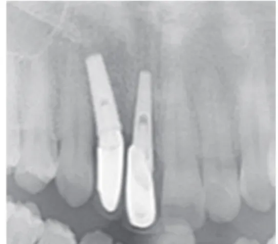
va, deep probing depth and bleeding on probing, and a scar formed in the labial gingiva due to the repeat- ed surgical history (Fig. 1). On the other hand, these implants were non-mobile. A radiograph showed that the alveolar bone around the implant was resorbed and the paths of the two implants were poor (Fig. 2).
Accordingly, it was diagnosed as peri-implantitis and proposed to require multidisciplinary treatment to resolve the inflammation and to improve the esthet- ics. First, the resolution of inflammation should be performed first followed by ridge augmentation with GBR and the fabrication of a new esthetic prosthesis.
Fig. 1. Initial clinical presentation. Note the long clinical crowns in comparison with the adjacent natural teeth and the inflamed gingiva and scar around the maxillary right lateral and central incisor implants. The area showed a lack of attached gingiva, gingival redness and bleeding tendency.
Fig. 2. Initial radiograph showing that the alveolar bone
around implants was resorbed and the paths of the two
implants were poor.
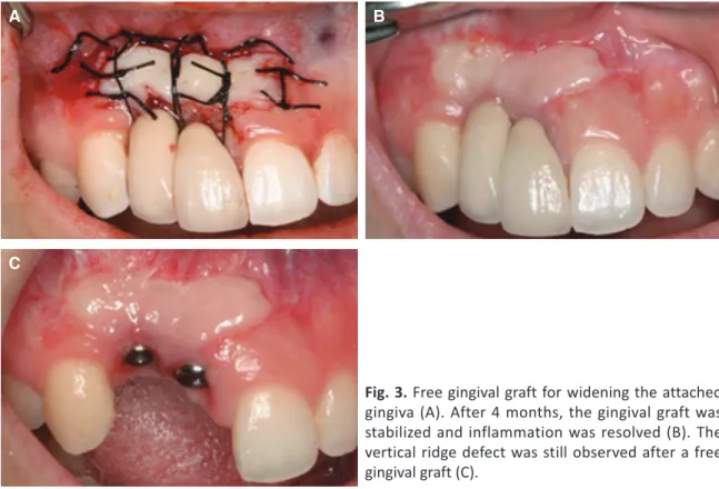
A free gingival graft (FGG) was performed to increase the amount of attached gingiva (Fig. 3A).
After 4 months, the gingival graft was stabilized and the signs of inflammation disappeared (Fig. 3B).
Improvement in the width and thickness of the soft tissue would be helpful for successful advanced ridge augmentation surgery (Fig. 3C).
The patient requested not only healthy soft tissue but also esthetic anterior restorations. Ridge augmen- tation with GBR had to be preceded in advance to achieve an aesthetic prosthesis and VIP-CTG was planned simultaneously for better soft tissue closure.
Ridge augmentation by GBR and VIP-CTG was per- formed 4 months after FGG. After administering the appropriate local anesthesia in the maxillary anterior area, horizontal sulcular incision and midcrestal inci- sion were made from the maxillary left central incisor to the maxillary right canine. Two vertical incisions were made at the distal of the left central incisor and distal of the right canine. After flap elevation, granulation tissues over the implant surface and the bony defect were removed carefully using a titanium curette (Osung MND, Seoul, Korea) (Fig. 4A). The
implant surfaces were cleaned carefully with cot- ton pellets soaked sequentially in normal saline and 0.12% chlorhexidine (CHX; Hexamedine
®, Bukwang pharm, Seoul, Korea) for detoxification of implant surfaces. They were then washed several times with normal saline and 0.12% CHX. After thorough de- bridement and implant surface detoxification, the bony defect was augmented with a bovine bone min- eral (Bio-Oss, Geistlich, Wolhusen, Swiss) and non- cross linked bioresorbable membrane (Bio-Gide, Geistlich) (Fig. 4B). VIP-CTG was prepared at the ipsilateral palatal area and the graft was anchored at the periosteum of the buccal flap by suturing with absorbable suture material (Vicryl, Ethicon, Johnson and Johnson, New Brunswick, NJ, USA) in advance (Fig. 4C). The overlying flap was advanced coronally and finally sutured to achieve primary closure (Fig.
4D). The patient was administered systemic antibi- otics (amoxicillin 250mg and metronidazole 250mg three times a day) for one week before surgery and two weeks after surgery. The patient was instructed to avoid mechanical cleaning in the surgical area and to rinse with 0.12% CHX solution twice a day for 14
Fig. 3. Free gingival graft for widening the attached gingiva (A). After 4 months, the gingival graft was stabilized and inflammation was resolved (B). The vertical ridge defect was still observed after a free gingival graft (C).
A B
C
days. All the sutures were removed at 2 weeks after surgery and healing was uneventful. The patient was recalled at 1 month, 3 months after surgery and the clinical follow-up showed improvement in the ridge deficiency (Fig. 5A-D).
Three kinds of prosthetic treatment options were given to the patient at 3 months after ridge augmen- tation surgery: (1) implant bridge supported by two implants, (2) submergence of the maxillary right lateral incisor implant and the implant cantilever bridge supported by a maxillary right central incisor implant, and (3) submergence of two implants and 4 unit fixed bridge from maxillary right canine and left central incisor. The patient did not adhere to the implant prosthesis and requested the most esthetic restorations. Therefore, Option 3 was chosen be- cause the prosthesis would be more esthetic. A fixed provisional restoration was placed at 3 months after
surgery. After a 6-month healing period, the final fixed prosthetic reconstructions were placed (Fig.
6A-C). The final prosthesis was an all ceramic res- toration (IPS Empress 2, Ivoclar Vivadent, Schaan, Liechtenstein) with a natural appearance in harmony with the adjacent teeth. The gingival tissue around the restoration was healthy and the restoration was esthetic.
Discussion
Dimensional alteration of the edentulous ridge occurs due to the remodeling of the buccal and lin- gual walls after extraction. This remodeling process also causes considerable buccal crestal bone loss and problem of esthetics of implant prosthesis.
7If it takes a long time to place dental implant after extraction, the amount of ridge alteration would be increased.
8In these cases, the timing of tooth extrac- Fig. 4. Ridge augmentation with guided bone regeneration and a vascularized interpositional periosteal connective tissue graft. Note the bony defect around the implants (A). Granulation tissue was removed and the implant surface was detoxified. Bovine bone minerals (Bio-Oss, Geistlich) and non-cross linked bioresorbable membrane (Bio- Gide, Geistlich) was applied (B). Vascularized interpositional periosteal connective tissue graft was rotated over the membrane (C). Overlying flap was sutured for achieving primary closure (D).
A B
C D
Fig. 5. Clinical follow-up showing the improvement of the ridge deficiency. At stich-out (A, B) and after 1 month (C, D).
A B
C D
Fig. 6. Final restoration after 6 months of ridge augmentation. Note the natural-looking prostheses in harmony with the adjacent teeth and periodontium reconstructed (A). Occlusal view showed well-reconstructed ridge contour (B).
Significant improvements in function, esthetics, and periodontal health were achieved (C).
A B
C
tion and implant placement should be considered carefully. The extraction site must be preserved as much as possible if you expect a lot of the resorp- tion of alveolar ridge. Forced eruption,
9socket preservation
9and root submerge technique
10could be used to preserve the alveolar ridge and develop a future implant site.
According to the patient’s dental history, a re- peated bone graft and soft tissue graft surgery failed.
Therefore, the attached gingiva was lost and the im- plants were surrounded by non-keratinized inflamed tissue. The presence of keratinized mucosal tissue collar around the implants plays a critical role for the longevity of the implant by providing a soft tissue seal that can cope with the bacterial challenge.
2FGG could be used to correct the soft tissue defects and provide the optimal peri-implant health to increase the long-term prognosis of the implant reconstruc- tion.
11In this case, after FGG, the gingival inflamma- tion was resolved and an attachment of the implant and the soft tissue was promoted.
A peri-implant bony defect could be restored with a regenerative treatment via GBR with membrane and bone substitute. First, decontamination of the implant surfaces is most important but very difficult to achieve effectively.
12In particular, the bacteria and plaque biofilm is difficult to remove completely in the rough surface of the implant surface.
13Many studies have suggested the various implant surface decontamination methods: air-abrasive instrument with glycine,
14irradiation with an Er:YAG laser,
13sandblasting,
14washing with 0.12% CHX and sa- line,
15using citric acid, 10% hydrogen peroxide or tetracycline hydrochloride, and cleaning with a cot- ton pellet soaked in normal saline.
16As no method is superior, simple and effective methods were adopted, i.e., washing with 0.12% CHX and normal saline and cleaning with cotton pellets soaked in normal saline and 0.12% CHX.
GBR using a barrier membrane and bone graft ma- terial showed more favorable clinical and histologi- cal results compared to a bone graft alone.
17,18Both resorbable and non-resorbable membranes could be useful for peri-implant bone regeneration and both have advantages respectively in special clinical situa-
tions. In this case, a resorbable membrane was used to avoid additional surgery and decrease the risk of unaesthetic scar formation because the maxillary an- terior dentition is esthetic sensitive area.
For successful ridge augmentation via GBR, suf- ficient soft tissue volume and primary wound closure were mandatory. A vascularized flap rather than free flap was recommended for reconstruction of the large ridge defect because of preservation of suffi- cient blood supply. VIP-CTG
19could be used to cor- rect an aesthetic deficiency and establish stable new peri-implant soft tissue contours as this technique allows the reconstruction of a large soft tissue defi- ciency, with little constriction postoperatively. More- over, the VIP-CTG would be useful for achieving the primary soft tissue coverage of the GBR site. In this case, we could reconstruct the large ridge defect using GBR technique combined with VIP-CTG and could achieve the primary flap closure without mem- brane exposure thanks to VIP-CTG.
It could be an alternative to non- esthetic implant fixed prosthesis and simplify a difficult prosthetic management to rehabilitate with a conventional fixed bridge restoration with submergence of dental im- plants, so called “sleeping” implants.
20It is important not to overlook the fact that a conventional fixed bridge is a more predictable form of aesthetic pros- thesis.
Conclusion
Perfect aesthetics of implant fixed prosthesis is difficult to achieve in the esthetic zone. Moreover, it is more difficult to correct the serious problem of a non-esthetic implant prosthesis using only a surgical or prosthetic approach. Therefore, a multidisciplinary approach using the surgical and prosthetic methods could be useful to enhance the aesthetics of non- esthetic implant prosthesis.
Acknowledgements
This study was supported by Clinical Research
Grant from Pusan National University Dental Hos-
pital (2015).
Orcid
Ji-Young Joo http://orcid.org/0000-0002-4050-5797 Jeomil Choi http://orcid.org/0000-0002-7491-6711 Ju-Youn Lee http://orcid.org/0000-0002-0772-033X
References
1. Froum SJ, Rosen PS. A proposed classification for peri-implantitis. Int J Periodontics Restorative Dent 2012;32:533-40.
2. Wennström JL, Bengazi F, Lekholm U. The in- fluence of the masticatory mucosa on the peri- implant soft tissue condition. Clin Oral Implants Res 1994;5:1-8.
3. Le B, Burstein J. Esthetic grafting for small volume hard and soft tissue contour defects for implant site development. Implant Dent 2008;17:136-41.
4. Jovanovic SA. The management of peri-implant breakdown around functioning osseointegrated dental implants. J Periodontol 1993;64:1176-83.
5. Lindhe J, Meyle J; Group D of European Work- shop on Periodontology. Peri-implant disease: Con- sensus Report of the Sixth European Workshop on Periodontology. J Clin Periodontol 2008;35:282-5.
6. Heitz-Mayfield LJ, Mombelli A. The therapy of peri-implantitis: a systematic review. Int J Oral Maxillofac Implants 2014;29:325-45.
7. Araújo MG, Lindhe J. Dimensional ridge alterations following tooth extraction. An experimental study in the dog. J Clin Periodontol 2005;32:212-8.
8. Buser D, Martin W, Belser UC. Optimizing esthet- ics for implant restorations in the anterior maxilla:
anatomic and surgical considerations. Int J Oral Maxillofac Implants 2004;19:43-61.
9. Salama H, Salama M. The role of orthodontic extrusive remodeling in the enhancement of soft and hard tissue profiles prior to implant placement:
a systematic approach to the management of ex- traction site defects. Int J Periodontics Restorative Dent 1993;13:312-33.
10. Harper KA. Submerging an endodontically treated root to preserve the alveolar ridge under a bridge-a case report. Dent Update 2002;29:200-3.
11. Simons AM, Darany DG, Giordano JR. The use of free gingival grafts in the treatment of peri-implant soft tissue complications: clinical report. Implant Dent 1993;2:27-30.
12. Steinemann SG. Titanium-the material of choice?
Periodontol 2000 1998;17:7-21.
13. Kreisler M, Kohnen W, Christoffers AB, Götz H, Jansen B, Duschner H, d’Hoedt B. In vitro evalu- ation of the biocompatibility of contaminated implant surfaces treated with an Er:YAG laser and an air powder system. Clin Oral Implants Res 2005;16:36-43.
14. Singh G, O’Neal RB, Brennan WA, Strong SL, Horner JA, Van Dyke TE. Surgical treatment of in- duced peri-implantitis in the micro pig: clinical and histological analysis. J Periodontol 1993;64:984-9.
15. Wetzel AC, Vlassis J, Caffesse RG, Hämmerle CH, Lang NP. Attempts to obtain re-osseointegration following experimental peri-implantitis in dogs.
Clin Oral Implants Res 1999;10:111-9.
16. Froum SJ, Froum SH, Rosen PS. Successful man- agement of peri-implantitis with a regenerative ap- proach: a consecutive series of 51 treated implants with 3- to 7.5-year follow-up. Int J Periodontics Restorative Dent 2012;32:11-20.
17. Hürzeler MB, Quiñones CR, Morrison EC, Caffesse RG. Treatment of peri-implantitis using guided bone regeneration and bone grafts, alone or in combination, in beagle dogs. Part 1: Clinical find- ings and histologic observations. Int J Oral Maxil- lofac Implants 1995;10:474-84.
18. Hürzeler MB, Quiñones CR, Schüpback P, Morri- son EC, Caffesse RG. Treatment of peri-implantitis using guided bone regeneration and bone grafts, alone or in combination, in beagle dogs. Part 2:
Histologic findings. Int J Oral Maxillofac Implants 1997;12:168-75.
19. Sclar AG. The vascularized interpositional perios- teal-connective tissue (VIP-CT) flap. In: Sclar AG, editor. Soft tissue and esthetic considerations in im- plant therapy. Florida; Quintessence; 2003. p. 205- 21.
20. Poqqio CE, Salvato A. Implant repositioning for
esthetic reasons: a clinical report. J Prosthet Dent
2001;86:126-9.
*교신저자: 이주연
(626-770) 경상남도 양산시 물금읍 범어리 부산대학교 치의학전문대학원 치주과학교실 Tel: 055-360-5202|Fax: 055-360-5194|E-mail: [email protected] 접수일: 2015년 3월 10일|수정일: 2015년 5월 7일|채택일: 2015년 5월 31일
비심미적 임플란트의 심미성 회복을 위한 다각적 접근법
주지영
1, 최점일
2, 이주연
2*
1
부산대학교치과병원 치주과
2