Nontraumatic spinal epidural hematoma (SEH) is a rare clinical entity and the majority of these conditions are thought to result from rupture of the epidural vascu- lar network. Hemorrhagic lumbar synovial cysts (1) and a hematoma occurring from the ligamentum flavum (2) or from a lumbar facet joint (3) have been reported as rare types of epidural hematoma. We report here on the radiologic and surgical findings of a case of chronic non- traumatic SEH in a patient who had spondylolytic spondylolisthesis at the L4-5 level.
Case Report
A previously healthy 44-year-old woman was admit-
ted to our hospital with a 3-month history of lower back pain along with tingling and radicular pain in her left lower limb. These symptoms had progressively wors- ened and were exaggerated upon walking. The neuro- logic examination revealed weakness during both leg flexion and plantar flexion, and the sensory examination was normal. She had no history of coagulopathy, previ- ous lumbar operation or epidural puncture. The lateral radiograph (Fig. 1) and CT scan (Fig. 2A) showed spondylolysis (arrow) at the L4 vertebra with a mild de- gree of spondylolisthesis at the L4-5 level. CT (Fig. 2), and the MRI (Fig. 3A-C) enabled us to make the correct preoperative diagnosis. These modalities revealed a nodular, well-circumscribed mass in the posterior epidural space at the L4-5 level. The signal intensity was high, but not homogeneous on the T1-weighted im- age, and the signal intensity was high with a low-signal intensity rim on the T2-weighted image. An axial image showed an epidural mass that was continuous with the left vertebral foramen at the L4-5 level and the signal intensity of the mass was consistent with chronic
J Korean Radiol Soc 2006;55:501-504
─ 501 ─
Chronic Lumbar Epidural Hematoma in a Patient Suffering With Spondylolytic Spondylolisthesis
at the L4-5 Level: A Case Report1
Hyeon Seon Park, M.D., Sang-Ho Lee, M.D.2, Wei Chiang Lie, M.D., Jee Young Park, M.D., Sang Yeun Lee, M.D.3
1Departments of Diagnostic Radiology and 2Neurosurgery, Wooridul Spine Hospital
3Department of Diagnostic Radiology, Chuk Spine Hospital Received June 22, 2006 ; Accepted September 28, 2006
Address reprint requests to : Hyeon Seon Park, M.D., Department of Diagnostic Radiology, Wooridul Spine Hospital, 47-7 Chungdam-dong Gangnam-gu, Seoul 135-100, Korea.
Tel. 82-2-513-8000 Fax. 82-2-513-8175 E-mail: [email protected]
Nontraumatic spinal epidural hematoma (SEH) is a rare condition and the exact cause of the hemorrhage in SEH has never been established. However, there have been a few recent reports on some types of the epidural hematoma with a detectable origin of hemorrhage. We encountered a case of chronic SEH in a patient who had spondylolytic spondylolisthesis, which is also a rare condition to be associated with SEH. We report here on the radiologic findings of a case of chronic epidural hematoma in a patient who had spondylolytic spondylolisthesis at the L4-5 level, and we include a review of the related literatures.
Index words :Spondylolysis
Magnetic resonance (MR) Hematoma
Spinal cord
hematoma. The patient underwent the left total facetec- tomy with fusion of L4 and L5; the operation revealed a nodular encapsulated black fibrotic mass at the L4-5 lev- el that compressed the dural sac forward. The mass was strongly adhered to the dura and there was severe adhe- sion around the spondylolysis. The mass was complete- ly extirpated under microscopic magnification. The cap- sule of the mass was hard and elastic, and it was filled with dark red-gray solid and liquid materials.
Examination of the histological sections of the mass re-
vealed an organizing hematoma with infiltration by the surrounding fibrous granulation tissue (Fig. 4)
The postoperative course was uneventful; the patient experienced rapid and complete relief from the pain.
Discussion
Nontraumatic SEH is a rare clinical entity and it can occur at any level, but it is most frequently found at the cervical and the thoracic spines and rarely at the lum- bosacral level (4). Hematomas below the level of the conus medullaris are more likely to be chronic because the spinal roots appear to better tolerate pressure than can the spinal cord, and the subarachnoid space of the lumbar spinal canal is larger than that of the cervical or thoracic spine (5-7).
The cause of epidural hematoma in the lumbar spine is not clear. The majority of these conditions are thought to result from rupture of the epidural vascular network.
A hemorrhagic lumbar synovical cyst (1) and a hematoma occurring from the ligamentum flavum (2) or from a lumbar facet joint (3) were recently reported as rare types of epidural hematoma. Nagata et al (8) report- ed the case of an epidural hematoma associated with spondylolysis in a seventeen-year-old rugby player.
They suspect that the hemorrhage occurred from the epidural veins under the pars interarticularis at the L3 level because of recurrent minor traumatic episodes during ruby training.
In the present case, the radiologic and surgical find- ings clearly suggested a chronic epidural hematoma in a patient who has spondylolysis at the L4 level along with
Hyeon Seon Park, et al : Chronic Lumbar Epidural Hematoma in a Patient Suffering With Spondylolytic Spondylolisthesis at the L4-5 Level
─ 502 ─
A B
Fig. 2. A, B. The transaxial CT scan demonstrates bilateral spondylolysis (arrows in A) with a high attenuated mass-like lesion (ar- rows in B) in the left posterolateral epidural space at the L4-5 level.
Fig. 1. A 44-year-old female with back pain and left radiculopa- thy. The lateral radiograph shows spondylolysis (arrow) at the L4 vertebra with a mild degree of spondylolisthesis at the L4-5 level.
mild spondylolisthesis at the L4-5 level. Spondylolysis is a common condition and it usually occurs at the L5 vertebra; it is caused by a fatigue fracture in patients who have a lower lumbar index (a trapezoidal lumbar vertebra) (9). The cause of spondylolysis at a more cau- dal level in the lumbar spine is considered to be related to a history of trauma. For our patient, we suspect that chronic stress over time in the lumbar spine could have developed the spondylolysis of L4 along with spondyli- olisthesis at the L4-5 level, as well as the weakness of the walls of the epidural veins and the rupture of the
veins. MRI is the generally used modality to diagnosis epidural hematoma. There are signal intensity changes over time for hemorrhage on MRI after the onset, ac- cording to the oxidation and deoxidation of hemoglobin, the hemolysis of erythrocytes and their phagocytosis.
Our patient’s lesion showed high signal intensity that wasn’t homogeneous on the T1-weighted image, and high signal intensity with a low-signal intensity rim was seen on the T2-weighted image. These findings suggest- ed an old hemorrhage. The differential diagnosis of chronic SEH should include other benign spinal epidur- al masses such as synovial or ligamentum flavum cysts, and both of these are prone to intralesional hemorrhage and epidural cavernous angiomas (7).
Excision of the mass and spinal decompression with fusion is considered to be the definite treatment for symptomatic epidural hematoma that occurs from spondylolytic spondylolisthesis.
In conclusion, we report here on a rare case of chronic SEH that occurred in a patient who had spondylolytic spondylolisthesis at the L4-5 level.
References
1. Ramieri A, Domenicucci M, Seferi A, Paolini S, Petrozza V, Delfini R. Lumbar hemorrhagic synovial cysts: diagnosis, pathogenesis, and treatment. Report of 3 cases. Surg Neurol 2006;65:385-390 2. Yuceer N, Baskaya MK, Smith P, Willis BK. Hematoma of the liga-
mentum flavum in the lumbar spine: case report. Surg Neurol 2000;53:598-600
J Korean Radiol Soc 2006;55:501-504
─ 503 ─ Fig. 4. Examination of the histological sections of the mass shows an organizing hematoma with infiltration by the sur- rounding fibrous granulation tissue (Hematoxyline and eosin,
×400).
A B C
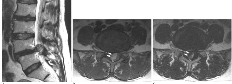
Fig. 3. A. The T2-weighted sagittal spin echo image shows a large oval mass (arrows) in the posterior epidural space of the L4-5 lev- el.
B, C. The T1 and T2-weighted axial spin echo images show an epidural mass (arrows) that’s continuous with the left vertebral fora- men at the L4-5 level. The signal intensity of the epidural mass is high on the T1-weighted axial image (B) and there is high signal intensity with a low-intensity rim on the T2-weighted image (C).
3. Nishida K, Iguchi T, Kurihara A, Doita M, Kasahara K, Yoshiya S.
Symptomatic hematoma of lumbar facet joint: joint apoplexy of the spine? Spine 2003;28:E206-208
4. Groen RJ, van Alphen HA. Operative treatment of spontaneous spinal epidural hematomas: a study of the factors determining postoperative outcome. Neurosurgery 1996;39:494-508
5. Boyd HR, Pear BL. Chronic spontaneous spinal epidural hematoma. Report of two cases. J Neurosurg 1972;36:239-342 6. Nakagami W, Yokota S, Ohishi Y, Ueda H, Takahashi Y, Sakuma
M, et al. Chronic spontaneous lumbar spinal epidural hematoma.
Spine 1992;17:1509-1511
7. Riffaud L, Morandi X, Chabert E, Brassier G. Spontaneous chronic spinal epidural hematoma of the lumbar spine. J Neuroradiol 1999;26:64-67
8. Nagata K, Ariyoshi M, Ishibashi K, Hashimoto S, Inoue A. Chronic lumbar epidural hematoma in a patient who had spondylolysis at the third lumbar vertebra. Report of a rare case involving a seven- teen-year-old adolescent. J Bone Joint Surg Am 1998;80:1515-1520 9. Saraste H. The etiology of spondylolysis. A retrospective radi-
ographic study. Acta Orthop Scand 1985;56:253-255
Hyeon Seon Park, et al : Chronic Lumbar Epidural Hematoma in a Patient Suffering With Spondylolytic Spondylolisthesis at the L4-5 Level
─ 504 ─
대한영상의학회지 2006;55:501-504
협부 결손성 전방전위증에서 발생한 만성 요추 경막 외 혈종: 증례 보고1
1우리들병원 영상의학과
2우리들병원 신경외과
3척병원 영상의학과
박현선・이상호2・유위강・박지영・이상윤3
비외상성 척추 경막 외 혈종은 매우 드물며, 주원인은 경막외 정맥총의 파열로 생각되고 있으나 아직 정립되지 않았다. 최근에는 경막외 혈종의 원인을 알 수 있는 증례들이 드물게 보고되고 있으며, 저자들은 제 요추 4-5번에 협부 결손성 전방전위증을 가진 환자에서 발생한 만성 요추 경막 외 혈종 1예를 경험하여 보고하고자 한다.