서 론
원위 상완골 관상면 관절 골절은 소두와 활차의 골절을 동반할 수 있으며 1996년 McKee 등1)이 활차를 포함한 관상면 전단골절 을 처음으로 보고하면서 이에 대한 중요성이 연구되어 왔다. 기
존의 소두 골절의 경우, 하나의 큰 소두 골편을 가지는 1형 Hahn- Steinthal 골절, 관절 연골과 얇은 연골하 골절을 포함하는 2형의 Kocher-Lorenz 골절, 그리고 분쇄 골절인 3형으로 분류되었다.
관상면 전단골절은 소두 골절 분류의 4형 골절로서, 최근 Sabo 등2)의 생역학적 연구에 의하면, 소두만을 침범한 골절에서 골편 을 절제한 경우 의미있는 불안정성이 유발되지 않았으나 활차를 포함한 골절에서 골편을 절제한 경우는 심각한 불안정성이 나타 내어 임상적 중요성이 주목을 받고 있다. 이는 주관절의 안정성 에 활차가 중요한 역할을 하기 때문에 골절이 활차를 포함하는 경우 골절의 골편을 절제하기 보다 골절을 정복하여 내고정하는
Copyright © 2016 by The Korean Orthopaedic Association
“This is an Open Access article distributed under the terms of the Creative Commons Attribution Non-Commercial License (http://creativecommons.org/licenses/by-nc/4.0/) which permits unrestricted non-commercial use, distribution, and reproduction in any medium, provided the original work is properly cited.”
The Journal of the Korean Orthopaedic Association Volume 51 Number 4 2016 Received December 7, 2015 Revised December 22, 2015
Accepted February 6, 2016
Correspondence to: Dong-Ju Shin, M.D.
Department of Orthopaedic Surgery, Daegu Fatima Hospital, 99 Ayang-ro, Dong-gu, Daegu 41199, Korea
TEL: +82-53-940-7320 FAX: +82-53-940-7417 E-mail: [email protected]
원위 상완골 관상면 관절 골절의 개방적 정복술 및 내고정술의 임상적 결과
변영수 • 신동주 • 단진명* • 이성만
†• 정대근 • 구태회 • 하성수
대구파티마병원 정형외과, *CHA 의과학대학교 구미차병원 정형외과, †굳센병원
Clinical Results of Open Reduction and Internal Fixation in the Coronal Plane Articular Fracture of the Distal Humerus
Young-Su Byun, M.D., Dong-Ju Shin, M.D. , Jin-Myoung Dan, M.D.*, Seong-Man Lee, M.D.
†, Dae-Geun Jeong, M.D., Tae-Hoe Gu, M.D., and Sung-Soo Ha, M.D.
Department of Orthopaedic Surgery, Daegu Fatima Hospital, Daegu, *Department of Orthopaedic Surgery, CHA Gumi Medical Center, CHA University, Gumi,
†Goodssen Hospital, Daegu, Korea
Purpose: The purpose of this study is to evaluate the surgical outcomes according to the Ring’s classification system in patients with the
distal humeral coronal plane articular fracture after treatment with open reduction and internal fixation (OR/IF).Materials and Methods: Patients with the distal humeral coronal plane articular fracture treated with OR/IF in the three hospitals were
reviewed retrospectively. The patients were evaluated clinically and radiographically according to the Ring’s classification system.Results: Eleven patients, including three males and eight female patients, with a mean age of 55 years (15–88 years) were enrolled in
this study. Average Mayo elbow performance score was 85 (60–100), four patients had excellent, four had good, and three had fair results.Fracture union was achieved in ten of 11 patients who underwent open reduction and internal fixation. In the analysis of the results according to Ring’s classification, patients presenting fracture of the posterior aspect of the lateral column showed worse clinical results than those who did not. It was the same for the patient presenting fracture of the posterior aspect of the trochlea.
Conclusion: The open reduction and internal fixation provides good clinical and radiologic outcomes for the distal humeral coronal plane
articular fracture. Our results suggest that the type of fracture involvement with posterior aspect of trochlear or capitellum can result in poor clinical outcomes.Key words: elbow, humerus, coronal fracture, Ring’s classification
302
Young-Su Byun, et al.
수술적 치료가 필요함을 보여준다.
Ring 등3)은 원위 상완골 관상면 관절 골절의 형태학적 특성을 이해하고 골절의 침범 부위에 따라 다섯 가지 형태로 분류하였 다. 1형은 소두와 활차를 침범한 경우, 2형은 1형에 외상과를 포 함한 경우, 3형은 외과의 후방을 포함한 경우, 4형은 활차의 후방 을 포함한 경우, 5형은 4형에 내상과를 포함한 경우로 분류하였다 (Fig. 1). 본 연구의 목적은 개방적 정복술 및 내고정술을 시행받 은 원위 상완골 관상면 관절 골절 환자를 Ring의 분류법에 따라 분류하고, 그 임상적 결과를 분석하는 것이다.
대상 및 방법
2007년 6월에서 2011년 11월까지 각기 다른 3개의 병원에서 수술
적 치료를 받은 13명의 관상면 관절 골절 환자들 중, 개방적 정복 술 및 내고정술을 시행받은 11명의 환자를 대상으로 하였다. 본 연구에서는 Ring의 연구와 같이 주 골절이 관절면에 있고 소두를 넘어선 경우 골간단부의 골절은 주두와의 기저부이거나 이보다 원위부에 있는 경우만을 포함하였다. 모든 환자에서 주관절의 전 후 사진 및 측면 방사선 사진과 함께, 컴퓨터 단층촬영을 시행하 였으며, 이를 통하여 골절 양상을 파악하여 Ring이 제시한 형태 학적 분류에 따라 후향적으로 분류하였다. 골절은 금속판, 나사 못, K-강선 등을 이용하여 개방적 정복술 및 내고정술이 시행되 었고, 내측 접근법, 양측 접근법, 그리고 경주두 도달법을 이용한 후방 접근법 중 하나가 선택됐다. 수술 후 임상적 평가는 주관절 의 통증, 운동 범위, 안정성, 기능을 통한 Mayo elbow performance score (MEPS) system으로 하였으며, 방사선적 경과 추시를 통하 여 유합의 정도와 무혈성 괴사 및 외상성 관절염의 정도를 조사 하였고, Ring의 골절분류 따른 운동범위, MEPS의 결과값을 비교 하여 치료 결과를 분석하였다. 통계적 분석은 Mann-Whitney U- test를 통하여 p값이 0.05보다 낮을 때 유의성을 인정하였다.
결 과
11명의 환자 중, 남자는 3명이었고 여자는 8명이었으며, 비우세 팔이 수상한 경우가 7예, 우세팔이 수상한 경우가 4예였다. 평균 추시 기간은 21개월(12-30개월)이었으며, 평균 연령은 55세(15- 88세)였고, 수상기전은 낙상이 8명, 교통사고가 2명, 추락 사고는 1명으로 저 에너지에 의한 손상의 경우가 많았다. 11예 중, Ring의 골절분류 1형은 없었으며, 2형은 3예, 3형은 2예, 4형은 4예, 5형은 2예였다(Table 1). 11예에서 개방적 정복술 및 내고정술이 시행되
1
2
3 4
5
Medial Lateral
Figure 1. Distal humerus articular surface, demonstrating the location of the five components of the articular fracture.
Table 1. Demographics of Patients
Patient
No.
Ring’s/AO classification
Age (yr)
/Sex Vector Approach ROM (
o) MEPS Treatment Olecranon
osteotomy
Complications/
Secondary operation
1 2/B3 75/F TA Lateral 125 100/E Plate & K-wire & screw X2 2/B3 16/M FG Lateral 135 100/E Plate & screw X
3 2/B3 15/M FG Lateral 150 100/E Plate & screw X Implant removal 4 3/B3 69/F FH Posterior 104 100/E Plate & K-wire O K-wire migration
→removal
5 3/B3 88/F FG Posterior 106 85/G Plate O
6 4/C3 58/F FG Lateral 78 75/G Plate & screw X
7 4/C3 40/M FG Lateral 135 85/G Plate & screw X Stiffness→ankylolysis 8 4/C3 56/F FG Bilateral 65 60/F K-wire X Nonunion &
avascular necrosis 9 4/C3 58/F FG Lateral 105 85/G Plate & screw & K-wire X
10 5/C3 66/F FG Posterior 75 70/F Plate & K-wire O Superficial infection 11 5/C3 63/F TA Posterior 75 70/F Plate & screw O
ROM, range of motion; MEPS, Mayo elbow performance score; F, female; M, male; TA, traffic accident; FG, fall on the ground; FH, fall from a height; E, excellent; G, good; F, fair.
었다(Fig. 2).
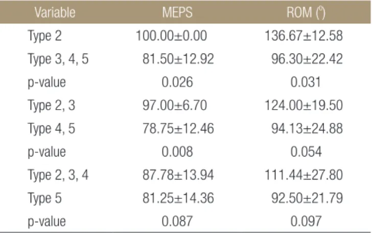
방사선적 경과 추시를 통하여 골절선이 소실되고 골소주가 연 결되는 것을 유합으로 정의하고 유합의 시기를 평가하였다. 골유 합은 1예를 제외한 10예에서 얻었고 평균 골유합 기간은 10.4주였 다. 기능적 평가에서 MEPS는 평균 85점(60-100점)으로 4예에서 최우수, 4예에서 우수, 3예에서 보통의 결과를 얻었다. 운동 범위 는 평균 105도(65-150도)였고, 굴곡 구축은 평균 18도(0-40도)였 다. 합병증으로 주관절의 구축, 불유합과 무혈성 괴사, K-강선의 이동, 그리고 표재성 감염이 각각 1예에서 확인되었으며, 주관절 구축에 대한 관절경적 관절 유리술을 시행한 1예, 그리고 내고정 물 제거를 한 2예 등으로 총 3예에서 2차 수술이 시행되었다. Ring 의 분류에 따른 결과의 분석에서, 외과 후방 골편을 침범한 3, 4, 5 형의 골절이, 외과 후방 골편이 없는 2형보다 MEPS와 운동 범위 에서 유의하게 좋지 않은 결과를 보였으며, 활차 후방 골편을 침 범한 4, 5형의 경우, 활차 후방 골편의 골절이 없는 2, 3형보다 통
A B
C D
A
Figure 2. A 16-year-old male fell on the ground and injured the left elbow. (A, B) Antero-posterior X-ray and lateral X-ray and computed tomography of a type 2 distal humerus capitellar coronal shear fracture. (C) Intraoperative photo of the fracture. (D) Twenty-month postoperative anteroposterior and lateral X-ray of the patient fixed with a plate and screws. The patient had an excellent result.
Table 2. Result of the MEPS, ROM Compared to the Type of Ring’s Classification
Variable MEPS ROM (
o)
Type 2 100.00±0.00 136.67±12.58 Type 3, 4, 5 81.50±12.92 96.30±22.42
p-value 0.026 0.031
Type 2, 3 97.00±6.70 124.00±19.50 Type 4, 5 78.75±12.46 94.13±24.88
p-value 0.008 0.054
Type 2, 3, 4 87.78±13.94 111.44±27.80 Type 5 81.25±14.36 92.50±21.79
p-value 0.087 0.097
Values are presented as mean±standard deviation. MEPS, Mayo elbow performance score; ROM, range of motion.
304
Young-Su Byun, et al.
계적으로 유의하게 좋지 못한 임상적 결과를 보였다. 하지만 내 상과의 포함여부에 따른 5형과 2, 3, 4형과의 비교에서는 임상적 결과의 유의성이 없었다(Table 2).
고 찰
현재까지 발표된 여러 연구들은 소두 골절의 발생은 여성에서 보 다 흔하고1,4-9) 비우세한 팔에서 더 잘 발생하며,9,10) 낙상과 같은 저 에너지 손상에 의한 경우가 많았다고 보고하고 있다.4,10) 여성의 골밀도가 남자보다 낮고, 폐경 이후 더 심해지며, 여성의 주관절 운반각이 좀 더 커서 신전된 상태에서 외과에 가해지는 힘이 더 욱 크게 된다는 점 등이 이유로 제시되기도 한다. 본 연구에서도 11예 중 여자가 8명으로 남자보다 많았고, 비우세 팔의 수상이 7 예로 보다 흔하였으며, 8예에서 낙상 등의 저 에너지에 의한 손상 으로 기존의 연구결과와 비슷한 소견을 보였다.
Ring의 분류에 따른 결과의 분석에서, 외과 후방 골편을 침범 한 3, 4, 5형이 그렇지 않은 2형보다 좋지 않은 임상적 결과를 보 였고, 활차 후방 골편을 침범한 4, 5형이 그렇지 않은 경우인 2, 3 형보다 좋지 못한 임상적 결과를 보였다. 활차 후방 골편을 침범 하는 3형 이상의 골절의 경우 대개 전방의 전단골편과 함께 외과 후방의 골간단부에 감입 또는 분쇄가 흔히 동반되며 이러한 단축 으로 골절의 정확한 정복이 쉽지 않고 또한 단순한 나사못만의 고정으로는 감입에 의한 단축을 교정할 수 없어 추가적인 금속판 고정이 요하게 되는 경우가 많다. O
’
Driscoll 등11)은 이러한 단축 을 간과한 경우 부정유합이 발생할 수 있으므로 주의하여야 한다 고 강조하였다. 활차 후방의 골편을 가지는 4형 이상의 골절에서 는 외측 접근법만으로 충분한 시야를 확보하기 어려운 경우가 많 아 보다 광범위한 경주두 도달법이 필요할 수 있다.3,8) 또한 활차 골절의 특성상 골절부의 대부분이 연골로 덮여 있어 고정이 더욱쉽지 않다. 따라서 이러한 이유로 외과 후방의 골절이 있는 경우 와 활차 후방의 골절이 있는 경우 임상적 결과가 좋지 못하였을 것으로 추정한다. Dubberley 등5)은 후방 분쇄가 동반된 경우 골이 식과 금속판 고정 또는 두 가지 모두가 필요하다고 하여 후방 분 쇄에 대한 적극적인 치료를 주장하였다. 하지만 2차원적인 측면 사진에서의 후방 분쇄만을 기준으로 정한 한계가 있어 분쇄 위치 에 대한 객관적인 확인에 한계가 있다. Ring의 분류법은 골편의 3 차원적 위치를 기준으로 분류함으로써 접근법의 선택, 골절의 정 복위치, 내고정물의 선택 등의 치료계획의 수립에 보다 구체적인 도움을 줄 수 있다. 또한 이 연구를 통하여 이러한 분류법이 임상 적 결과와 밀접한 관계가 있으므로 치료 방법의 선택에서 중요한 역할을 할 것으로 판단한다.
분쇄가 심한 관상면 관절 골절에서는 개방적 정복술 및 내고정 술보다는 인공관절 치환술을 고려할 수 있다. 본 연구에서는 제 외되었지만, 저자들도 활차 골편의 분쇄가 심하였던 두 예에서 인공관절 치환술을 시행하여 MEPS 평가상 최우수와 우수의 임 상 결과를 얻어 단기 추시 결과 만족스런 결과를 얻었다(Fig. 3).
이는 고령의 골다공증 환자의 심한 분쇄가 있는 원위 상완골 골 절에서 내고정의 제한이 있는 경우 인공 관절 전치환술을 제안하 는 기존의 연구와12-14) 부합하는 결과라 볼 수 있다. 하지만 젊고 활동력 있는 환자의 경우에는 인공 관절 치환술을 하기보다 적 극적인 개방적 정복술 및 내고정술이 우선적으로 고려되어야 한 다.12)
증례 8번 환자의 경우 4형 골절로 양측으로 접근하여 K-강선 만을 이용하여 내고정을 시행한 경우로 불유합과 무혈성 괴사가 나타나는 등 임상적으로 좋지 못한 결과를 얻었다(Fig. 4). K-강 선은 나사로 고정을 할 수 없는 작은 골연골 골편의 고정에는 용 이하나15) 강한 힘으로 고정이 필요한 경우에는 고정력의 제한으 로 골유합에 좋지 않은 영향을 미칠 수 있으며, 양측 접근으로 인
A B C D
Figure 3. A 69-year-old female fell on the ground and injuried the left elbow. (A, B) X-ray and computed tomography of a type 5 distal humerus capitellar coronal shear fracture. (C, D) Postoperative anteroposterior X-ray of patient treated with the total elbow arthroplasty. The patient had good result.
한 연부조직의 박리 또한 불유합과 무혈성 괴사에 영향을 미쳤 을 것으로 추정해 볼 수 있다.
이 연구는 고정 방법의 선호도가 서로 다른 3개의 병원에서 수 술을 시행하고 후향적인 방법으로 시행한 연구로서 연령대가 넓 게 분포하고 증례의 수가 적어 연구의 한계가 있으나 비교적 드 문 골절에 대하여 내고정술의 치료결과를 분석하였고 비록 적은 증례임에도 통계적으로 유의성 있는 임상적 결과를 얻었다는 점 에서 의미 있는 연구라 할 수 있겠다.
결 론
원위 상완골 관상면 골절 환자에서 개방적 정복술 및 내고정술은 비교적 우수한 임상적 결과를 나타내었으며, 외과나 활차 후면까 지 골절이 침범할 경우 불량한 예후가 예측되어 치료에 주의를 요한다.
CONFLICTS OF INTEREST
The authors have nothing to disclose.
REFERENCES
1. McKee MD, Jupiter JB, Bamberger HB. Coronal shear frac- tures of the distal end of the humerus. J Bone Joint Surg Am.
1996;78:49-54.
2. Sabo MT, Fay K, McDonald CP, Ferreira LM, Johnson JA, King GJ. Effect of coronal shear fractures of the distal humer- us on elbow kinematics and stability. J Shoulder Elbow Surg.
2010;19:670-80.
3. Ring D, Jupiter JB, Gulotta L. Articular fractures of the distal part of the humerus. J Bone Joint Surg Am. 2003;85:232-8.
4. Brouwer KM, Jupiter JB, Ring D. Nonunion of operatively treated capitellum and trochlear fractures. J Hand Surg Am.
2011;36:804-7.
5. Dubberley JH, Faber KJ, Macdermid JC, Patterson SD, King GJ. Outcome after open reduction and internal fixation of capitellar and trochlear fractures. J Bone Joint Surg Am. 2006;
88:46-54.
6. Goodman HJ, Choueka J. Complex coronal shear fractures of the distal humerus. Bull Hosp Jt Dis. 2005;62:85-9.
7. Mosheiff R, Liebergall M, Elyashuv O, Mattan Y, Segal D.
Surgical treatment of fractures of the capitellum in adults: a modified technique. J Orthop Trauma. 1991;5:297-300.
8. Sano S, Rokkaku T, Saito S, Tokunaga S, Abe Y, Moriya H.
Herbert screw fixation of capitellar fractures. J Shoulder El- bow Surg. 2005;14:307-11.
9. Stamatis E, Paxinos O. The treatment and functional out- come of type IV coronal shear fractures of the distal hu-
A B C D
Figure 4. A 56-year-old female fell on the ground and injured the left elbow. (A, B) Anteroposterior X-ray and computed tomography of a type 4 distal humerus capitellar coronal shear fracture. (C) Postoperative anteroposterior X-ray of the patient fixed with multiple K-wires. K-wires were removed after one-year. (D) Anteroposterior X-ray taken after ten-months after removal of K-wires shows the non-union of fracture.
306
Young-Su Byun, et al.
merus: a retrospective review of five cases. J Orthop Trauma.
2003;17:279-84.
10. Mighell M, Virani NA, Shannon R, Echols EL Jr, Badman BL, Keating CJ. Large coronal shear fractures of the capitel- lum and trochlea treated with headless compression screws. J Shoulder Elbow Surg. 2010;19:38-45.
11. O'Driscoll SW, Jupiter JB, Cohen MS, Ring D, McKee MD.
Difficult elbow fractures: pearls and pitfalls. Instr Course Lect. 2003;52:113-34.
12. Lapner M, King GJ. Elbow arthroplasty for distal humeral fractures. Instr Course Lect. 2014;63:15-26.
13. Ali A, Shahane S, Stanley D. Total elbow arthroplasty for distal humeral fractures: indications, surgical approach, tech- nical tips, and outcome. J Shoulder Elbow Surg. 2010;19 2 Suppl:53-8.
14. Kamineni S, Morrey BF. Distal humeral fractures treated with noncustom total elbow replacement. J Bone Joint Surg Am.
2004;86:940-7.
15. Sen RK, Tripahty SK, Goyal T, Aggarwal S. Coronal shear fracture of the humeral trochlea. J Orthop Surg (Hong Kong).
2013;21:82-6.
원위 상완골 관상면 관절 골절의 개방적 정복술 및 내고정술의 임상적 결과
변영수 • 신동주 • 단진명* • 이성만
†• 정대근 • 구태회 • 하성수
대구파티마병원 정형외과, *CHA 의과학대학교 구미차병원 정형외과, †굳센병원
목적: 개방적 정복술 및 내고정술을 시행한 원위 상완골 관상면 관절 골절 환자를 Ring의 분류법에 따라 분류하고, 임상적 결과를 분 석하였다.
대상 및 방법: 세 곳의 각기 다른 병원에서 원위 상완골 관상면 관절 골절로 개방적 정복술 및 내고정술을 시행받은 11명을 후향적 방법으로 분석하였다. Ring의 분류법에 따라 분류하고, Mayo elbow performance score (MEPS)로 임상적 결과를 평가하고, 골유합 및 합병증 발생 여부를 분석하였다.
결과: 평균 나이 55세(15-88세)였고, 남자 3명, 여자 8명이었다. 평균 MEPS는 85점(60-100점), 최우수 4예, 우수 4예, 보통 3예의 결과를 보였으며, 1예를 제외한 10예에서 골절이 유합되었다, Ring 분류법에 따라 외과 후면 골절이 동반될 경우에 그렇지 않은 경우 보다, 또한 활차 후면 골절이 동반될 경우에 그렇지 않은 경우보다 MEPS와 운동 범위가 유의하게 좋지 않은 결과를 보였다(p<0.05).
결론: 원위 상완골 관상면 골절 환자에서 개방적 정복술 및 내고정술은 비교적 우수한 임상적 결과를 나타내었으며, 외과나 활차 후 면까지 골절이 침범할 경우 불량한 예후가 예측되어 치료에 주의를 요한다.
색인단어: 주관절, 상완골, 관상면 골절, Ring의 분류법
접수일 2015년 12월 7일 수정일 2015년 12월 22일 게재확정일 2016년 2월 6일 책임저자 신동주
41199, 대구시 동구 아양로 99, 대구파티마병원 정형외과
TEL 053-940-7320, FAX 053-940-7417, E-mail [email protected]
Copyright © 2016 by The Korean Orthopaedic Association
“This is an Open Access article distributed under the terms of the Creative Commons Attribution Non-Commercial License (http://creativecommons.org/licenses/by-nc/4.0/) which permits unrestricted non-commercial use, distribution, and reproduction in any medium, provided the original work is properly cited.”