been developed to measure strain (є) and strain rate (SR).
These parameters are closely related to the intrinsic function of the myocardium,7) and are relatively independent of myo- cardial loading.8)9) Analyses of є and SR values have been per- formed in other illnesses affecting the RV10-13) and in ASD.14-20) In ASD patients, no differences have been found between pre- and post-closure RV deformation values determined at the mid-segment of the RV free wall.14)15) However, more re- cent studies have applied a segmental analysis approach and divided the RV free wall into two or three segments.16-18)20) In ASD patients prior to defect closure, RV apical є and SR were higher than in controls,16)17)20) and these values decreased after Introduction
Atrial septal defect (ASD) is a common congenital heart defect that constitutes 7.5–10% of all congenital heart defects found in children,1)2) and is one of the most common congenital heart le- sions presenting in adult life.3) The hemodynamic mechanism of ASD is a left-to-right shunt, which induces a volume overload to the right ventricle (RV). Although this volume overload can be well tolerated,4) if it is sustained it can be detrimental and can cause adverse outcomes.5)6) During follow up of patients with ASD, functional parameters that are able to detect subtle chang- es in RV function are needed to evaluate the status of patients.
Myocardial deformation imaging techniques have recently
ORIGINAL ARTICLE J Cardiovasc Ultrasound 2014;22(4):182-188
Segmental Analysis of Right Ventricular Longitudinal Deformation in Children
before and after Percutaneous Closure of Atrial Septal Defect
Hong Ki Ko, MD1, Jeong Jin Yu, MD2, Eun Kyung Cho, RN2, So Yeon Kang, MD2,
Chang Deok Seo, MD2, Jae Suk Baek, MD2, Young-Hwue Kim, MD2, and Jae-Kon Ko, MD2
1Department of Pediatrics, Ajou University School of Medicine, Suwon, Korea
2Division of Pediatric Cardiology, College of Medicine, University of Ulsan, Seoul, Korea
Background: The aim of study is to identify the dependence of right ventricular (RV) free wall longitudinal deformation on ventricular loading through segmental approach in relatively large number of patients with atrial septal defect (ASD).
Methods: Patients with ASD (n = 114) and age matched healthy children (n = 60) were echocardiographically examined the day before percutaneous device closure and within 24 hours afterwards. RV free wall deformation parameters, strain (є) and strain rate (SR), were analyzed in the apical (єA, SRA) and basal (єB, SRB) segments. Measured deformation parameters were adjusted for RV size (єAL, SRAL, єBL, SRBL) by multiplying by body surface area indexed RV longitudinal dimension. Regression analyses determined the relationships of these deformation parameters with RV loading parameters that were measured by catheterization.
Results: єBL and SRBL were not different between pre-closure patients and controls (p = 0.245, p = 0.866), and were decreased post-closure (p = 0.001, p = 0.018). Post-closure єBL was lower than in controls (p = 0.001). Pre-closure єAL and SRAL were higher than in controls (p = 0.001, p < 0.001), but decreased after closure (all p < 0.001). The pulmonary to systemic flow ratio was related to procedural differences of єBL (p = 0.017) and of SRBL (p = 0.019). RV end diastolic pressure was negatively related to post-closure єBL (p = 0.020) and post-closure SRBL (p = 0.012), and the procedural SRBL difference (p = 0.027).
Conclusion: The longitudinal deformation of the RV basal segment is dependent and its remodeling is also dependent on volume loading in children with ASD.
KEY WORDS: Atrial septal defect · Right ventricular function · Deformation imaging · Strain · Strain rate.
• Received: September 1, 2014 • Revised: October 23, 2014 • Accepted: November 27, 2014
• Address for Correspondence: Jeong Jin Yu, Division of Pediatric Cardiology, Asan Medical Center, 88 Olympic-ro 43-gil, Songpa-gu, Seoul 138-736, Korea Tel: +82-2-3010-3924, Fax: +82-2-473-3725, E-mail: [email protected]
• This is an Open Access article distributed under the terms of the Creative Commons Attribution Non-Commercial License (http://creativecommons.org/licenses/by-nc/3.0) which permits unrestricted non-commercial use, distribution, and reproduction in any medium, provided the original work is properly cited.
day before and within 24 hours of the percutaneous closure of defects in 114 subjects with ASD. The subjects were prepared for examination in the left lateral decubitus position and a sed- ative (chloral hydrate) was administered as required. An iE33 (Philips, Bothell, WA, USA) equipped with a 5-MHz trans- ducer was used. Still images from subcostal, apical and para- sternal views, and moving images from modified apical four- chamber view (3 cardiac cycles, 61–72 frames/s) were stored.
All offline measurements were performed by one author blinded to the demographic and catheterization data of the subjects. Two dimensional (2D) measurements were performed using Image-Arena version 4.6 (TomTec Imaging Systems, Un- terschleissheim, Germany). RV longitudinal dimension (RVLD) was measured according to the guidelines of the American So- ciety of Echocardiography,8) and indexed RV longitudinal di- mension (iRVLD) by division with the body surface area which was calculated using the Haycock formula.21)
Two segment analyses of RV free wall deformation
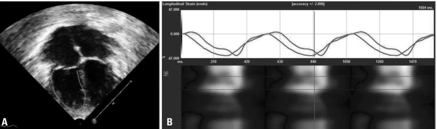
Analyses were performed using a 2D Cardiac Performance Analysis Version 1.2.3 (TomTec Imaging Systems, Unter- schleissheim, Germany) by the single examiner. Longitudinal systolic є and SR were measured from the stored moving im- ages of the modified apical four-chamber view, using a two seg- mental approach, following the structure of the RV free wall with a smooth inlet and trabeculated apex, as has been de- scribed previously.10)16) The left ventricular algorithm was ap- plied to the RV. The endocardial border was manually traced at end diastole starting from the tricuspid annulus to the RV apex (Fig. 1A). Tracking was performed automatically. Values of є and SR were obtained at two points, which were selected as the middle positions of the respective basal (єB, SRB) and apical (єA, SRA) segments (Fig. 1B). The values from the best tracked cycle out of three were selected as the data to be used for analysis, and were converted to absolute values.
To account for the decrease in the RV deformation seen in a bigger ventricle, we adjusted the deformation values to the lon- defect closure.16)18)20) These results suggest that in patients
with ASD, the deformation of the RV apical segment is de- pendent on the loading of the RV.
Studies analyzing RV deformation in ASD patients are of- ten limited by the small number of available subjects, and the lack of an analysis method to assess the correlation between deformation values and parameters that are linked to RV load- ing, measured using cardiac catheterization.14-20) This study aims to identify the dependence of the RV free wall longitudi- nal deformation on RV loading, by examination of the apical and basal segments of the RV free wall in a relatively large number of ASD patients.
Methods
Between March 2009 and December 2012, 146 children with ASD were admitted to the Department of Pediatrics, Asan Medical Center, Korea, for percutaneous device closure of defect. One hundred fourteen among them had qualified for echocardiographic images to analyze RV free wall deformation before and after device closure and also had complete results from catheterization studies, and so were enrolled as subjects in the present study. Five patients with major cardiac anomalies and 27 patients with incomplete echocardiographic image data were excluded. As a control group, 60 age matched healthy children were enrolled. These children had been referred for evaluation of a heart murmur or for nonspecific chest pain dur- ing the aforementioned period, and clinical and echocardio- graphic examinations had shown no evidence of cardiac disease.
There were no additional cardiac anomalies in patients, except for one patient with persistent left superior vena cava. Associ- ated systemic problems were Down syndrome in two patients, CATCH22 in one patient, and Kallmann syndrome in one patient. The institutional review boards of the Asan Medical Center approved this retrospective study (2013-0614) and waived the need for patient consent.
Echocardiographic examination
Echocardiographic examinations were performed both the
Fig. 1. A: The endocardial border was manually traced, starting from the tricuspid annulus to the right ventricular apex, in a moving image of a modified apical four-chamber view. Two points were selected as the middle positions of the respective basal and apical segments. B: Strain (є) values of the two points were presented after automatic tracking. Peak systolic strains (єB, єA) were measured manually in this window.
A B
dural differences (pre-closure values minus post-closure values), and the parameters obtained from the catheterization results.
Chi-square tests were performed to analyze gender distribu- tions. To detect variables which present with gender differenc- es, unpaired t-tests were performed between genders in the control subjects.
Results
Twenty seven excluded patient with incomplete echocardio- graphic images were not different from 114 subjects with ASD in demographic findings including age (p = 0.644), and in data of catheterization including Qp/Qs (p = 0.584) and RVEDP (p = 0.545). The demographic findings of the pa- tients and controls are presented in Table 1. There is a signifi- cantly greater number of females in the patient group (p = 0.003). However, no another demographic or echocardiographic variables were significantly different between two genders (data not presented).
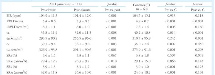
The results of the echocardiographic examinations of the patients, pre- and post-closure, and the controls are presented in Table 2 and Fig. 2. In ASD patients, mean iRVLD was re- duced post-closure (p < 0.0001), and there was no significant difference between the mean iRVLD in post-closure ASD pa- tients and controls (p = 0.160).
The mean values of єBL and SRBL were not different between pre-closure patients and controls (p = 0.245, p = 0.866); how- ever, in patients, these values were decreased after closure (p = 0.001, p = 0.018). In ASD patients, the mean values for єAL and SRAL were also decreased after closure (all p < 0.001);
however, these values pre-closure were higher than those in controls (p = 0.001, p < 0.001). Mean єBL in post-closure ASD patients was lower than that in controls (p = 0.001). Compari- sons of unadjusted deformation values between groups showed similar tendencies of values, except several different results of statistical significances.
Using an analysis of covariance adjusted for heart rate which undergone due to the difference of heart rate between pre- and post-closure ASD patients, the mean values of SRB (p = 0.021), SRBL (p = 0.016), and SRAL (p = 0.045) remained significantly different between pre- and post-closure ASD patients.
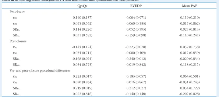
Qp/Qs was 2.71 ± 1.09, RVEDP was 8.6 ± 3.2 mm Hg, and mean pulmonary artery pressure was 18.9 ± 4.2 mm Hg in ASD patients. Qp/Qs was significantly associated with the gitudinal RV length, according to the approach by Dragulescu
et al.17) However, we substituted iRVLD for RVLD in this ad- justment to avoid an overestimation of the resultant values in older subjects. We think that iRVLD is more appropriate than RVLD which should be dependent on the growth of children, not only dependent on a remodeling from the volume overload.
In detail, RV longitudinal length adjusted deformation indexes were calculated by multiplying values of є and SR with iRV- LD as єBL = єB × iRVLD; єAL = єA × iRVLD; SRBL = SRB × iR- VLD; SRAL = SRA × iRVLD.
We randomly selected 16 subjects to determine the intra- and inter-observer variability in the deformation value mea- surements. The deformation values were measured by one ob- server on two occasions and by two observers on two occasions, and the percent precisions were calculated.22) The percent pre- cision of intra-observer for єB, єA, SRB, and SRA measurements was 3.7%, 4.1%, 7.4%, and 8.9%, respectively; the percent precision of inter-observer was 8.5%, 7.2%, 13.7%, and 6.6%, respectively.
Cardiac catheterization
Cardiac catheterization was done under general anesthesia.
In all ASD patients prior to defect closure, mean pulmonary arterial pressure and RV end diastolic pressure (RVEDP) had been measured. Pulmonary to systemic flow ratios (Qp/Qs) were calculated, according to Fick’s principle. These catheter- ization results were used as independent variables in the analy- ses of the deformation values.
Defects were successfully closed by an insertion of an appro- priately selected Amplatzer Septal Occluder (AGA Medical Corporation, Golden Valley, MN, USA).
Statistics
All numerical data are presented as means ± standard devia- tions. All statistical analyses were performed using SPSS 21.0 (SPSS Korea Data Solutions, Seoul, Korea). Statistical signifi- cance was defined as a p-value lower than 0.05. Comparisons of numerical variables between groups were performed using the unpaired t-test. In comparisons between the pre- and post-clo- sure states of patients, paired t-tests were used, and additional- ly, analysis of covariance adjusted for heart rate was performed.
Simple linear regression analyses were used to determine any correlations between the deformation values and their proce-
Table 1. Demographics of patients and controls
ASD patients (n = 114) Controls (n = 60) p-value
Age (years) 4.08 ± 1.57 3.66 ± 2.32 0.162
Males 31 (27.2%) 30 (50%) 0.003
Height (cm) 100.8 ± 11.0 97.5 ± 18.9 0.151
Weight (kg) 15.9 ± 3.9 15.4 ± 6.1 0.503
Body surface area (m2) 0.67 ± 0.11 0.64 ± 0.19 0.314
Data shown are means ± standard deviations, and the number of male subjects (%). ASD: atrial septal defect
RV deformation would reflect immediate preload dependence of RV in ASD patients. Resultantly, RV deformation values in pre-closure state would reflect both RV remodeling from chronic volume overload and immediate preload dependence of RV. Regression analysis showed that Qp/Qs was correlated with procedural differences of RV basal deformation and that RVEDP was correlated with post-closure basal deformation.
These results suggest the dependence of the deformation of RV basal free wall and its remodeling on RV preload in spite of low correlation coefficient values.
In this study, we used a two segmental approach, by dividing the RV free wall into apical and basal segments. This two seg- mental approach has been applied in another studies,10)16)19) and seems to be reasonable in a longitudinal segmental analysis of RV deformation because anatomically the RV has three parts, the inlet, the trabecula, and the infundibulum of which two can be examined on an apical four-chamber view. In healthy indi- viduals, the deformation of the two segments of the RV free procedural differences of єBL (p = 0.017) (Table 3) and SRBL (p
= 0.019). RVEDP was significantly negatively associated with post-closure єBL (p = 0.020) and post-closure SRBL (p = 0.012), and it was positively associated with the SRBL procedural dif- ference (p = 0.027) (Fig. 3). Mean pulmonary arterial pressure was negatively associated with the procedural difference of SRAL (p = 0.028).
Discussion
Myocardial deformation is not only dependent on myocardi- al contractility, but also dependent on ventricular dimension and preload/afterload.9)17)23) In this study, we adjusted measured values of RV deformation to indexed longitudinal RV length and we expected the correction of the effect of increased RV di- mension. We assumed RV deformation values in post-closure state in which RV preload completely removed would reflect RV contractility and remodeling from chronic volume over- load. Additionally, we assumed that procedural differences of
Table 2. Echocardiographic measurements in patients and controls
ASD patients (n = 114) p-value Controls (C)
(n = 60)
p-value p-value
Pre-closure Post-closure Pre vs. post Pre vs. C Post vs. C
HR (bpm) 104.9 ± 11.3 101.4 ± 12.0 0.001 104.7 ± 15.1 0.913 0.118
RVLD (cm) 5.4 ± 0.6 5.3 ± 0.5 < 0.001 4.8 ± 0.7 < 0.001 < 0.001
iRVLD (cm/m2) 8.3 ± 1.1 8.0 ± 1.0 < 0.001 7.8 ± 1.4 0.008 0.160
єB 35.8 ± 11.4 32.0 ± 11.3 0.008 40.2 ± 10.8 0.014 < 0.001
єBL (cm/m2) 293.5 ± 90.2 256.5 ± 96.6 0.001 310.7 ± 95.8 0.245 0.001
єA 39.3 ± 9.4 36.1 ± 9.8 0.003 35.0 ± 7.4 0.002 0.458
єAL (cm/m2) 326.9 ± 93.8 291.1 ± 90.6 < 0.001 275.9 ± 91.6 0.001 0.298
SRB (/s) 3.6 ± 1.5 3.3 ± 1.1 0.067 3.8 ± 1.8 0.507 0.039
SRBL (cm/m2/s) 29.4 ± 12.2 26.3 ± 9.7 0.018 29.1 ± 15.0 0.866 0.145
SRA (/s) 3.9 ± 1.3 3.3 ± 1.2 < 0.001 3.0 ± 1.0 < 0.001 0.123
SRAL (cm/m2/s) 32.0 ± 11.8 26.6 ± 10.0 < 0.001 24.0 ± 10.2 < 0.001 0.103 Data shown are means ± standard deviations. Right ventricular free wall deformation parameters: є: strain, SR: strain rate, B: basal, A: apical, L: a value that has been multiplied by iRVLD. ASD: atrial septal defect, HR: heart rate, bpm: beats per minute, RVLD: right ventricular longitudinal dimension, iRVLD: indexed RVLD
Fig. 2. Corrected RV free wall deformation parameters. Mean values and 95% confidence intervals are shown in pre- and post-closure ASD patients and controls. A: Corrected peak systolic strains at basal lateral RV wall. B: Corrected peak systolic strain at apical lateral RV wall. C: Corrected strain rate at basal lateral RV wall. *p < 0.05, pre-closure vs. post-closure, †p < 0.05, vs. controls. є: strain, SR: strain rate, B: basal, A: apical, L: corrected by multiplication by indexed right ventricular longitudinal dimension, RV: right ventricle, ASD: atrial septal defect.
A B C
100.0 100.0 10.0
200.0 200.0 20.0
300.0 300.0 30.0
400.0 400.0 40.0
0.0 0.0 0.0
Pre-closure Post-closure Control Pre-closure Post-closure Control Pre-closure Post-closure Control
Group Group Group
єBL (cm/m2 ) єAL (cm/m2 ) SRBL (cm/m2 /s)
*
*,†
*
†
Table 3. Simple regression analyses of RV free wall deformation parameters in ASD patients
Qp/Qs RVEDP Mean PAP
Pre-closure
єBL 0.140 (0.137) 0.004 (0.971) 0.119 (0.210)
єAL 0.055 (0.562) -0.060 (0.533) -0.017 (0.862)
SRBL 0.114 (0.226) 0.052 (0.593) 0.023 (0.813)
SRAL 0.051 (0.592) -0.159 (0.098) -0.110 (0.247)
Post-closure
єBL -0.145 (0.124) -0.223 (0.020) 0.032 (0.738)
єAL 0.035 (0.711) -0.080 (0.409) 0.017 (0.859)
SRBL -0.168 (0.074) -0.240 (0.012) -0.020 (0.834)
SRAL 0.034 (0.723) -0.019 (0.842) 0.118 (0.215)
Pre- and post-closure procedural differences
єBL 0.223 (0.017) 0.183 (0.057) 0.064 (0.501)
єAL 0.020 (0.834) 0.016 (0.867) -0.031 (0.743)
SRBL 0.219 (0.019) 0.212 (0.027) 0.034 (0.722)
SRAL 0.022 (0.816) -0.140 (0.148) -0.207 (0.028)
Data shown are Pearson correlation coefficients (R) and (p-values). Right ventricular free wall deformation parameters: є: strain, SR: strain rate, B: basal, A: apical, L: a value that has been multiplied by iRVLD. RV: right ventricle, ASD: atrial septal defect, Qp/Qs: pulmonary to systemic flow ratio, RVEDP: right ventricular end diastolic pressure, PAP: pulmonary arterial pressure, iRVLD: indexed right ventricular longitudinal dimension
Fig. 3. Relationships between RV free wall deformation parameters and RVEDP. A: Relationship between RVEDP and post-closure єBL. B:
Relationship between RVEDP and post-closure SRBL. C: Relationship between RVEDP and procedural difference of єBL (Post-Pre). D: Relationship between RVEDP and procedural difference of SRBL (Post-Pre). є: strain, SR: strain rate, B: basal, A: apical, L: corrected by multiplication by indexed right ventricular longitudinal dimension, RVEDP: right ventricular end diastolic pressure, RV: right ventricle.
C D
B
0.0 100.0
-200.0 200.0 300.0
0.0 400.0
200.0 500.0 600.0
400.0 0.0
0.0
5.0
5.0
10.0
10.0
15.0
15.0
20.0
20.0 RVEDP (mm Hg)
RVEDP (mm Hg) Post-closure єBL (cm/m2 )Procedural difference of єBL (cm/m2 )
10.0
-40.0 20.0 30.0
-20.0 40.0
0.0 50.0 60.0
20.0 70.0
40.0 0.0
0.0
5.0
5.0
10.0
10.0
15.0
15.0
20.0
20.0 RVEDP (mm Hg)
RVEDP (mm Hg) Post-closure SRBL (cm/m2 /s)Procedural difference of SRBL (cm/m2 /s)
A
sure SR values using analysis of covariance adjusted for heart rate although which cannot support a comparison of paired data.
This study had meaningful limitations. Thus far, several ex- planations for the basal-apical RV deformation differences in ASD patients has been suggested in one previous study; dif- ferent wall thickness between the two segments, altered ge- ometry of RV, and altered distribution of adrenergic receptors in RV.16) However, this study cannot add any further details to the above explanations. To investigate the differential behav- iors of the two segments of the RV in ASD patients, we rec- ommend a study using cardiac magnetic resonance imaging with which an accurate investigation of segmental volumes and wall motions is possible.
Different sedative conditions between in echocardiography and in catheterization also a limitation.
In conclusion, the longitudinal deformation of the RV basal segment is dependent and its remodeling is also dependent on volume loading in children with ASD.
References
1. Hoffman JI, Kaplan S. The incidence of congenital heart disease. J Am Coll Cardiol 2002;39:1890-900.
2. Jeong JA, Kim YM, Lee HS, Kwon TC, Kang CM. Statistical study on congenital heart disease. Korean Circ J 1989;19:89-96.
3. Hoffman JI, Christianson R. Congenital heart disease in a cohort of 19,502 births with long-term follow-up. Am J Cardiol 1978;42:641-7.
4. Craig RJ, Selzer A. Natural history and prognosis of atrial septal defect.
Circulation 1968;37:805-15.
5. Attie F, Rosas M, Granados N, Zabal C, Buendía A, Calderón J.
Surgical treatment for secundum atrial septal defects in patients >40 years old. A randomized clinical trial. J Am Coll Cardiol 2001;38:2035-42.
6. Konstantinides S, Geibel A, Olschewski M, Görnandt L, Roskamm H, Spillner G, Just H, Kasper W. A comparison of surgical and medical therapy for atrial septal defect in adults. N Engl J Med 1995;333:469- 73.
7. D’hooge J, Heimdal A, Jamal F, Kukulski T, Bijnens B, Rademak- ers F, Hatle L, Suetens P, Sutherland GR. Regional strain and strain rate measurements by cardiac ultrasound: principles, implementation and limitations. Eur J Echocardiogr 2000;1:154-70.
8. Rudski LG, Lai WW, Afilalo J, Hua L, Handschumacher MD, Chandrasekaran K, Solomon SD, Louie EK, Schiller NB. Guidelines for the echocardiographic assessment of the right heart in adults: a report from the American Society of Echocardiography endorsed by the European Association of Echocardiography, a registered branch of the European Society of Cardiology, and the Canadian Society of Echocardiography. J Am Soc Echocardiogr 2010;23:685-713; quiz 786-8.
9. Missant C, Rex S, Claus P, Mertens L, Wouters PF. Load-sensitivity of regional tissue deformation in the right ventricle: isovolumic versus ejection- phase indices of contractility. Heart 2008;94:e15.
10. Dambrauskaite V, Delcroix M, Claus P, Herbots L, D’hooge J, Bij- nens B, Rademakers F, Sutherland GR. Regional right ventricular dys- function in chronic pulmonary hypertension. J Am Soc Echocardiogr 2007;20:1172-80.
11. Sutherland GR, Di Salvo G, Claus P, D’hooge J, Bijnens B. Strain and strain rate imaging: a new clinical approach to quantifying regional myocardial function. J Am Soc Echocardiogr 2004;17:788-802.
12. Kjaergaard J, Sogaard P, Hassager C. Right ventricular strain in pul- wall is not different.24) However, the behaviors of the two seg-
ments were found to be different in this study in common with other reported study design which segmental approach used for analyzing RV deformation in patients with RV illnesses.10)16)17)19)
The basal segment deformation values in pre-closure ASD patients were higher than those in post-closure, and were not different from those of controls. The lower post-closure values may be the result of RV basal free wall remodeling, and the higher pre-closure values may be due to compensation caused by the increased preload. Decreased deformation of RV basal segments has been observed in extreme athletes.25) This might be the result of adaptation to chronic loading conditions, and seems to be equivalent to the results seen in post-closure pa- tients in this study. Di Salvo et al.19) also reported decreased RV basal segment deformation in post-closure patients, and reverse remodeling of the basal segment might not be achieved until 6 months post-closure as all their subjects underwent ASD clo- sure more than 6 months before their study.
Increased deformation of the RV apical segment in pre-clo- sure ASD patients has been reported, and has been assumed to be the result of RV loading.16-18)20) Van De Bruaene et al.16) showed that RV apical segment deformation was significantly correlated with preload-linked echocardiographic variables. In this study, apical segment deformation values in pre-closure patients were found to be higher than those in post-closure, and were also higher than in controls. However apical defor- mation values in post-closure patients were not found to be different from those of controls, as has been described in other studies where percutaneous device insertion was used for ASD closure.19)20) In one study, decreased post-closure apical defor- mation was observed only in patients who had undergone sur- gical closure.19) Therefore we assumed that the remodeling of RV apical segment from chronic volume overload is not sig- nificant in ASD patients, and that the increased deformation values in apical segment may be from immediate preload de- pendence. Unfortunately, we could not found significant cor- relation between apical segment deformation values and the preload-linked variables that were determined by catheteriza- tion. However, a significant correlation was observed between procedural difference of SRAL and mean pulmonary arterial pressure. This relation suggests a possibility of the dependence of RV apical segment on the afterload at least partly in ASD patients. In patients with pulmonary hypertension, Dam- brauskaite et al.10) demonstrated a prominent dependence of apical deformation on the afterload. The dependence of RV apical segment on afterload possibly deranged the identifica- tion of correlation between the deformation of RV apical seg- ment and preload in this study.
In this study, we observed that the mean heart rate was dif- ferent between pre- and post-closure ASD patients. However, this confounding factor may not invalidate the interpretation of the procedural differences of RV deformation results, as sim- ilar results were obtained in comparisons of pre- and post-clo-
strain rate imaging for quantitative evaluation of regional left and right ventricular function after surgical versus percutaneous closure of atrial septal defect. Am J Cardiol 2005;96:299-302.
20. Jategaonkar SR, Scholtz W, Butz T, Bogunovic N, Faber L, Horst- kotte D. Two-dimensional strain and strain rate imaging of the right ven- tricle in adult patients before and after percutaneous closure of atrial septal defects. Eur J Echocardiogr 2009;10:499-502.
21. Haycock GB, Schwartz GJ, Wisotsky DH. Geometric method for mea- suring body surface area: a height-weight formula validated in infants, children, and adults. J Pediatr 1978;93:62-6.
22. Galderisi M, Benjamin EJ, Evans JC, D’Agostino RB, Fuller DL, Lehman B, Wolf PA, Levy D. Intra- and interobserver reproducibility of Doppler-assessed indexes of left ventricular diastolic function in a popula- tion-based study (the Framingham Heart Study). Am J Cardiol 1992;70:
1341-6.
23. Marciniak A, Claus P, Sutherland GR, Marciniak M, Karu T, Balta- baeva A, Merli E, Bijnens B, Jahangiri M. Changes in systolic left ven- tricular function in isolated mitral regurgitation. A strain rate imaging study. Eur Heart J 2007;28:2627-36.
24. Naito H, Arisawa J, Harada K, Yamagami H, Kozuka T, Tamura S.
Assessment of right ventricular regional contraction and comparison with the left ventricle in normal humans: a cine magnetic resonance study with pre- saturation myocardial tagging. Br Heart J 1995;74:186-91.
25. Teske AJ, Prakken NH, De Boeck BW, Velthuis BK, Martens EP, Doevendans PA, Cramer MJ. Echocardiographic tissue deformation im- aging of right ventricular systolic function in endurance athletes. Eur Heart J 2009;30:969-77.
monary embolism by Doppler tissue echocardiography. J Am Soc Echocar- diogr 2004;17:1210-2.
13. Chow PC, Liang XC, Cheung EW, Lam WW, Cheung YF. New two-dimensional global longitudinal strain and strain rate imaging for as- sessment of systemic right ventricular function. Heart 2008;94:855-9.
14. Eyskens B, Ganame J, Claus P, Boshoff D, Gewillig M, Mertens L.
Ultrasonic strain rate and strain imaging of the right ventricle in children before and after percutaneous closure of an atrial septal defect. J Am Soc Echocardiogr 2006;19:994-1000.
15. Abd El Rahman MY, Hui W, Timme J, Ewert P, Berger F, Dsebis- sowa F, Hetzer R, Lange PE, Abdul-Khaliq H. Analysis of atrial and ventricular performance by tissue Doppler imaging in patients with atrial septal defects before and after surgical and catheter closure. Echocardiography 2005;22:579-85.
16. Van De Bruaene A, Buys R, Vanhees L, Delcroix M, Voigt JU, Budts W. Regional right ventricular deformation in patients with open and closed atrial septal defect. Eur J Echocardiogr 2011;12:206-13.
17. Dragulescu A, Grosse-Wortmann L, Redington A, Friedberg MK, Mertens L. Differential effect of right ventricular dilatation on myocardial deformation in patients with atrial septal defects and patients after tetralogy of Fallot repair. Int J Cardiol 2013;168:803-10.
18. Bussadori C, Oliveira P, Arcidiacono C, Saracino A, Nicolosi E, Ne- gura D, Piazza L, Micheletti A, Chessa M, Butera G, Dua JS, Car- minati M. Right and left ventricular strain and strain rate in young adults before and after percutaneous atrial septal defect closure. Echocardiog- raphy 2011;28:730-7.
19. Di Salvo G, Drago M, Pacileo G, Carrozza M, Santoro G, Bigazzi MC, Caso P, Russo MG, Carminati M, Calabró R. Comparison of