Introduction
Nonalcoholic fatty liver disease (NAFLD) is the most com- mon cause of abnormal liver function tests in adults and al- most one third of the population in the Korean has hepatic steatosis.1)2) NAFLD is commonly associated with visceral obesity, dyslipidemia, insulin resistance, and type 2 diabetes and may represent another component of the metabolic syn- drome (MetS),3-5) a condition associated with a high cardiovas- cular risk and, in particular, an increased prevalence of carotid lesions.6) It is important to determine whether NAFLD is an
independent predictor of cardiovascular morbidity and mor- tality and several studies have suggested that there is an asso- ciation between NAFLD and cardiovascular disease.6-11) Non- invasively detected increased carotid intima media thickness (IMT) is generally accepted as an early indicator of generalized atherosclerosis and has been related to cardiovascular risk fac- tors, and cardiovascular disease including incidence of myocar- dial infarction and stroke.12-14) Some case-controlled and cross- sectional studies6-8) showed a relationship between NAFLD and carotid IMT.7)11)15) Indeed, it is hypothesized that NAFLD ORIGINAL ARTICLE J Cardiovasc Ultrasound 2012;20(3):126-133
Relationship between Nonalcoholic Fatty Liver Disease and Carotid Artery
Atherosclerosis Beyond Metabolic Disorders in Non-Diabetic Patients
Ji Hoon Kang, MD, Kyoung Im Cho, MD, Seong Man Kim, MD, Ja Young Lee, MD, Jae Joon Kim, MD, Ja Jun Goo, MD, Kyoung Nyoun Kim, MD, Joon Hyung Jhi, MD, Dong Jun Kim, MD, Hyeon Gook Lee, MD and Tae Ik Kim, MD
Division of Cardiology, Department of Internal Medicine, Maryknoll Medical Center, Busan, Korea
Background: The objective of this study was to investigate the association between nonalcoholic fatty liver disease (NAFLD) and carotid artery atherosclerosis beyond metabolic disorders.
Methods: We studied 320 non-diabetic patients with ultrasonographically diagnosed NAFLD and 313 non-diabetic patients without NAFLD who have less than 40 g alcohol/week drinking history. Carotid atherosclerotic burden was assessed by carotid intima-media thickness (IMT) and plaque. All subjects were divided to the metabolic syndrome (MetS) according to International Diabetes Federation criteria.
Results: NAFLD patients had a significantly increased mean carotid IMT (0.79 ± 0.18 vs. 0.73 ± 0.13 mm; p < 0.001) than those without the condition. The prevalence of increased IMT, defined as IMT ≥ 1 mm, and carotid plaque were 52.5% and 34.1% in the patients with NAFLD vs. 35.8% and 18.8% in the patients without this condition (p < 0.001). The difference in IMT and prevalence of plaque was also significant even in patients without MetS as well as those with MetS (all p < 0.05).
NAFLD-associated adjusted odds ratio for increased IMT was 1.236 [95% confidence interval (CI), 1.023-1.467, p = 0.016]
without MetS and 1.178 (95% CI, 1.059-1.311, p = 0.003) with MetS. NAFLD-associated adjusted odds ratio of carotid plaque was 1.583 (95% CI, 1.309-1.857, p = 0.024) without MetS and 1.536 (95% CI, 0.512-4.604, p = 0.444) with MetS.
Conclusion: NAFLD is significantly associated with carotid atherosclerosis in non-diabetic outpatients even without MetS.
Carotid screening for NAFLD might be beneficial for assessment of future atherosclerotic complications.
KEY WORDS: Fatty liver · Carotid intima media thickness · Metabolic syndrome.
• Received: July 23, 2012 • Revised: July 27, 2012 • Accepted: August 8, 2012
• Address for Correspondence: Kyoung Im Cho, Division of Cardiology, Department of Internal Medicine, Maryknoll Medical Center, 121 Junggu-ro, Jung-gu, Busan 600-730, Korea Tel: +82-51-461-2384, Fax: +82-51-441-6950, E-mail: [email protected]
• This is an Open Access article distributed under the terms of the Creative Commons Attribution Non-Commercial License (http://creativecommons.org/licenses/by-nc/3.0) which permits unrestricted non-commercial use, distribution, and reproduction in any medium, provided the original work is properly cited.
is not merely a marker of cardiovascular disease but may also be involved in its pathogenesis.8) A possible relationship be- tween NAFLD and carotid lesions might have important practical consequences, considering the frequent incidental finding of hepatic steatosis in subjects undergoing abdominal ultrasound (US) for any reason. In these subjects, an US assess- ment of carotid arteries might also be advisable. However, some degree of variability about the mean carotid IMT values has been observed among all the published reports that result in a difficult evaluation of the magnitude of the observation.
For instance, among the different studies, mean carotid IMT values in NAFLD patients range from 0.64 ± 0.10 mm to 1.24 ± 0.13 mm.7)15) It seems to be important to decide wheth- er further recommendations with regard to carotid atheroscle- rosis screening should be implemented in all NAFLD pa- tients, as currently available epidemiological data indicate that a value of carotid IMT at or above 1 mm at any age is associat- ed with a significantly increased risk of myocardial infarction and/or cerebrovascular disease.14) Thus, we studied a random group of consecutive outpatients undergoing abdominal US to establish the strength of the increased carotid IMT defined
≥ 1 mm and carotid plaque observed in NAFLD patients. In addition, we evaluated whether such an association is indepen- dent of classical risk factors and MetS features.
Methods
Study population
From January 2010 to December 2010, we performed a cross- sectional analysis on consecutive 320 non-diabetic NAFLD out- patients (192 men and 128 women, ages 53.5 ± 9.25 years) who received abdominal and carotid US assessment in Maryk- noll Medical Center for health screening. The NAFLD diag- nosis was based on abdominal US and exclusion of other known etiologic factors of chronic liver disease (alcohol abuse or in- take > 40 g/week, viral hepatitis, autoimmune hepatitis, and use of hepato-toxic drugs). No patients had clinical evidence of diabetes mellitus (DM), advanced liver or renal disease, car- diovascular events, or recent history of acute illness. The control group, also recruited from health screening centers consisted of 313 apparently healthy subjects comparable for age and sex with normal liver US/liver function tests and drinking less than 40 g alcohol/week. The study protocol was approved by the eth- ics review boards of Maryknoll Medical Center, and written in- formed consent was obtained from all patients enrolled.
Clinical measurements and laboratory procedures
Body mass index (BMI) was calculated by dividing weight in kilograms by the square of height in meters. Waist circum- ference was measured in a standing position at the level of the umbilicus. Blood pressure (BP) was measured with a standard mercury manometer. Subjects were considered to have hyper-
tension if their BP was ≥ 140/≥ 90 mmHg as recommended by the Joint National Committee VII or if they were on treat- ment for hypertension. Information on daily alcohol consump- tion and other lifestyle characteristics were obtained from all participants by questionnaire.4) Venous blood was drawn in the morning after an overnight fast. Plasma liver function tests and glucose, total cholesterol, triglycerides, low density lipoprotein (LDL) and high density lipoprotein (HDL) were determined by standard laboratory procedures. Normal ranges for aspartate aminotransferase, alanine aminotransferase (ALT), and γ-glutamyltransferase (γ-GTP), in our laboratory, were 10-35 units/L for female subjects and 10-50 units/L for male subjects, respectively. The American Diabetes Association cri- teria were used to define DM and we considered a subject to have DM when the fasting plasma glucose levels were ≥ 126 mg/dL in 2 consecutive assessments or if they were on treat- ment for DM. The presence of dyslipidemia was assumed if subjects were taking lipid-lowering drugs, or high cholesterol level. Smoking was categorized into the 3 following levels:
never smoked, up to 20 pack-years and more than 20 pack- years. Adopting international diabetes federation criteria,16) the metabolic syndrome in South Korean adults was defined by central obesity (waist circumference ≥ 90 cm for men and ≥ 85 cm for women)17) plus two of the following four factors: se- rum triglyceride ≥ 150 mg/dL, HDL cholesterol < 40 mg/dL for men and < 50 mg/dL for women, systolic BP ≥ 130 mmHg or diastolic BP ≥ 85 mmHg, and fasting plasma glucose ≥ 100 mg/dL.
Ultrasound evaluation
All subjects underwent abdominal and carotid US in order to assess hepatic steatosis and carotid IMT measurement or analy- sis for the presence of plaques. We used Accuson Sequoia (Sie- mens, Mountain View, CA, USA), with convex probes (2.5-5 MHz) to scan the liver, and Vivid 7 (GE Medical System, Mil- waukee, WI, USA) equipped with a 7 to 12-Mhz linear-array scanner, with a limit of detection of < 0.1 mm to scan carotid arteries. All investigations were performed by two experienced operators (for abdominal and carotid US), blinded to each other regarding the respective US measurements and unaware of pa- tients’ clinical data. Following the American gastroenterological association classification of NAFLD,18) NAFLD was defined as the presence of diffuse hyperechoic echo-texture, bright liver,19) increased liver echo-texture compared with the kidneys, vascu- lar blurring and deep attenuation of the ultrasonic beam.
For carotid US, all subjects were examined in a supine posi- tion, neck extended, and the chin facing the counter lateral side. Carotid arteries were examined bilaterally in the longitu- dinal and transversal planes. The common, internal, and exter- nal carotid arteries were examined for evidence of atheroscle- rotic lesions as seen in thickness of the IMT of the common carotid artery, internal carotid artery and external carotid ar- tery and plaque presence. After placing the region of interests
in the far wall of the common carotid artery (CCA), mean IMT was estimated in a region free of atherosclerotic plaques with the use of an automatic tracking system.20) Mean IMT was averaged from mean CCA IMT in both far wall, and max- imum IMT was defined as the thickest IMT regardless of sites. The increased IMT was considered as ≥ 1.0 mm in either or both carotid arteries and the presence of atherosclerotic plaque was defined as localized lesions with protrusion into the arterial lumen or IMT greater than 1.5 mm.21) Carotid ste- nosis was not included in the analysis, because no subjects had severe stenosis (≥ 50%).
Statistical analysis
Data were expressed as mean values ± standard deviation and frequencies were expressed as percentages. All analyses were per- formed using a SPSS 13.0 package program (IBM corp., Ar- monk, NY, USA). Statistical analysis between the groups was performed using student’s t-test for continuous variables and the chi-square test for categorical data. Statistical correlations
were determined by the nonparametric Spearman test. Multiple linear regression analysis was performed with respect to carotid IMT and age, BP, BMI, waist circumference, lipid profile, liver enzymes, and the presence of NAFLD were included as covari- ates. The independence of the associations of variables with ab- normal IMT and presence of plaque, considered as the depen- dent variable, was also assessed by binary logistic regression analyses and age, BP, BMI, waist circumference, lipid profile, liver enzymes, and the presence of NAFLD were included as co- variates. Separate regression models were tested in two groups of patients according to the presence of MetS. Probability levels lower than 0.05 were considered significant.
Results
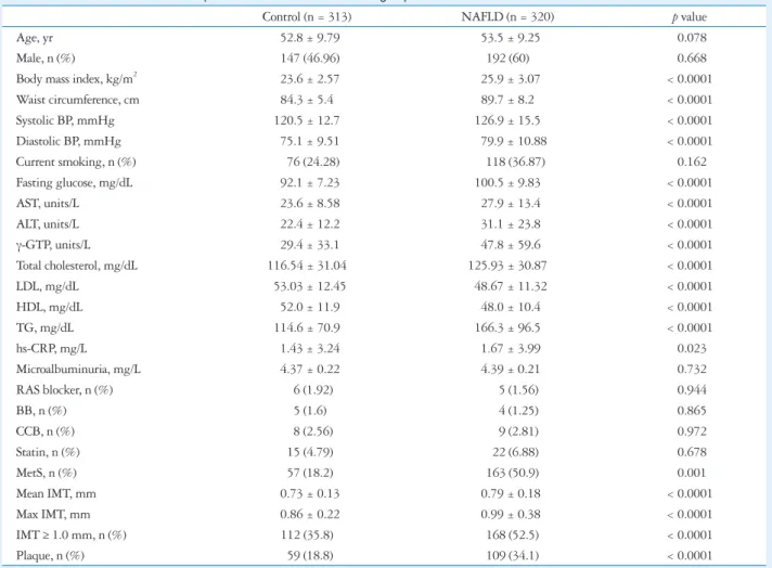
The baseline characteristics of participants are shown in Ta- ble 1. Because of the study design, NAFLD and control sub- jects were comparable in terms of age and sex. Significantly higher BMI, BP, liver enzymes and high sensitivity C-reactive protein (hs-CRP), lipid profiles were found in the subjects
Table 1. Clinical characteristics of the patients with NAFLD and control groups
Control (n = 313) NAFLD (n = 320) p value
Age, yr 52.8 ± 9.79 53.5 ± 9.25 0.078
Male, n (%) 147 (46.96) 192 (60) 0.668
Body mass index, kg/m2 23.6 ± 2.57 25.9 ± 3.07 < 0.0001
Waist circumference, cm 84.3 ± 5.4 89.7 ± 8.2 < 0.0001
Systolic BP, mmHg 120.5 ± 12.7 126.9 ± 15.5 < 0.0001
Diastolic BP, mmHg 75.1 ± 9.51 79.9 ± 10.88 < 0.0001
Current smoking, n (%) 76 (24.28) 118 (36.87) 0.162
Fasting glucose, mg/dL 92.1 ± 7.23 100.5 ± 9.83 < 0.0001
AST, units/L 23.6 ± 8.58 27.9 ± 13.4 < 0.0001
ALT, units/L 22.4 ± 12.2 31.1 ± 23.8 < 0.0001
γ-GTP, units/L 29.4 ± 33.1 47.8 ± 59.6 < 0.0001
Total cholesterol, mg/dL 116.54 ± 31.04 125.93 ± 30.87 < 0.0001
LDL, mg/dL 53.03 ± 12.45 48.67 ± 11.32 < 0.0001
HDL, mg/dL 52.0 ± 11.9 48.0 ± 10.4 < 0.0001
TG, mg/dL 114.6 ± 70.9 166.3 ± 96.5 < 0.0001
hs-CRP, mg/L 1.43 ± 3.24 1.67 ± 3.99 0.023
Microalbuminuria, mg/L 4.37 ± 0.22 4.39 ± 0.21 0.732
RAS blocker, n (%) 6 (1.92) 5 (1.56) 0.944
BB, n (%) 5 (1.6) 4 (1.25) 0.865
CCB, n (%) 8 (2.56) 9 (2.81) 0.972
Statin, n (%) 15 (4.79) 22 (6.88) 0.678
MetS, n (%) 57 (18.2) 163 (50.9) 0.001
Mean IMT, mm 0.73 ± 0.13 0.79 ± 0.18 < 0.0001
Max IMT, mm 0.86 ± 0.22 0.99 ± 0.38 < 0.0001
IMT ≥ 1.0 mm, n (%) 112 (35.8) 168 (52.5) < 0.0001
Plaque, n (%) 59 (18.8) 109 (34.1) < 0.0001
All values are presented as the mean ± SD.
NAFLD: non alcoholic fatty liver disease, BP: blood pressure, AST: aspartate aminotransferase, ALT: alanine aminotransferase, GTP: glutamyltransferase, LDL:
low density lipoprotein cholesterol, HDL: high density lipoprotein cholesterol, TG: triglycerides, hs-CRP: high sensitivity C-reactive protein, RAS: rennin- angiotensin system, BB: beta blocker, CCB: calcium channer blocker, MetS: metabolic syndrome, IMT: intima media thickness
with NAFLD. Smoking history, microalbuminuria, and med- ications did not differ between the groups. NAFLD patients had a significantly increased carotid IMT (mean IMT: 0.79 ± 0.18 vs. 0.73 ± 0.13 mm, maximal IMT: 0.99 ± 0.38 vs. 0.86
± 0.22 mm; all p < 0.001) and the prevalence of MetS (50.9%
vs. 18.2%, p < 0.001) than those without the condition. The prevalence of increased IMT and carotid plaque were 52.5%
and 34.1% in the patients with NAFLD vs. 35.8% and 18.8%
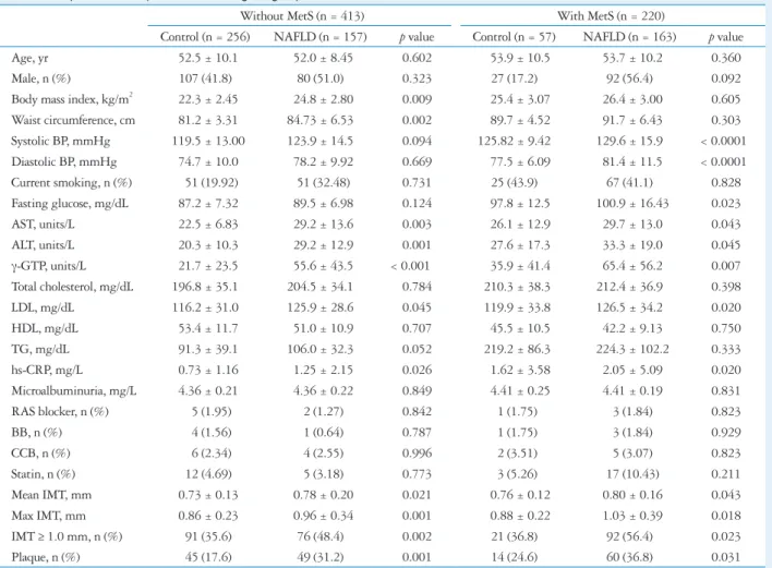
in the patients without this condition (p < 0.001). As shown in Table 2, the difference in IMT and prevalence of plaque were also significant even without MetS as well as subjects with MetS (all p < 0.05). The lowest level of carotid IMT was found in con- trol subjects without MetS, intermediate in NAFLD patients with without MetS, and highest in those with NAFLD pa- tients with MetS (Table 2).
Association between the NAFLD and carotid atherosclerosis
Age was strongly correlated with mean IMT (r = 0.420, p <
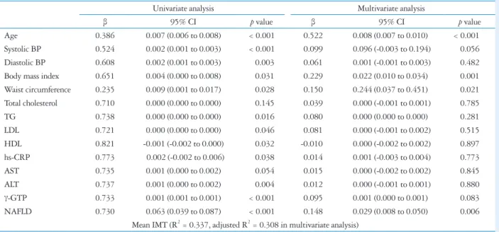
0.001) and maximal IMT (r = 0.402, p < 0.001). Systolic and diastolic BP, BMI, waist circumference, triglycerides, LDL cho- lesterol and hs-CRP showed modest correlation with mean IMT (Table 3). IMT was also positively correlated with liver en- zymes such as ALT and γ-GTP and was inversely associated with HDL cholesterol (all p < 0.05) (Table 3). In multiple linear regression analysis, the presence of NAFLD was significantly as- sociated with carotid IMT after adjustment of age, BP, BMI, waist circumference, lipid profile, liver enzymes and hs-CRP (all p < 0.05) (Table 4).
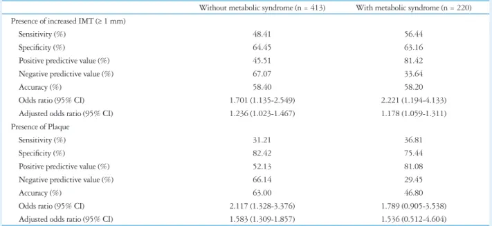
The utility of NAFLD findings in identifying patients with carotid artery atherosclerosis with or without MetS was as- sessed further by determining the sensitivity, specificity, posi- tive predictive value, and odds ratio (Table 5). By binary linear logistic regression analysis, NAFLD-associated adjusted odds ratio for increased IMT was 1.236 [95% confidence interval (CI), 1.023-1.467, p = 0.016] without MetS (R2 = 0.299, ad- justed R2 = 0.222) and 1.178 (95% CI, 1.059-1.311, p = 0.003) with MetS (R2 = 0.351, adjusted R2 = 0.263) after ad-
Table 2. Comparison of the parameters among the groups
Without MetS (n = 413) With MetS (n = 220)
Control (n = 256) NAFLD (n = 157) p value Control (n = 57) NAFLD (n = 163) p value
Age, yr 52.5 ± 10.1 52.0 ± 8.45 0.602 53.9 ± 10.5 53.7 ± 10.2 0.360
Male, n (%) 107 (41.8) 80 (51.0) 0.323 27 (17.2) 92 (56.4) 0.092
Body mass index, kg/m2 22.3 ± 2.45 24.8 ± 2.80 0.009 25.4 ± 3.07 26.4 ± 3.00 0.605 Waist circumference, cm 81.2 ± 3.31 84.73 ± 6.53 0.002 89.7 ± 4.52 91.7 ± 6.43 0.303
Systolic BP, mmHg 119.5 ± 13.00 123.9 ± 14.5 0.094 125.82 ± 9.42 129.6 ± 15.9 < 0.0001
Diastolic BP, mmHg 74.7 ± 10.0 78.2 ± 9.92 0.669 77.5 ± 6.09 81.4 ± 11.5 < 0.0001
Current smoking, n (%) 51 (19.92) 51 (32.48) 0.731 25 (43.9) 67 (41.1) 0.828
Fasting glucose, mg/dL 87.2 ± 7.32 89.5 ± 6.98 0.124 97.8 ± 12.5 100.9 ± 16.43 0.023
AST, units/L 22.5 ± 6.83 29.2 ± 13.6 0.003 26.1 ± 12.9 29.7 ± 13.0 0.043
ALT, units/L 20.3 ± 10.3 29.2 ± 12.9 0.001 27.6 ± 17.3 33.3 ± 19.0 0.045
γ-GTP, units/L 21.7 ± 23.5 55.6 ± 43.5 < 0.001 35.9 ± 41.4 65.4 ± 56.2 0.007
Total cholesterol, mg/dL 196.8 ± 35.1 204.5 ± 34.1 0.784 210.3 ± 38.3 212.4 ± 36.9 0.398
LDL, mg/dL 116.2 ± 31.0 125.9 ± 28.6 0.045 119.9 ± 33.8 126.5 ± 34.2 0.020
HDL, mg/dL 53.4 ± 11.7 51.0 ± 10.9 0.707 45.5 ± 10.5 42.2 ± 9.13 0.750
TG, mg/dL 91.3 ± 39.1 106.0 ± 32.3 0.052 219.2 ± 86.3 224.3 ± 102.2 0.333
hs-CRP, mg/L 0.73 ± 1.16 1.25 ± 2.15 0.026 1.62 ± 3.58 2.05 ± 5.09 0.020
Microalbuminuria, mg/L 4.36 ± 0.21 4.36 ± 0.22 0.849 4.41 ± 0.25 4.41 ± 0.19 0.831
RAS blocker, n (%) 5 (1.95) 2 (1.27) 0.842 1 (1.75) 3 (1.84) 0.823
BB, n (%) 4 (1.56) 1 (0.64) 0.787 1 (1.75) 3 (1.84) 0.929
CCB, n (%) 6 (2.34) 4 (2.55) 0.996 2 (3.51) 5 (3.07) 0.823
Statin, n (%) 12 (4.69) 5 (3.18) 0.773 3 (5.26) 17 (10.43) 0.211
Mean IMT, mm 0.73 ± 0.13 0.78 ± 0.20 0.021 0.76 ± 0.12 0.80 ± 0.16 0.043
Max IMT, mm 0.86 ± 0.23 0.96 ± 0.34 0.001 0.88 ± 0.22 1.03 ± 0.39 0.018
IMT ≥ 1.0 mm, n (%) 91 (35.6) 76 (48.4) 0.002 21 (36.8) 92 (56.4) 0.023
Plaque, n (%) 45 (17.6) 49 (31.2) 0.001 14 (24.6) 60 (36.8) 0.031
All values are presented as the mean ± SD.
MetS: metabolic syndrome, NAFLD: non alcoholic fatty liver disease, BP: blood pressure, AST: aspartate aminotransferase, ALT: alanine aminotransferase, GTP: glutamyltransferase, LDL: low density lipoprotein cholesterol, HDL: high density lipoprotein cholesterol, TG: triglycerides, hs-CRP: high sensitivity C-reactive protein, RAS: rennin-angiotensin system, BB: beta blocker, CCB: calcium channer blocker, IMT: intima media thickness
justment of age, BP, BMI, waist circumference, lipid profile, liver enzymes. NAFLD-associated adjusted odds ratio of ca- rotid plaque was 1.583 (95% CI, 1.309-1.857, p = 0.024) without MetS (R2 = 0.281, adjusted R2 = 0.192) and 1.536 (95% CI, 0.512-4.604, p = 0.444) with MetS (R2 = 0.270, adjusted R2 = 0.196). The value of variation inflation factor was less than 10 in age, BP, BMI, waist circumference, lipid profile, and liver enzymes in every cases.
Discussion
Our study demonstrated that an incidental finding of NAFLD is associated with carotid artery atherosclerosis in non-diabetic outpatients undergoing abdominal US assess- ment for health screening, even without MetS, after adjust- ment for a broad spectrum of potential confounders. These findings not only support the view of NAFLD as a hepatic manifestation of MetS,9) but also suggest that hepatic fat accu- mulation is atherogenic beyond its association with insulin re- sistance.
An association between NAFLD and carotid IMT has al- ready been reported in some previous studies,6-11)15) and even in children.22) Although Targher et al.6) found a significant in- crease in carotid IMT in the presence of NAFLD in non-obese healthy volunteers, the other study reported that the associa- tion between NAFLD and carotid IMT concerned only the patients with MetS.11) The same relationship is absent or pres- ent but largely explained by insulin resistance, in type 2 dia- betic patients,23)24) and Volzke et al.15) described an indepen- dent association of hepatic steatosis with carotid plaques, but not with carotid IMT. In the present study, we found that al-
though both MetS and NAFLD were independently associat- ed with carotid IMT, the presence of NAFLD showed inde- pendent affect on carotid IMT and plaque in patients without MetS. Also, there was significant positive correlation between ALT and γ-GTP and carotid IMT. These results are supported by previous prospective studies reporting strong associations between elevated serum liver enzymes as surrogate markers of
Table 3. Correlation coefficients between carotid artery IMT and clinical parameters in study groups
Mean IMT
r p value
Age 0.420 < 0.001
Systolic BP 0.187 < 0.001
Diastolic BP 0.130 0.005
Body mass index 0.085 0.035
Waist circumference 0.086 0.064
Fasting glucose 0.072 0.285
Total cholesterol 0.054 0.177
TG 0.091 0.023
LDL 0.068 0.092
HDL -0.091 0.023
hs-CRP 0.102 0.033
AST 0.057 0.158
ALT 0.093 0.020
γ-GTP 0.130 0.002
IMT: intima media thickness, BP: blood pressure, AST: aspartate amino- transferase, ALT: alanine aminotransferase, GTP: glutamyltransferase, LDL: low density lipoprotein cholesterol, HDL: high density lipoprotein cholesterol, TG: triglycerides, hs-CRP: high sensitivity C-reactive protein
Table 4. Multiple linear regression analysis of carotid IMT and clinical parameters in study groups
Univariate analysis Multivariate analysis
β 95% CI p value β 95% CI p value
Age 0.386 0.007 (0.006 to 0.008) < 0.001 0.522 0.008 (0.007 to 0.010) < 0.001
Systolic BP 0.524 0.002 (0.001 to 0.003) < 0.001 0.099 0.096 (-0.003 to 0.194) 0.056
Diastolic BP 0.608 0.002 (0.001 to 0.003) 0.003 0.061 0.001 (-0.001 to 0.003) 0.482
Body mass index 0.651 0.004 (0.000 to 0.008) 0.031 0.229 0.022 (0.010 to 0.034) 0.001
Waist circumference 0.235 0.009 (0.001 to 0.017) 0.028 0.150 0.244 (0.037 to 0.451) 0.021 Total cholesterol 0.710 0.000 (0.000 to 0.000) 0.145 0.039 0.000 (-0.001 to 0.001) 0.785
TG 0.738 0.000 (0.000 to 0.000) 0.016 0.080 0.000 (0.000 to 0.000) 0.281
LDL 0.721 0.000 (0.000 to 0.000) 0.046 0.081 0.000 (-0.001 to 0.002) 0.515
HDL 0.821 -0.001 (-0.002 to 0.000) 0.032 -0.010 0.000 (-0.002 to 0.002) 0.897
hs-CRP 0.773 0.002 (-0.002 to 0.006) 0.038 0.014 0.001 (-0.003 to 0.004) 0.773
AST 0.735 0.001 (0.000 to 0.002) 0.054 0.015 0.000 (-0.002 to 0.002) 0.845
ALT 0.737 0.001 (0.000 to 0.002) 0.004 0.012 0.000 (-0.001 to 0.001) 0.880
γ-GTP 0.733 0.001 (0.001 to 0.001) < 0.001 0.095 0.001 (0.000 to 0.001) 0.083
NAFLD 0.730 0.063 (0.039 to 0.087) < 0.001 0.148 0.029 (0.008 to 0.050) 0.006
Mean IMT (R2 = 0.337, adjusted R2 = 0.308 in multivariate analysis)
IMT: intima media thickness, CI: confidence interval, BP: blood pressure, TG: triglycerides, LDL: low density lipoprotein cholesterol, HDL: high density lipoprotein cholesterol, hs-CRP: high sensitivity C-reactive protein, AST: aspartate aminotransferase, ALT: alanine aminotransferase, GTP: glutamyltransferase, NAFLD: non alcoholic fatty liver disease
NAFLD2-5) and the incidence of cardiovascular disease (CVD) in both non diabetic and diabetic individuals.25)26) Our results are also supported by recent cross-sectional observations docu- menting a significant increase in carotid IMT among patients with ultrasonographically diagnosed NAFLD.6)15)27)
Because our study was only designed to ascertain whether an incidental finding of NAFLD in outpatients may suggest the search for carotid lesions, the role of NAFLD in the forma- tion of initial or advanced carotid lesions is not explained. The biological mechanisms of accelerated atherosclerosis contrib- uted by NAFLD are still poorly understood. NAFLD itself might act as a stimulus for further increased whole-body insu- lin resistance and dyslipidemia, leading to accelerated athero- sclerosis. Recent prospective studies demonstrated that raised liver enzymes independently predict the development of the MetS,28)29) implicating that patients with more severe fatty liv- er disease are those who showed elevated liver function test re- sults. Our result also showed that GTP was associated with carotid atherosclerosis, although the significance was disap- peared after adjustment of other confounding factors. Howev- er, NAFLD was associated with increased carotid IMT inde- pendently classical risk factors and MetS, it is conceivable that other atherogenic mechanisms could be involved. One hy- pothesis could be a direct link between fatty liver and dyslip- idemia, endothelial dysfunction, or oxidative stress, and thus atherosclerosis.30) A strong association between NAFLD and endothelial dysfunction as measured by brachial artery flow mediated vasodilatation, a reliable marker of early atheroscle- rosis, was also recently described.31)32)
Although the association between NAFLD and early or ad- vanced carotid lesions is not new, we demonstrated this associa- tion for the first time in a random group of non-diabetic outpa-
tients undergoing abdominal US for health screening. Despite several previous studies demonstrated the association between NAFLD and carotid IMT and/or carotid plaque, no general consensus exists on the systematic screening of carotid athero- sclerosis in patients with fatty liver disease. Our study sug- gests that an incidental finding of NAFLD was significantly associated with increased carotid IMT (≥ 1 mm) or plaque, which may represent a new indication for performing an as- sessment to search for silent arterial lesions. Thus, our findings might have important clinical and public health implications, emphasizing the importance of evaluating the CVD risk in patients diagnosed with NAFLD. Currently, it is not known whether improving NAFLD will ultimately prevent the de- velopment of CVD. In fact, the only general recommendation for management of NAFLD patients to date is related to life- style changes and an attempt at gradual weight loss along with appropriate control of serum glucose and lipid levels.3)33) However, patients with NAFLD having increased carotid IMT could be candidates not only for aggressive treatment of the liver disease, but also for cholesterol lowering and aggres- sive treatment of underlying CVD risk factors; this would help to modify and potentially decrease the global CVD risk of these patients.
Study limitations
Because our study was cross-sectional, the causative nature of the associations cannot be established. Prospective studies will be required to sort out the time sequence of events. More- over, carotid US was the optional test in our health screening center, so the patients who received both abdominal and ca- rotid US assessment would have more traditional risk factor for atherosclerosis, which may explain the high prevalence of
Table 5. Predictive value of NAFLD in identification of increased IMT or presence of plaque
Without metabolic syndrome (n = 413) With metabolic syndrome (n = 220) Presence of increased IMT (≥ 1 mm)
Sensitivity (%) 48.41 56.44
Specificity (%) 64.45 63.16
Positive predictive value (%) 45.51 81.42
Negative predictive value (%) 67.07 33.64
Accuracy (%) 58.40 58.20
Odds ratio (95% CI) 1.701 (1.135-2.549) 2.221 (1.194-4.133)
Adjusted odds ratio (95% CI) 1.236 (1.023-1.467) 1.178 (1.059-1.311)
Presence of Plaque
Sensitivity (%) 31.21 36.81
Specificity (%) 82.42 75.44
Positive predictive value (%) 52.13 81.08
Negative predictive value (%) 66.14 29.45
Accuracy (%) 63.00 46.80
Odds ratio (95% CI) 2.117 (1.328-3.376) 1.789 (0.905-3.538)
Adjusted odds ratio (95% CI) 1.583 (1.309-1.857) 1.536 (0.512-4.604)
NAFLD: non alcoholic fatty liver disease, IMT: intima media thickness, CI: confidence interval
carotid atherosclerosis in our study subjects. However, it is important to emphasize that the evidence from this strongly supports the possibility that NAFLD could also be atherogen- ic among NAFLD patients without diabetes.
Another limitation of this study was that the diagnosis of NAFLD was based on the exclusion of known etiologic factors of liver disease and on US examination but was not confirmed by liver biopsy for ethical reasons. Thus, currently it is uncer- tain whether there is a significant association between early ca- rotid atherosclerosis and the severity of liver histology among NAFLD patients. Clarification of this aspect may help to ex- plain the underlying mechanisms and may be of clinical im- portance in planning preventive and therapeutic strategies.
However, US examination is by far the commonest way of diag- nosing NAFLD in clinical practice19) and the presence of > 33%
fat on liver biopsy was optimal for radiological detection of steatosis.34) Although US is highly operator-dependent, and the diagnosis of fatty liver is based mainly on the subjective assessment of liver echogenicity, the reliability of US for the detection of fatty liver showed kappa statistics ranging from 0.54 to 0.92 for intrarater reliability and from 0.44 to 1.00 for interrater reliability.35)
In conclusion, NAFLD is significantly associated with ca- rotid atherosclerosis in non-diabetic outpatients even without MetS. Carotid screening for NAFLD might be beneficial for assessment of future atherosclerotic complications, because NAFLD might be a marker of increased carotid IMT and of the presence of carotid plaque in outpatients undergoing ab- dominal US.
References
1. Park SH, Jeon WK, Kim SH, Kim HJ, Park DI, Cho YK, Sung IK, Sohn CI, Keum DK, Kim BI. Prevalence and risk factors of non- alcoholic fatty liver disease among Korean adults. J Gastroenterol Hepatol 2006;21(1 Pt 1):138-43.
2. Choi SY, Kim D, Kang JH, Park MJ, Kim YS, Lim SH, Kim CH, Lee HS. [Nonalcoholic fatty liver disease as a risk factor of cardiovascular disease: relation of non-alcoholic fatty liver disease to carotid atherosclerosis].
Korean J Hepatol 2008;14:77-88.
3. Angulo P. Nonalcoholic fatty liver disease. N Engl J Med 2002;346:
1221-31.
4. Adams LA, Angulo P. Recent concepts in non-alcoholic fatty liver disease.
Diabet Med 2005;22:1129-33.
5. McCullough AJ. The clinical features, diagnosis and natural history of nonalcoholic fatty liver disease. Clin Liver Dis 2004;8:521-33, viii.
6. Targher G, Bertolini L, Padovani R, Zenari L, Zoppini G, Falezza G.
Relation of nonalcoholic hepatic steatosis to early carotid atherosclerosis in healthy men: role of visceral fat accumulation. Diabetes Care 2004;27:
2498-500.
7. Aygun C, Kocaman O, Sahin T, Uraz S, Eminler AT, Celebi A, Sen- turk O, Hulagu S. Evaluation of metabolic syndrome frequency and carot- id artery intima-media thickness as risk factors for atherosclerosis in patients with nonalcoholic fatty liver disease. Dig Dis Sci 2008;53:1352-7.
8. Targher G, Bertolini L, Rodella S, Tessari R, Zenari L, Lippi G, Ar- caro G. Nonalcoholic fatty liver disease is independently associated with an increased incidence of cardiovascular events in type 2 diabetic patients. Dia- betes Care 2007;30:2119-21.
9. Musso G, Gambino R, Bo S, Uberti B, Biroli G, Pagano G, Cassad- er M. Should nonalcoholic fatty liver disease be included in the definition of metabolic syndrome? A cross-sectional comparison with Adult Treatment Panel III criteria in nonobese nondiabetic subjects. Diabetes Care 2008;
31:562-8.
10. Marchesini G, Brizi M, Bianchi G, Tomassetti S, Bugianesi E, Lenzi M, McCullough AJ, Natale S, Forlani G, Melchionda N. Nonalco- holic fatty liver disease: a feature of the metabolic syndrome. Diabetes 2001;50:1844-50.
11. Kim HC, Kim DJ, Huh KB. Association between nonalcoholic fatty liv- er disease and carotid intima-media thickness according to the presence of metabolic syndrome. Atherosclerosis 2009;204:521-5.
12. Jeong JW. Intima-media thickness of the carotid artery: non-invasive marker of atherosclerosis. J Korean Soc Echocardiogr 2002;10:8-12.
13. O’Leary DH, Polak JF, Kronmal RA, Manolio TA, Burke GL, Wolfson SK Jr. Carotid-artery intima and media thickness as a risk fac- tor for myocardial infarction and stroke in older adults. Cardiovascular Health Study Collaborative Research Group. N Engl J Med 1999;340:
14-22.
14. Park BH, Yoon GH, Park JH, Choi CS, Kook H, Yoo NJ, Oh SG, Jung JW, Park YG, Park OG. Relation of carotid artery intima-media thickness and atherosclerotic plaque with the extent of coronary artery steno- sis. J Korean Soc Echocardiogr 2000;8:45-53.
15. Volzke H, Robinson DM, Kleine V, Deutscher R, Hoffmann W, Ludemann J, Schminke U, Kessler C, John U. Hepatic steatosis is as- sociated with an increased risk of carotid atherosclerosis. World J Gastroen- terol 2005;11:1848-53.
16. International Diabetes Federation Press Conference. The IDF consen- sus worldwide definition of the metabolic syndrome [Internet]. 2006. Avail- able form: http://www.idf.org/webdata/docs/MetS_def_update2006.pdf.
17. Korean Society for the Study of Obesity. Report on cut-off point of body mass index and waist circumference for criteria of obesity and abdominal obesity among Korean. In Proceedings of the Symposium for Criteria of Obe- sity and Abdominal Obesity Among Korean, Seoul, Korea, 2005. Korean Society for the Study of Obesity;2005. p.2-3.
18. Sanyal AJ; American Gastroenterological Association. AGA techni- cal review on nonalcoholic fatty liver disease. Gastroenterology 2002;123:
1705-25.
19. Palmentieri B, de Sio I, La Mura V, Masarone M, Vecchione R, Bru- no S, Torella R, Persico M. The role of bright liver echo pattern on ultra- sound B-mode examination in the diagnosis of liver steatosis. Dig Liver Dis 2006;38:485-9.
20. Vermeersch SJ, Rietzschel ER, De Buyzere ML, Van Bortel LM, D’Asseler Y, Gillebert TC, Verdonck PR, Segers P. Validation of a new automated IMT measurement algorithm. J Hum Hypertens 2007;21:976-8.
21. Barth JD. An update on carotid ultrasound measurement of intima-media thickness. Am J Cardiol 2002;89:32B-8B; discussion 38B-9B.
22. Pacifico L, Cantisani V, Ricci P, Osborn JF, Schiavo E, Anania C, Ferrara E, Dvisic G, Chiesa C. Nonalcoholic fatty liver disease and ca- rotid atherosclerosis in children. Pediatr Res 2008;63:423-7.
23. Petit JM, Guiu B, Terriat B, Loffroy R, Robin I, Petit V, Bouillet B, Brindisi MC, Duvillard L, Hillon P, Cercueil JP, Verges B. Nonalco- holic fatty liver is not associated with carotid intima-media thickness in type 2 diabetic patients. J Clin Endocrinol Metab 2009;94:4103-6.
24. Targher G, Bertolini L, Padovani R, Poli F, Scala L, Zenari L, Zop- pini G, Falezza G. Non-alcoholic fatty liver disease is associated with ca- rotid artery wall thickness in diet-controlled type 2 diabetic patients. J En- docrinol Invest 2006;29:55-60.
25. Wannamethee G, Ebrahim S, Shaper AG. Gamma-glutamyltransfer- ase: determinants and association with mortality from ischemic heart disease and all causes. Am J Epidemiol 1995;142:699-708.
26. Jousilahti P, Rastenyte D, Tuomilehto J. Serum gamma-glutamyl
transferase, self-reported alcohol drinking, and the risk of stroke. Stroke 2000;31:1851-5.
27. Brea A, Mosquera D, Martín E, Arizti A, Cordero JL, Ros E. Nonal- coholic fatty liver disease is associated with carotid atherosclerosis: a case- control study. Arterioscler Thromb Vasc Biol 2005;25:1045-50.
28. Nannipieri M, Gonzales C, Baldi S, Posadas R, Williams K, Haff- ner SM, Stern MP, Ferrannini E; Mexico City diabetes study. Liver enzymes, the metabolic syndrome, and incident diabetes: the Mexico City di- abetes study. Diabetes Care 2005;28:1757-62.
29. Hanley AJ, Williams K, Festa A, Wagenknecht LE, D’Agostino RB Jr, Haffner SM. Liver markers and development of the metabolic syndrome:
the insulin resistance atherosclerosis study. Diabetes 2005;54:3140-7.
30. Stefan N, Kantartzis K, Häring HU. Causes and metabolic consequences of Fatty liver. Endocr Rev 2008;29:939-60.
31. Villanova N, Moscatiello S, Ramilli S, Bugianesi E, Magalotti D, Vanni E, Zoli M, Marchesini G. Endothelial dysfunction and cardiovas-
cular risk profile in nonalcoholic fatty liver disease. Hepatology 2005;
42:473-80.
32. Schindhelm RK, Diamant M, Bakker SJ, van Dijk RA, Scheffer PG, Teerlink T, Kostense PJ, Heine RJ. Liver alanine aminotransfer- ase, insulin resistance and endothelial dysfunction in normotriglyceridaemic subjects with type 2 diabetes mellitus. Eur J Clin Invest 2005;35:369-74.
33. Neuschwander-Tetri BA, Caldwell SH. Nonalcoholic steatohepatitis:
summary of an AASLD Single Topic Conference. Hepatology 2003;37:
1202-19.
34. Saadeh S, Younossi ZM, Remer EM, Gramlich T, Ong JP, Hurley M, Mullen KD, Cooper JN, Sheridan MJ. The utility of radiological imaging in nonalcoholic fatty liver disease. Gastroenterology 2002;123:
745-50.
35. Hernaez R, Lazo M, Bonekamp S, Kamel I, Brancati FL, Guallar E, Clark JM. Diagnostic accuracy and reliability of ultrasonography for the detection of fatty liver: a meta-analysis. Hepatology 2011;54:1082-90.