Copyright © 2017 Korean Neurological Association 205
Atypical Acute Motor Axonal Neuropathy
with Cerebrospinal Pleocytosis Mimicking Myelitis
Dear Editor,
Acute motor axonal neuropathy (AMAN) is an axonal variant of Guillain-Barré syn- drome (GBS) that is distinguished from acute inflammatory demyelinating polyradiculo- neuropathy (AIDP) by both the electrophysiological pattern and clinical presentation.
Because AMAN selectively involves motor nerves, sensory and autonomic disturbances are generally not observed. Here we report an atypical case of AMAN that presented with sudden-onset quadriparesis that was worse in the lower extremities, severe acroparesthe- sia, abrupt-onset bladder dysfunction, and cerebrospinal fluid (CSF) pleocytosis.
A 53-year-old woman experienced severe diarrhea for 10 days that was followed by muscle weakness (grade 3 or 4 in the upper extremities and grade 1 or 2 in the lower ex- tremities according to Medical Research Council criteria) and pain and numbness in all limbs. Difficulty with urination appeared suddenly and almost simultaneously. Cranial nerve function was intact. Deep tendon reflexes were normal in the arm but reduced in the legs. Pathological reflexes and sensory level could not be clearly observed. Blood lab- oratory findings were within normal ranges. CSF analysis revealed a red blood cell count of 5/mm3, a white blood cell count of 101/mm3 (comprising 86% lymphocytes and 14%
monocytes), 67.1 mg/dL total protein, and 51 mg/dL glucose. Brain and spine MRI find- ings were unremarkable (Supplementary Fig. 1 in the online-only Data Supplement). So- matosensory-evoked potentials at the median and tibial nerves were also normal.
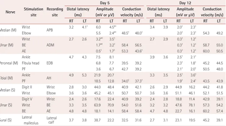
Nerve conduction studies performed on days 5 and 12 after the onset of weakness re- vealed reduced amplitudes in the distal nerves, with partial conduction block as well as mild slowing of the motor conduction velocity and prolonged distal latencies, which con- firmed the diagnosis of AMAN (Table 1) (Supplementary Fig. 2 in the online-only Data Supplement). Furthermore, the finding of an enzyme-linked immunosorbent assay test for serum IgG autoantibodies to ganglioside GM1 was strongly positive (219.93%). The findings of extensive CSF and serological analyses for infectious diseases (syphilis, HIV, fungus, bacterium, and mycobacterium) and viral agents (herpes simplex, varicella zos- ter, cytomegalovirus, and Epstein-Barr virus) as well as CSF oligoclonal band and CSF malignant cell tests were all negative. The patient was treated with intravenous immuno- globulin, but her clinical improvement was slow.
AMAN is a motor-nerve-selective and axonal variant of GBS. The characteristic elec- trophysiological pattern is low-amplitude or absent compound muscle action potentials with normal sensory nerve action potentials and reversible conduction block. Unlike AIDP, AMAN rarely manifests with sensory loss, pain, paresthesia, or autonomic distur- bance.1 The presence of certain symptoms such as bladder/bowel disturbance at symptom onset, the presence of a sensory level and CSF pleocytosis, and marked asymmetry of weakness cast doubt on a GBS diagnosis and may indicate a spinal cord lesion.2 This list of symptoms, which has been provided to clinicians to avoid misdiagnosis of GBS, should Jiwon Yang
Yeong-Bae Lee Kwang-Woo Lee Hyeon-Mi Park
Department of Neurology, Gachon University Gil Medical Center, Incheon, Korea
pISSN 1738-6586 / eISSN 2005-5013 / J Clin Neurol 2017;13(2):205-206 / https://doi.org/10.3988/jcn.2017.13.2.205
Received September 20, 2016 Revised October 10, 2016 Accepted October 13, 2016 Correspondence Hyeon-Mi Park, MD, PhD Department of Neurology, Gachon University Gil Medical Center, 21 Namdong-daero, 774beon-gil, Namdong-gu, Incheon 21565, Korea Tel +82-32-460-3346 Fax +82-32-460-3344
E-mail [email protected]
cc This is an Open Access article distributed under the terms of the Creative Commons Attribution Non-Com- mercial License (http://creativecommons.org/licenses/by-nc/4.0) which permits unrestricted non-commercial use, distribution, and reproduction in any medium, provided the original work is properly cited.
JCN
Open Access LETTER TO THE EDITOR206 J Clin Neurol 2017;13(2):205-206
Atypical AMAN with CSF Pleocytosis
JCN
also be considered in the diagnosis of AMAN.
To the best of our knowledge, the present AMAN patient represents the first published case of AMAN mimicking a spinal cord lesion with pain, neurogenic bladder, and CSF pleocytosis. Only two cases of concomitant transverse my- elitis and AMAN in one adult and one adolescent, respec- tively, have been reported previously.3,4 In these previous cases, sensory and bladder symptoms as well as CSF pleo- cytosis–which are atypical in AMAN–were due to concom- itant myelopathy. In our case there was no evidence of spi- nal cord involvement in MRI, which suggests that the etiology of the atypical features was due to AMAN.
Our case implies that a patient with AMAN may exhibit several atypical manifestations that could lead to a misdiag- nosis with spinal cord disease. Several studies therefore need to be applied to patients who present with the features outlined in this case in order to exclude diseases with simi-
lar presentations.
Supplementary Materials
The online-only Data Supplement is available with this arti- cle at https://doi.org/10.3988/jcn.2017.13.2.205
Conflicts of Interest
The authors have no financial conflicts of interest.
REFERENCES
1. Kuwabara S, Yuki N. Axonal Guillain-Barré syndrome: concepts and controversies. Lancet Neurol 2013;12:1180-1188.
2. Asbury AK, Cornblath DR. Assessment of current diagnostic criteria for Guillain-Barré syndrome. Ann Neurol 1990;27 Suppl:S21-S24.
3. Saidha S, Renganathan R, Spillane J, McNamara B, Fanning N, Ryan AM. Simultaneous transverse myelitis and acute motor axonal neu- ropathy in an adult. J Neurol Neurosurg Psychiatry 2008;79:1302-1303.
4. Howell KB, Wanigasinghe J, Leventer RJ, Ryan MM. Concomitant transverse myelitis and acute motor axonal neuropathy in an adoles- cent. Pediatr Neurol 2007;37:378-381.
Table 1. Results of nerve conduction studies (NCSs). The first NCS revealed (1) reduced CMAP amplitudes in the left median nerve and bilateral ul- nar nerves with partial conduction block, (2) mild slowing of the motor conduction velocity in the bilateral median and tibial nerves, and (3) pro- longed or absent F-wave and H-reflex latencies (data not shown). All of the sensory nerves were normal. Follow-up NCS showed remarkable CMAP amplitude reduction in all motor nerves (bilateral median, ulnar, peroneal, and tibial nerves) and still-preserved sensory nerve functions
Nerve Stimulation site
Recording site
Day 5 Day 12
Distal latency (ms)
Amplitude (mV or μV)
Conduction velocity (m/s)
Distal latency (ms)
Amplitude (mV or μV)
Conduction velocity (m/s)
RT LT RT LT RT LT RT LT RT LT RT LT
Median (M) Wrist
APB 3.2 4.1† 6.0 4.5*† 3.4 3.9 2.0† 2.3†
Elbow 5.5 2.4*† 48.5† 48.0† 2.0† 2.3† 54.3 49.2
Ulnar (M)
Wrist
ADM
2.7 2.6 3.2*† 3.5† 2.7 2.9 0.3† 1.2†
BE 1.7*† 3.2† 58.4 56.5 0.3† 1.2† 58.7 55.0
AE 0.5† 1.7† 53.3 43.6† 0.3† 1.2† 60.0 50.5
Peroneal (M) Ankle
EDB
4.7 4.3 7.5 8.1 3.9 3.6 2.5† 2.1†
Fibula head 6.8 7.7 39.5 39.2 2.3† 1.8† 45.2 44.5
PF 3.6 6.7 42.7 39.2 2.1† 2.0† 50.5 48.0
Tibial (M) Ankle
AH 4.9 5.3 21.9 20.7 3.3 3.5 2.5† 3.6†
PF 18.5 12.8 34.0† 37.3† 1.9† 2.4† 43.5 43.9
Median (S) Digit II Wrist 2.8 3.0 44.0 48.4 40.9 42.1 2.6 2.9 44.9 16.2 44.2 41.8
Wrist Elbow 3.6 3.6 45.2 45.1 50.7 50.7 3.6 3.6 51.1 46.1 52.1 51.5
Ulnar (S)
Digit V Wrist 2.4 2.6 17.6 22.4 40.9 39.2 2.4 2.8 18.8 11.4 42.9 39.1
Wrist BE 3.3 3.5 63.9 70.9 54.0 51.6 3.2 3.2 47.6 78.1 57.3 54.2
BE AE 4.8 4.8 18.1 14.1 58.4 58.4 4.7 4.8 22.7 16.1 60.2 57.4
Sural (S) Lateral malleolus
Lateral
calf 3.7 3.8 38.7 22.2 32.5 31.6 2.7 3.1 23.1 19.5 45.2 39.1
*Conduction block, †Abnormal values.
ADM: abductor digiti minimi, AE: above elbow, AH: abductor hallucis, APB: abductor pollicis brevis, BE: below elbow, CMAP: compound muscle action potential, LT: left, M: motor, PF: popliteal fossa, RT: right, S: sensory.