INTRODUCTION
An ameloblastoma is a fairly common and highly aggressive odontogenic tumor of epithelial origin commonly found in pos- terior mandible and treated with the surgical excision.1As a gen- eral rule, the resection of a portion of the mandible without loss of mandibular continuity is usually not as debilitating as a resec- tion that compromises mandibular continuity.2 Loss of mandibular continuity causes deviation of remaining mandibu- lar segment(s) towards the defect and rotation of the mandibu- lar occlusal plane inferiorly. When surgery includes a segmental mandibulectomy, masticatory function is compromised because of muscular imbalance that results from unilateral muscle removal, altered maxillomandibular relationship, and decreased tooth-to-tooth contacts. Although immediate mandibular reconstruction aims to restore facial symmetry, arch alignment, and stable occlusion, masticatory function often remains compromised.3,4In 1990, a review5of 32 articles described out- comes of various mandibular reconstruction techniques and indi- cated that functional outcomes were provided for only 4% of the 782 patients evaluated. Prosthetic rehabilitation was pre-
sented for only 16 patients (2%) of all mandibular reconstruc- tions. Significant strides in microvascular surgical approach- es during the past decade have permitted predictable restora- tion of bony and soft tissue orofacial defects.6-10However, lim- ited studies indicate only varying degrees of improvement in terms of esthetics, speech intelligibility, swallowing, and masticatory performance.11-17 The longitudinal prospective study was designed by Garrett et al.18to determine whether con- ventional prostheses or implant-supported prostheses and current surgical reconstructive procedures restore patients’oral functions and quality of life to their status prior to segmental mandibulectomy with immediate fibula free-flap recon- struction. They concluded that 72% (33/46) of the subjects enrolled were able and willing to complete treatment with con- ventional prosthesis, and only 35% (16/46) with completed implant-supported prostheses treatment. Guide flange prosthesis (GFP) is a mandibular conventional prosthesis designed for the patient who is able to achieve an appropriate mediolateral posi- tion of the mandible but is unable to repeat this position consistently for adequate mastication.19 This case report describes early prosthodontic management (during initial
Guide flange prosthesis for early management of reconstructed hemimandibulectomy: a case report
Pravinkumar Gajanan Patil1*, MDS, Smita Pravinkumar Patil2, BDS
1Department of Prosthodontics, Government Dental College and Hospital, Nagpur, Maharashtra,
2Department of Orthodontics and Dentofacial Orthopedics, SDKS Dental College and Hospital, Wanadongri, Nagpur, Maharashtra, India
Surgical resection of the mandible due to presence of benign or malignant tumor is the most common cause of the mandibular deviation. Depending upon the location and extent of the tumor in the mandible, various surgical treatment modalities like marginal, segmental, hemi, subtotal, or total mandibulectomy can be performed. The clinicians must wait for extensive period of time for completion of healing and acceptance of the osseous graft before considering the definitive prosthesis. During this initial healing period prosthodontic intervention is required for preventing the mandibular deviation. This case report describes early prosthodontic management of a patient who has undergone a reconstructed hemi-mandibulec- tomy with modified mandibular guide flange prosthesis. The prosthesis helps patient moving the mandible normally without deviation during functions like speech and mastication. [J Adv Prosthodont 2011;3:172-6]
KEY WORDS: Ameloblastoma; Dental prosthesis design; Fibula free-flap; Mandibular prosthesis; Maxillofacial prosthesis; Segmental mandibulectomy
Corresponding author: Pravinkumar Gajanan Patil
Department of Prosthodontics, Government Dental College & Hospital GMC Campus, Medical Square, Nagpur (Maharashtra) 440003, India Tel. 91 9923294699: e-mail, [email protected]
Received May 11, 2011 / Last Revison June 13, 2011 / Accepted June 17, 2011
ⓒ 2011 The Korean Academy of Prosthodontics
This is an Open Access article distributed under the terms of the Creative Commons Attribution Non-Commercial License (http://creativecommons.org/licenses/by- nc/3.0) which permits unrestricted non-commercial use, distribution, and reproduction in any medium, provided the original work is properly cited.
healing period of the reconstructed mandible) of a patient who has undergone a hemimandibulectomy (from left condyle to left parasymphyseal region) and reconstructed with an auto- genous microvascular fibula free-flap (FFF). Modified GFP and the maxillary stabilization plate were fabricated to treat the patient.
CASE REPORT
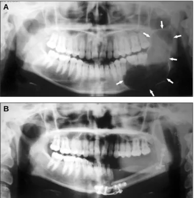
A 17 year old girl was referred to the Department of Prosthodontics (Government Dental College and Hospital, Nagpur, Maharashtra, India) for prosthetic rehabilitation fol- lowing a hemi-mandibulectomy reconstructed with FFF. A detailed case history revealed that the patient was diagnosed with the follicular ameloblastoma of the left mandible 6 months back. A pre-surgical panoramic radiograph revealed extensive radiolucency in the entire left ramus (including a coro- noid process) and left body of the mandible up to the first pre- molar region (Fig. 1A). The patient had undergone hemi- mandibulectomy (from the left condyle to the left parasymphyseal region) and the resultant defect was immediately recon- structed with the FFF 4 months back. A post-surgical panoram- ic radiograph revealed reconstruction bone-plates in the ante- rior region joining the right half of the normal mandible to the horizontally aligned FFF (Fig. 1B). The reconstruction bone- plate was also observed in the left mandibular angle region join-
ing the horizontal and vertical segments of the FFF (Fig.
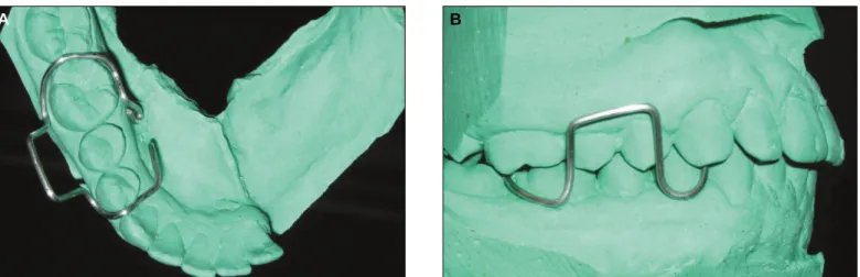
1B). Condylar end of the FFF was not coinciding with the condy- lar fossa but observed to be shifted anterior to the articular emi- nence. Intraoral examination revealed thick, freely movable soft tissues with scar formation, loss of alveolar ridge and obliteration of buccal and lingual sulci in the left half of mandibular region (distal to left lateral incisor) (Fig. 2). The deviation of mandible was observed towards the reconstructed (left) side (about 10 - 12 mm from midline on 40 mm of mouth opening) on opening due to the effect of the normal right mandibular depressor muscles action (Fig. 2). The patient was able to achieve an appropriate mediolateral position of the mandible but was unable to repeat this position consistently for adequate mas- tication. On the basis of clinical and radiographic examination the patient was classified as Class IV (severely compro- mised) according to Prosthodontic Diagnostic Index Resources for partial edentulous patients as described by McGarry et al.20 A stainless steel stock edentulous tray (modified by trimming buccal flange of left half) and irreversible hydrocolloid (Dentalgin; Prime dental products, Mumbai, India) were used to record preliminary impression of the mandibular arch. Maxillary impression was also made with irreversible hydrocolloid. The impressions were poured with Type III gypsum material (Kalstone; Kalabhai Karson, Mumbai, India) and casts were retrieved. A 19 gauge hard, round, stainless steel orthodontic wire (KC Smith and Co, Monmouth, UK) was manipulated (as shown in Figs 3A and B) to fabricate a sub- structure for the modified GFP. The vestibular (buccal and lin- gual) flanges and the mandibular guide-flange were waxed-up with modeling wax (Modeling wax; Deepti Dental Products, Ratnagiri, India) around the wire substructure by keeping a max- illary cast in occlusion and subsequently acrylized into the clear heat-polymerized acrylic resin (DPI Heat cure clear; Dental prod- ucts of India, Mumbai, India) to make the GFP (Fig. 4). A 19
Fig. 1. A: Pre-operative panoramic radiograph showing extensive radi- olucent area in left mandible, B: Post-surgical panoramic radiograph show- ing reconstructed left mandible with osseous graft.
A
B
Fig. 2. Intraoral view showing loss of alveolar ridge on left side with vestibu- lar obliteration. Note arrows indicating thick freely movable soft tissues.
Note the deviation of the mandible on defect (left) side on opening.
gauge hard, round, stainless steel orthodontic wire was manip- ulated to fabricate C clasps on the first premolars and first molars on both the sides of the maxillary cast. A single thickness mod- eling wax was adapted on the maxillary cast covering entire hard palate and the occlusal surfaces of the left posterior teeth and subsequently acrylized into the heat-polymerized clear acrylic resin to make the maxillary stabilization plate (Fig. 5).
The acrylic resin extended over the occlusal surfaces of the left maxillary posterior teeth prevents the possible extrusion of the teeth till replacement of mandibular teeth on defect side.
The GFP and the maxillary stabilization plate were finished and polished in usual manner.
The GFP was tried in patient’s mouth and the initial stabil- ity and retention was checked. The inclination of the guide-flange was adjusted by selectively trimming the teeth-contacting surface or adding the auto-polymerizing clear acrylic resin (DPI Cold cure clear; Dental products of India, Mumbai, India). Thus
the smooth gliding flange surface was developed intraorally to guide the mandible in a definite closing point (rather than the area) in occlusion. Care should be taken to preserve the buc- cal-surface indentations of the opposing maxillary teeth which were guiding the mandible in a final definite closing point during mastication. The flange height was adjusted in such a way that it guided the mandible from large opening position (in practical limits of the height of the buccal vestibule) to the maximum intercuspation in a smooth and unhindered path (Fig.
6 A, B and C). The prosthesis was delivered and post-insertion instructions were given. The patient was followed up at the reg- ular interval of two months for next one year. Patient’s last recall visit was after one and half year of the reconstruction. The patient was pleased with the overall performance of the prosthesis and successfully speaks and masticates without clinically signif- icant deviation. No clinically significant extrusion was observed with maxillary teeth.
Fig. 3. A: Wire substructure: occlusal view, B: Wire substructure: buccal view.
A B
Fig. 4. Completed modified guide flange prosthesis. Note arrows indi- cating the buccal indentations of opposing maxillary teeth in occlusion to guide the mandible in a definite closing point.
Fig. 5. Completed maxillary stabilization plate.
DISCUSSION
Depending upon the location and extent of the tumor in the mandible, various surgical treatment modalities like margin- al, segmental, hemi, subtotal, or total mandibulectomy can be performed.2Loss of mandibular continuity causes deviation of remaining mandibular segment(s) towards the defect and rotation of the mandibular occlusal plane inferiorly. Mandibular deviation toward the defect side occurs primarily because of the loss of tissue involved in the surgical resection.2A verti- cal extension from the buccal aspect of a mandibular prosthesis extends to contact the buccal surface of the opposing maxil- lary teeth. This extension maintains the mandible in the prop- er mediolateral position for vertical chewing, but little, if any, lateral movement is possible. When a segment of the mandible is removed, immediate reconstruction is usually recommended to improve both facial symmetry and masticatory function. Although techniques for reconstructive surgery and prosthodontic rehabilitation have advanced, more than 50% of reconstructed head and neck cancer patients still report impaired masticatory function.3,4 Recent advancements in facial reconstructive surgery and osseointegrated dental implants provide a treatment modality that may adequately reha- bilitate oral cancer patients so that they can return to a healthy, productive life.
Though osseointegrated dental implants is the final solution for replacing the missing teeth for reconstructed mandibulec- tomy patients, the clinicians must wait for extensive period of time (more than a year) for completion of healing and accep- tance of the osseous graft. During this initial healing period ear- ly prosthodontic intervention by mandibular guide flange and maxillary stabilization prosthesis serve the purpose of reduc- ing the mandibular deviation, preventing extrusion of the maxillary teeth and improving the masticatory efficiency.
The patient presented in this article was a teenager female who was very esthetic conscious. Our principal aim was to main- tain her esthetics during mandibular movements. Hence the GFP was fabricated in clear acrylic resin and the retentive wire com-
ponents were kept distal to the mandibular canine to minimize the prosthesis display. Another purpose to keep the prosthe- sis components distally was to prevent any disturbance of the GFP in anterior area where the FFF was attached to the nor- mal mandible with the bone plates. The retentive compo- nents were modified and incorporated into the prosthesis as a wire substructure. Because of amount of force which can be generated by the flange against the maxillary teeth the max- illary stabilizing plate was provided to resist their palatal orthodontic movement. The purpose of occlusal extensions on maxillary left posteriors was to prevent the extrusion of the teeth because of missing opposing mandibular teeth. The retentive components of the maxillary stabilization plate were also kept distal to the canines to minimize the display during functions.
The flange of GFP was localized to three teeth (two premolars and a first molar) to avoid possible dislodging forces in the ante- rior lingual sulcus area (i.e. junction between mandible and fibu- la) and to minimize the display due to the esthetic concerns.
Though the lingual flange of the GFP was short in length, it was sufficient to stabilize the GFP (in this particular patient) as the deviation force was lesser than the stability of the GFP. The lin- gual flange extension on the entire lingual surfaces of the three teeth and deep in the lingual sulcus also helps increasing the stability of the prosthesis. Orthodontic teeth movement of these teeth was one of the concerns. We did not find any clin- ically significant teeth movement on one and half year follow- up-visit may be due to extension of the flanges of the GFP over entire alveolar segment along with the teeth. However the GFP can be extended as long as possible to improve the stability of the appliance as esthetics and comfort permits. The stress result- ed from the GFP can affect healed junction between mandible and grafted fibula, and healed mandibular angle portion between fibula and fibula. In such situation the prosthetic inter- vention can only be started after complete healing of the graft. Care must be taken not to extend the prosthesis in the junc- tional area of the graft and the normal tissue.
Support for the GFP is no different from that of any other Fig. 6. A: Prosthesis in place during large mouth opening. Note occlusal extensions of the maxillary stabilization plate on right posteriors, B: Prosthesis during mandibular movements, C: Prosthesis guided the mandible in a definite closing point.
A B C
removable prosthesis, the natural teeth and the residual alve- olar ridge being the primary sources. Multiple retentive clasps in widely distributed areas of the arch would be the best approach, but actual placement would be determined by the posi- tion of the teeth. Retentive elements should be no more rigid than necessary, but they require a more rigidity with a decreas- ing number of teeth.19In the presented case retentive components were modified and incorporated into the prosthesis as a wire substructure. The buccal and lingual vestibular flanges can be brought closer by bending the occlusal cross-over wire com- ponents with a universal orthodontic plier to improve the retention.
The GFP can be regarded as a training type of prosthesis. If the patient can successfully repeat the mediolateral position, the GFP can often be discontinued. Some patient, however, may continue indefinitely with a guide flange, and the stress gen- erated to the remaining teeth must then be carefully monitored.
REFERENCES
1. Shafer WG, Hine MK, Levy BM, Tomich CE. A textbook of oral pathology. 4thed. Philadelphia; WB Saunders; 1993. p. 86-229.
2. Taylor TD. Diagnostic considerations for prosthodontic reha- bilitation of the mandibulectomy patient. In: Taylor TD, editor.
Clinical maxillofacial prosthetics. Chicago; Quintessence Publishing; 2000. p. 155-70.
3. Olson ML, Shedd DP. Disability and rehabilitation in head and neck cancer patients after treatment. Head Neck Surg 1978;1:52-8.
4. Curtis DA, Plesh O, Miller AJ, Curtis TA, Sharma A, Schweitzer R, Hilsinger RL, Schour L, Singer M. A comparison of masti- catory function in patients with or without reconstruction of the mandible. Head Neck 1997;19:287-96.
5. Komisar A. The functional result of mandibular reconstruction.
Laryngoscope 1990;100:364-74.
6. Hidalgo DA. Fibula free flap: a new method of mandible re- construction. Plast Reconstr Surg 1989;84:71-9.
7. Hidalgo DA. Aesthetic improvements in free-flap mandible reconstruction. Plast Reconstr Surg 1991;88:574-85.
8. Jewer DD, Boyd JB, Manktelow RT, Zuker RM, Rosen IB, Gullane PJ, Rotstein LE, Freeman JE. Orofacial and mandibu-
lar reconstruction with the iliac crest free flap: a review of 60 cas- es and a new method of classification. Plast Reconstr Surg 1989;84:391-403.
9. Soutar DS, Widdowson WP. Immediate reconstruction of the mandible using a vascularized segment of radius. Head Neck Surg 1986;8:232-46.
10. Matloub HS, Larson DL, Kuhn JC, Yousif NJ, Sanger JR.
Lateral arm free flap in oral cavity reconstruction: a function- al evaluation. Head Neck 1989;11:205-11.
11. Marunick MT, Mathes BE, Klein BB. Masticatory function in hemimandibulectomy patients. J Oral Rehabil 1992;19:289-95.
12. McConnel FM, Pauloski BR, Logemann JA, Rademaker AW, Colangelo L, Shedd D, Carroll W, Lewin J, Johnson J. Functional results of primary closure vs flaps in oropharyngeal recon- struction: a prospective study of speech and swallowing. Arch Otolaryngol Head Neck Surg 1998;124:625-30.
13. Hsiao HT, Leu YS, Lin CC. Primary closure versus radial forearm flap reconstruction after hemiglossectomy: functional assessment of swallowing and speech. Ann Plast Surg 2002;49:
612-6.
14. McConnel FM, Teichgraeber JF, Adler RK. A comparison of three methods of oral reconstruction. Arch Otolaryngol Head Neck Surg 1987;113:496-500.
15. Pauloski BR, Rademaker AW, Logemann JA, McConnel FM, Heiser MA, Cardinale S, Lazarus CL, Pelzer H, Stein D, Beery Q. Surgical variables affecting swallowing in patients treated for oral/oropharyngeal cancer. Head Neck 2004;26:625-36.
16. Hsiao HT, Leu YS, Chang SH, Lee JT. Swallowing function in patients who underwent hemiglossectomy: comparison of pri- mary closure and free radial forearm flap reconstruction with vide- ofluoroscopy. Ann Plast Surg 2003;50:450-5.
17. Wagner JD, Coleman JJ 3rd, Weisberger E, Righi PD, Radpour S, McGarvey S, Bayler A, Chen J, Crow H. Predictive factors for functional recovery after free tissue transfer oromandibular reconstruction. Am J Surg 1998;176:430-5.
18. Garrett N, Roumanas ED, Blackwell KE, Freymiller E, Abemayor E, Wong WK, Gerratt B, Berke G, Beumer J 3rd, Kapur KK.
Efficacy of conventional and implant-supported mandibular resection prostheses: study overview and treatment outcomes.
J Prosthet Dent 2006;96:13-24.
19. Desjardins RP. Relating examination findings to treatment procedures. In: Laney WR. Maxillofacial prosthetics. Littleton;
PSG Publishing; 1979. p. 69-114.
20. McGarry TJ, Nimmo A, Skiba JF, Ahlstrom RH, Smith CR, Koumjian JH, Arbree NS. Classification system for partial edentulism. J Prosthodont 2002;11:181-93.