The Occurrence of Degenerative Change in the Mandibular Condyles of Korean Patients with Temporomandibular
Disorders
Jung-Hwan Jo, D.D.S.,M.S.D., Min-Woo Park, D.D.S., Young-Ku Kim, D.D.S.,Ph.D., Jeong-Yun Lee, D.D.S.,Ph.D.
Department of Oral Medicine & Oral Diagnosis, School of Dentistry & Dental Research Institute, Seoul National University, Yeongeon-Dong 28, Jongno-Gu, Seoul 110-749, Korea (ROK)
Osteoarthritis (OA), the most common form of arthritis, is a result of both mechanical and biological events that destabilize the normal coupling of degradation and synthesis of articular cartilage chondrocytes and extracelluar matrix, and subchondral bone. Although it is likely that the molecular basis of pathogenesis is similar to that of other joints, additional study of the temporomandibular joint (TMJ) is required due to its unique structure and function. This study was carried out to evaluate the epidemiologic characteristics of TMJ osteoarthritis.
The purpose of this study was to investigate the prevalence of TMJ OA in Patients with temporomandibular disorders (1405 men and 2922 women whose mean age was 30.2 ± 15.4 and 33.1 ± 15.2 years, respectively) who had visited the TMJ and Orofacial Pain Clinic of Seoul National University Dental Hospital in 2007. Orthopantomograms, TMJ tomograms and transcranial radiographs were used to evaluate degenerative change of the mandibular condyle.
The obtained results were as follows:
1. Degenerative change of the mandibular condyle was observed in 883 (20.4%) of 4327 subjects. The prevalence was significantly higher in women (706 patients, 24.1%) than in men (177 patients, 12.6%), and this significant difference between genders was observed in all age groups.
2. The prevalence of degenerative change of the mandibular condyle in TMD patients showed a gentle increase along with age. Such increase was statistically significant in women (P < 0.001), but not in men.
3. Sclerosis was observed the most frequently in all age groups and the mean age of the patients with osteophyte was the highest among four types of degenerative change.
4. Although men showed degenerative change in the left side more often and women showed degenerative change more frequently in both sides, the difference of distribution in sides between genders was not significant.
In conclusion, the prevalence of degenerative change of the mandibular condyle in TMD patients is higher in women than in men, and increases steadily with aging, but not as dramatically as in other joints that show a steep increase in prevalence around the age of 45 years. It can be suggested that the epidemiologic characteristic of OA of the TMJ differs from those of other joints, and that a more extensive study based on the general population is necessary.
Key words: Temporomandibular joint, degenerative joint diseases, osteoarthritis, prevalence
Corresponding author: Dr. Jeong-Yun Lee
Assistant Professor, Dept. of Oral Medicine and Oral Diagnosis School of Dentistry & Dental Research Institute
Seoul National University Yeongeon-Dong 28, Jongno-Gu Seoul 110-749 Korea (ROK)
Tel: +82-2-2072-0212 Fax: +82-2-744-9135 E-mail: [email protected] Received: 2011-01-25 Accepted: 2011-03-11
Ⅰ. INTRODUCTION
Osteoarthritis (OA), the most common form of arthritis, is a consequence of mechanical and biological events that destabilize the normal coupling of degradation and synthesis of articular cartilage chondrocytes, extracellular matrix, and subchondral bone. Generally OA involves the knee, hip, hand, and spinal joints that are positioned to bear physical load. OA results from systemic factors such as gender, age, genetics, and ethinicity, and mechanical factors such as obesity, injury, and muscle weakness1).
Epidemiological studies of OA have been performed using population-based radiographic surveys for the last several decades. The prevalence of OA increases with age at all joint sites, with a noticeable increase after 45 years of age2). Another feature of OA is the higher prevalence and more generalized distribution in women than in men. For most joints, there is no significant difference between genders before the age of 50, but after 50 years, the prevalence and incidence of disease is significantly greater in women than in men2,3). Previous studies have provided estimates of the prevalence of degenerative temporomandibular joint (TMJ) disease ranging from 8% to 35%4-6). Examination of TMJ specimens through autopsy revealed changes consistent with OA in 22% to 40%7-9). The general consensus is that degenerative change in the TMJ becomes more common with aging, but most previous studies are based on relatively small sized smalples or samples of insufficient age range to establish the correlation between age and the prevalence of degenerative change of the TMJ. Interestingly, some studies have shown that degenerative change of the mandibular condyle can begin at a young age10,11). This suggests that the epidemiological characteristics of TMJ OA may differ from those of other joints which show an obvious increase of prevalence after the middle age.
Many clinical studies indicate that women have a higher prevalence of OA in the TMJ12,13), but other investigators have reported that there is no
significant gender difference in the actual incidence of changes in joint morphology8,14). Some investigators have even reported a higher incidence of OA in men9,15).
This study was aimed to provide clinical information concerning the prevalence of degenerative change of the mandibular condyle based on the radiographic data from initial-visit- patients with temporomandibular disorders (TMDs), and so to raise an issue on the epidemiological characteristics and pathophysiology of TMJ OA.
Ⅱ. MATERIALS AND METHODS 1. Subjects
A retrospective cross-sectional study was performed on radiographs taken of 4,327 patients (1,405 men and 2,922 women, mean age 30.2 ± 15.4 and 33.1 ± 15.2 years, respectively), who visited the TMJ & Orofacial Pain Clinic of Seoul National University Dental Hospital for the first time to have their TMD symptoms examined in 2007. The total number of patients who complained or were referred for examination of any type of TMD-related signs or symptoms in 2007 were 5,739, and the radiographs of 4,327 patients were finally chosen by exclusion of the patients who had previous examinations of TMD-related signs and symptoms during the past 5 years and patients with major deformity, TMJ fracture, and systemic diseases known to affect the TMJ, such as rheumatoid arthritis. The research protocol was approved by the Institutional Review Board of the University Hospital (#CRI08025).
2. Radiographic Evaluation
Degenerative change of the mandibular condyle was evaluated by reviewing the radiographs including standard panoramic, TMJ panoramic, and transcranial radiographs retrospectively. All three radiographs were routinely taken in the TMD diagnostic procedure. The radiographs were taken
into the image plates by OrthopantomographⓇ OP 100 (Instrumentarium Dental, Finland) for standard panoramic and TMJ panoramic radiographs and DXG-100N (Listem, Korea) for transcranial radiographs. Images were digitalized by scanning of image plates with FCR XG5000 (Fujifilm, Japan).
TMJ panoramic radiographs were taken according to the optimum standards provided by the manufacturer, with modified focal trough locating to the TMJ in mouth-opening position
Degenerative changes of the mandibular condyle were classified according to morphological change and cortical delineation of the mandibular condyle.
This resulted in four classifications: sclerosis, erosion, flattening, and osteophyte formation. When at least one of the changes was observed, in other words the normal bony condition of the condyle was disrupted, the condyle was diagnosed as having a degenerative change. For the consistency of reading, two authors who specialized in orofacial pain and temporomandibular disorders had a mutual calibration period reading radiographs together and reaching agreement on determining the type of bony change. After an agreement had been established, all measurements were carried out by a single examiner to guarantee higher reliability.
3. Statistical Analysis
To evaluate intraexaminer reliability within a single examiner, 30 randomly selected radiographs were examined twice by the same examiner at four week intervals. Cohen’s κ statistics were used to assess intraexaminer reproducibility.
Chi-square tests were performed to analyze the relationship between the frequency of degenerative change and age with regard to gender and side.
Odds ratios of gender and age groups for degenerative changes of the mandibular condyles were calculated by a binary logistic regression model. All statistical analyses were performed using SPSS 12.0 for Windows.
Ⅲ. RESULTS
Cohen’s κ value was 0.87 for determining OA and 0.79 for the types of OA (P < 0.001). This represented excellent and substantial reliability of examination, respectively16).
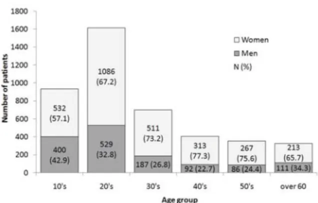
As shown in Figure 1, approximately twice as many women visited the clinic for an initial TMJ examination as men, and most patients were in their 20s. Of all patients, 4,255 (98.3%) patients complained of at least one symptom including jaw pain, joint noise, mouth opening limitation, jaw stiffness, occlusal discomfort, bruxism, joint swelling, subluxation, jaw asymmetry, headache, and phonetic problems. The rest of the patients were referred by other clinicians because of signs of TMDs that patients did not recognize, such as asymptomatic bone change of the mandibular condyles or mouth opening limitation. Jaw pain was the most frequent complaint (64.4%) followed by joint noise (43.7%), mouth opening limitation (19.3%), and jaw stiffness (10.8%). Among the clinical indices of the 8,654 TMJs of 4,327 patients including pain on palpation, joint noise, and subjective pain, only the incidence of joint noise was significantly related to the occurrence of degenerative change of the mandibular condyle (P
< 0.001, data not shown). Degenerative change of the mandibular condyle was observed in 883 (20.4%) of the 4,327 patients. The incidence was significantly higher in women (706 women, 24.1%)
Fig. 1. Age and gender distribution of the patients.
N (%)
Men Women††† Total†††
Degenerative
change Intact Degenerative
change Intact Degenerative
change Intact
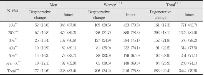
10’s** 52 (13.0) 348 (87.0) 109 (20.5) 423 (79.5) 161 (17.3) 771 (82.7) 20’s*** 57 (10.8) 472 (89.2) 236 (21.7) 850 (78.3) 293 (18.1) 1322 (81.9) 30’s** 25 (13.4) 162 (86.6) 127 (24.9) 384 (75.1) 152 (21.8) 546 (78.2) 40’s** 10 (10.9) 82 (89.1) 81 (25.9) 232 (74.1) 91 (22.5) 314 (77.5) 50’s** 14 (16.3) 72 (83.7) 88 (33.0) 179 (67.0) 102 (28.9) 251 (71.1) over 60** 19 (17.1) 92 (82.9) 65 (30.5) 148 (69.5) 84 (25.9) 240 (74.1) Total*** 177 (12.6) 1228 (87.4) 706 (24.2) 2216 (75.8) 883 (20.4) 3444 (79.6)
*: Gender difference by chi-square test, **: P < 0.01,***:P < 0.001
†: Difference according to age group by Chi-square test, †††: P < 0.001
Table 1. Age and gender distribution of patients with degenerative change of the mandibular condyle.
Fig. 2. Occurrence of degenerative change according to age and gender. The occurrence of degenerative change increased with age, and this increase was statistically significant in women (P < 0.001), but not in men, analyzed by chi-square test.
than in men (177 men, 12.6%) (P < 0.001). The prevalence of degenerative change of the mandibular condyle in TMD patients according to gender and age distribution is shown in Table 1 and Figure 2, and was significantly higher in women than in men in all age groups (P < 0.01). The occurrence of degenerative change increased with age, and this increase was statistically significant in women (P <
0.001), but not in men. Odds ratios of gender and
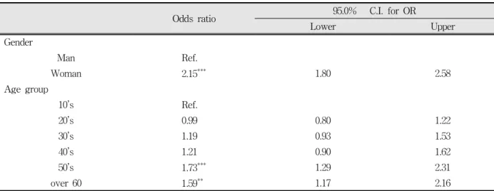
age groups for degenerative changes of the mandibular condyle are shown in Table 2. The odds ratios of being a woman (2.154, P < 0.001), aged 50’s (1.73, P < 0.001), and over 60 years (1.59, P < 0.01) were significant. Figure 3 shows the ubiquity of degenerative change of the mandibular condyle according to types of degenerative change. Sclerosis was observed most frequently in all age groups and the percentage of sclerosis in patients with degenerative change was 84.6% (747 among 883 patients). The presence of osteophytes significantly increased with age in both genders (P < 0.001) and erosion significantly increased with age in women (P < 0.001). Sclerosis and flattening did not show a significant increase with age in either gender. The mean age of patients with osteophytes was the highest (44.8 ± 16.6 years) among the four types of degenerative change in both genders; 35.2 ± 16.6 years for sclerosis, 38.0 ± 18.0 years for erosion, and 33.6 ± 16.1 years for flattening (analyzed by ANOVA, P < 0.01). The distribution of degenerative change of the mandibular condyle according to side was also analyzed. Although the left side was the most frequently affected in men (P < 0.05) and bilateral change was the most frequent in women (P
< 0.001), the overall pattern of distribution of affected sides was not different between genders.
Odds ratio 95.0% C.I. for OR
Lower Upper
Gender
Man Ref.
Woman 2.15*** 1.80 2.58
Age group
10’s Ref.
20’s 0.99 0.80 1.22
30’s 1.19 0.93 1.53
40’s 1.21 0.90 1.62
50’s 1.73*** 1.29 2.31
over 60 1.59** 1.17 2.16
*: determined by binary logistic regression model, **: P < 0.01, ***: P < 0.001 Model x2=109.07, df=6,P=0.000, R2=0.025
Table 2. Odds ratios of gender and age groups for degenerative change of the mandibular condyle in a binary logistic regression model.
Fig. 3. Occurrence of types of degenerative change according to age. Sclerosis was observed most frequently in all age groups. The presence of osteophytes significantly increased with age in both genders (P <
0.001) and erosion significantly increased with age in women (P < 0.001). analyzed by chi-square test.
Sclerosis and flattening did not show a significant increase with age in either gender.
Ⅳ. DISCUSSION
Although the results of epidemiological studies of TMJ OA vary according to diagnostic criteria4-9), TMJ OA is generally considered as an age-related joint disease as those of other joints of the body.
But, it is also true that there is no established epidemiological evidence based on a large scale sample of general population to support such a relationship or a difference compared to other joints between age and TMJ OA. There are several thousands of patients a year who visit the TMJ and Orofacial Pain Clinic of Seoul National University Dental Hospital seeking TMJ examination. They complain of TMD-related symptoms for themselves or are referred by other clinicians because of objective TMD-related signs regardless of their own discomfort. Through clinical evaluation, panoramic radiograph and interviews, the patients with non-TMD conditions such as primary headache, infection, neuralgia or salivary gland diseases are ruled out, and the remaining patient who are clinically diagnosed as TMDs are processed with additional radiographs including TMJ panoramic and transcranial radiographs. Such patients were 5,739 in 2007 and the clinical records and all three types of radiographs of them were reviewed for this study. Based on the clinical experience with such a large number of TMD patients, TMJ OA seems to commonly occur in young patients in their second to forth decade. This study was performed to report these clinical findings of Korea and raise an issue to activate an epidemiological study on the realistic prevalence of TMJ OA. For this purpose, we retrospectively reviewed radiographs taken of 4,327 patients who had had examinations of their TMJ for the first time in 2007, excluding the patients who had ever visited for the examination of TMD-related signs and symptoms during the past 5 years and patients with major deformity, TMJ fracture, and systemic diseases known to affect the TMJ, such as rheumatoid arthritis from the total of 5,739 patients.
Radiographic examination is of great value for
detecting degenerative changes in osseous components. In the present study, degenerative change of the mandibular condyle was evaluated using all three types of radiographs consisting of standard panoramic, TMJ panoramic, and transcranial radiographs routinely taken for the diagnosis of TMD. Although computed tomography (CT) and magnetic resonance imaging (MRI) are the most valuable radiographic tools in diagnosing OA17,18), they technically cannot be performed on every single patient due to the high cost or risks of high dose radiation exposure. TMJ panoramic radiographs taken with modified focal trough locating to the TMJ in the mouth-open position can give more accurate images for detecting structural changes than standard panoramic radiographs by avoiding the overlapping of the mandibular condyles with other adjacent structures. Moreover, since a TMJ panoramic radiograph can show the medial side of the condylar head of the TMJ while a transcranial radiograph can show the lateral side, these two techniques can be used together to more completely evaluate changes of the TMJ.
To determine degenerative change of the TMJ, four types of bony change were differentiated in this study. Although the clinical significance of sclerosis and flattening is doubtful, and minimal flattening is common in many asymptomatic persons6), the joint with at least one of any type of bony change was determined to have degenerative change because it was more reliable to determine whether a joint was normal or not than the degree of abnormality. The procedure of determining what significant sclerosis or flattening is may cause more bias than deciding the presence of sclerosis or flattening. The plain radiographs do not have a high enough sensitivity to apply complicated criteria of decision or overestimate the bony change significantly19). To reduce possible errors due to the inaccuracy of plain radiographs, it would be better to simplify the criteria and increase the sample size as in this study.
In previous studies, radiographic evidence of TMJ OA was found in 14% to 44% of symptom-free individuals20), and histological evidence of the
disease was present in 40% to 60% of studied populations22,23). Clinical evidence of disease occurred in only 8% to 16% of the population24,25), due to the fact that OA is not always accompanied by clinical symptoms. Principal clinical findings in OA of the TMJ including pain, reduced range of motion, joint tenderness on palpation and crepitation do not differ from those of other diseases, and are therefore not pathognomonic for TMJ OA. Also, crepitation has been shown to have low sensitivity as a diagnostic sign25,26). In this study, pain on palpation and subjective pain of the joint were not associated with the occurrence of degenerative change of the mandibular condyles as described in previous studies, but the incidence of joint noise showed significant relations (P < 0.001). This implies that, although its ubiquity makes it hard to be considered as a predictor of degenerative change of the mandibular condyles and treatment aimed directly at its elimination may not be necessary in most cases, joint noises should not be neglected simply because other signs or symptoms are not present or because the joint noise is not always pathologic. Patients with TMJ noise should be motivated and advised to reduce the load on their joint and should be observed closely during follow-up.
The increase in frequency of degenerative change of the mandibular condyle with age in TMD patients that was detected in this study is consistent with the results of previous studies27-29). However, it is noteworthy that, dividing the subjects in this study into two groups by age of 45 years, compared to the 18.6% increase in prevalence in subjects under the age 45, the 26.7% increase in prevalence after the age of 45 is very subtle and clearly distinguishable from those of other load-bearing joints which have been described as showing a steep increase of prevalence after the age of 45 in previous epidemiological studies. Van Saase and colleagues2) reported that the frequency of radiographic knee OA rose from 1-4% in women 24 to 45 years of age and to 53-55% in women 80 years of age and older. In men, the occurrence rises from 1-6% in those 45 years and younger to 22-33% in those 80 years of
age and older. In the results of this study, odds ratios of age groups with 10’s age group as a reference did not exceed 2.00 and were significant only in 50’s and over 60 years’ age group. The frequency of degenerative change of the mandibular condyle in young patients was as high as 17-20%.
This is consistent with the results of previous studies30), in which OA was observed before adolescence, and the symptoms of OA were found to occur mainly in the third decade of life, and degenerative change of the mandibular condyle was seen to start at a young age10,11). This low correlation between age and occurrence of degenerative change of the TMJ imply that there may be some difference in pathophysiology between the TMJ and other joints. Such difference may arise from the fact that the TMJ has a different mechanical load-bearing system compared to other weight-bearing joints, not that it has a biochemically different pathophysiology. In other words, although the biochemical or cellular tissue responses of the loaded TMJ are the same as those of other joints, the way in which the TMJ endure functional load is totally different. The functional load of the TMJ is much more complicated in a manner that depends more on how the joint is used actively by the host rather than what weight and how long it is passively applied to the joint. If heavy functional load overwhelming the adaptive capacity of the TMJ is given to the joint by any factors caused by change of muscle activity or jaw movement, it can bring about some degenerative change to the TMJ regardless of the chronologic age of the joint. Such functional aspects of the TMJ must have caused the differences in occurrence of degenerative change showing in this study, not the uniqueness of the histological and anatomical structure itself, nor unknown, if any, unique biochemical pathophysiology.
Considering the comprehensive meaning of the degeneration of the joint involving most abnormal tissue response to functional overload such as change of the position or shape of the TMJ disk, loosening of the ligamental tension, and denaturation
of the synovial component31), it is logical to think that bony degeneration can as easily occur even in a young age as other common TMD symptoms depending on how much functional load the joint should endure. On the same point of view, the relatively low prevalence in old age compared to the major joints suggests that the occurrence of degenerative changes of the TMJ is more related to the functional aspect rather than the chronological aspect.
Compared to the results of previous autopsy- based studies, the results of the present study reveal a relatively low prevalence of degenerative change in the elderly. This may be due to the high sensitivity of postmortem or autopsy examinations in evaluating degenerative change9,30). It is known from postmortem studies that the active pathological process of OA takes place several years before radiological detection of the disorder. The consideration of bony changes due strictly to aging, and not from pathological processes, would lead to the overestimation of OA in postmortem studies.
Haskin and colleagues32) noted that contemporary studies of the TMJ have not adequately distinguished age-related changes in joint structure and function from pathological changes associated with degenerative arthritis, including OA. With the anticipation that new diagnostic techniques will someday permit clinical researchers to probe the human TMJ at the molecular level, the ability to distinguish pathological states from the normal aging processes will be essential33).
The female predominance in degenerative change of the mandibular condyle observed in the present study (odds ratio=2.15, P < 0.001) is consistent with the results of previous studies12,13,27). The reason for this phenomenon is unclear, but it may be due to the differences of sex hormones, pain perception, responses to stress, and psychological factors.
Several studies have not found any gender differences in degenerative change of the TMJ8,14), while others have reported a higher prevalence in men9,15).
Among the four types of degenerative change,
sclerosis was observed the most frequently in all age groups. This may be because sclerosis is found in early stages of degenerative change as the thickening of the subchondral bony plate. Generally, as the disease progresses, flattening and marginal lipping of the condyle and flattening of the articular eminence may take place. In the late stages of the disease, changes are observed in the form of erosion of the cortical plate, osteophyte formation, or both34). This is in agreement with the result where the mean age of patients with osteophytes was the highest among the four types of degenerative change in both genders.
The results showed that in both genders, subjects more frequently had degenerative change on the left side, and that the gender differences of distribution of the affected side were not significant. Unilateral involvement was observed more frequently than bilateral involvement of degenerative change of the mandibular condyle. Gray35) states that OA is generally unilateral, and when bilateral involvement does occur, one side usually exhibits greater severity. The reasons for this phenomena are not clear, but some studies have reported a relationship between length of the mandibular ramus, unilateral chewing, and parafunctional habits36).
The major limitations of this study are that the study sample included patients who visited our clinic for TMJ examination rather than a randomly selected sample from the general population, and that the determination of the degenerative change was made only by plain radiographs. More extensive studies based on the general population and more advanced radiographic examinations, such as cone beam computed tomography, should be performed in order to gain comprehensive knowledge in understanding the epidemiological characteristics of OA of the TMJ. It is also true that social, political, and economical problems are obstacles. Considering the fact that the prevalence of TMD itself increases with age in the young population and then decreases in the elderly37,38) after peaking during middle age, the high prevalence of degenerative change of TMJ in the young TMD patients observed in this study
should not be underestimated due to the limitations of the samples or radiographic examinations used in this study. In other words, if TMD symptoms were generally rare in young adults, the high prevalence of degenerative change of young TMD patients should not be so high when interpolated into the general population. However, because TMD symptoms are very common in young adults, it can be postulated that the degenerative change of TMJ is as common in the young population as it was in the young TMD patients of this study, or more on the contrary. If any overestimation or underestimation of the incidence of degenerative changes had occurred because of the inaccuracy of the radiographic examinations used in this study, it would have equally affected all age groups of this large sample. So, at least, the trend of the high incidence in young adults and slight increase according to aging in this study should not be neglected just because of the limitation of radiographic examinations.
V. CONCLUSIONS
The prevalence of degenerative change of the mandibular condyle in Korean TMD patients is higher in women than in men, and relatively high in young adults. Its increase along with age is not as dramatic as in other joints that show a steep increase around middle age. Therefore, it can be suggested that the epidemiological characteristics of OA of the TMJ differ from those of other joints, and that a more expansive study to elucidate the exact epidemiological and biological characteristics of TMJ OA based on the general population is required.
REFERENCES
1. Kuettner KE, Goldberg VM. Introduction. In Kuettner KE, Goldberg VM (Eds). Osteoarthritic disorders.
Rosemont, 1995, American Academy of Orthopaedic Surgeons, pp. 21-25.
2. Van Saase JL, van Romunde LK, Cats A, Vandenbroucke JP, Valkenburg HA. Epidemiology of osteoarthritis: Zoetermeer survey. Comparison of
radiological osteoarthritis in a Dutch population with that in 10 other populations. Annals of the Rheumatic Diseases 1989;48:271-280.
3. Felson DT. An update on the pathogenesis and epidemiology of osteoarthritis. Radiol Clin N Am 2004;42:1-9.
4. Toller PA. Osteoarthrosis of the mandibular condyle.
Brit Dent J 1973;134:223-230.
5. Helöe B, Hel?e LA. Characteristics of a group of patients with temporomandibular joint disorders.
Community Dent. Oral Epidemiol 1975;3:72-79.
6. Brooks SL, Westesson PL, Eriksson L, Hansson LG, Barsotti JB. Prevalence of osseous changes in the temporomandibular joint of asymptomatic persons without internal derangement. Oral Surg Oral Med Oral Pathol 1992;73:118-122.
7. Axelsson S. Human and experimental osteoarthrosis of the temporomandibular joint. Morphological and biochemical studies. Swed Dent J 1993;92:1-45.
8. Widmalm SE, Westesson PL, Kim IK, Pereira FJ Jr, Lundh H, Tasaki MM. Temporomandibular joint pathosis related to sex, age, and dentition in autopsy material. Oral Surg Oral med Oral Pathol 1994;78:
416-425.
9. Magnusson C, Ernberg M, Magnusson T. A description of a contemporary human skull material in respect of age gender, temporomandibular joint changes, and some dental vaiables. Swed Dent J 2008;32:69-83.
10. Dibbets JM, van der Weele LT. Prevelance of structural bony change in the mandibular condyle. J Craniomandib Disord 1992;6:254-259.
11. Susami T, Kuroda T, Yano Y, Nakamura T. Growth changes and orthdontic treatment in a patient with condylolysis. Am J Orthodo Dentofac Orthop 1992;102:295-301.
12. Kamelchuk LS, Major PW. Degenerative disease of the temporomandibular joint. J Orofac Pain1995;9:
168-180.
13. Rasmussen OC. Description of population and progress of symptoms in a longitudinal study of temporomandibular arthropathy. Scand J Dent Res 1981;89:196-203.
14. Axelsson S, Fitins D, Hellsing G, Holmlund A.
Arthrotic changes and deviation in form of the temporomandibular joint-an autopsy study. Swed Dent J 1987;11:195-200.
15. Richards LC. Degenerative changes in the temporo- mandibular joint in two Australian Aboriginal
populations. J Dent Res 1988;67:1529-1533.
16. Landis JR, Koch GG. The measurement of observer agreement for categorical data. Biometrics 1977;33:
159-174.
17. Alexiou K, Stamatakis H, Tsiklakis K. Evaluation of the severity of temporomandibular joint osteoarthritic changes related to age using cone beam computed tomography. Dentomaxillofac Radiol 2009;38:141-147.
18. Tasaki MM, Westesson PL. Temporomandibular joint: diagnostic accuracy with sagittal and coronal MR imaging. Radiol 1993;186:723-729.
19. Larheim TA, Johannessen S, Tveito L. Abnormalities of the temporomandibular joint in adults with rheumatic disease: a comparison of panoramic, transcranial, and transpharyngeal radiography with tomography. Dentomaxillofac Radiol 1988;17:109-113.
20. Kellgren JH, Moore R. Generalized osteoarthritis and Heberden s nodes. Br Med J 1952;1:181-187.
21. Macalister AD. A microscopic study of the human temporomandibular joint. N Z Dent J 1954;50:161-172.
22. Dijkgraaf LC, Liem RS, de Bont LG. Synovial membrane involvement in osteoarthritic temporo- mandibular joints: A light microscopic study. Oral Surg Oral Med Oral Pathol Oral Radiol Endod 1997;83:373-386.
23. Mejersjo C. Therapeutic and prognostic consider- ations in TMJ osteoarthrosis: A literature review and a long term study in 11 subjects. Cranio 1987;5:69-78.
24. Toller P. Temporomandibular arthropathy. Proc R Soc Med 1974;67:153-159.
25. Rohlin M, Westesson P-L, Ericksson L. The correlation of temporomandibualar joint sounds with morphology in fifty-five autopsy specimens. J Oral Maxillofac Surg 1985;43:194-200.
26. Anderson QN, Katzberg RW. Pathologic evaluation of disc dysfunction and ossous abnormalities of the temporomandibular joint. J Oral Maxillofac Surg 1985;43:947-951.
27. Oberg T, Carlsson GE, Fajers CM. The temporoman- dibular joint. A morphologic study on a human autopsy material. Acta Odontol Scand 1971;29:349- 384.
28. Kopp S. Topographical distribution of sulfated glyscosaminoglycans in the surface layers of the human temporomandibular joint. A histochemical study of an autopsy material. J Oral Pathol 1978;7:
283-294.
29. Akerman S. Morphologic, radiologic and thermometric assessment of degenerative and inflammatory temporomandibular joint disease. An autopsy and clinical study. Swed Dent J 1987;52:1-110.
30. Bates RE Jr, Gremillion HA, Stewart CM.
Degenerative joint disease. Part I: Diagnosis and management considerations. Cranio 1993;11:284-290.
31. Stegenga B: Osteoarthritis of the temporomandibular joint organ and its relationship to disc displacement.
J Orofac Pain 15:193, 2001
32. Haskin CL, Milam SB, Cmeron IL. Pathogenesis of degenerative joint disease in the human temporomandibular joint. Crit Rev Oral Biol Med 1995; 6: 248-277.
33. Milam SB. TMJ Osteoarthritis. In Laskin DM, Greene CS, Hylander WL, editors. Temporomandibular disorders. An evidence-based approach to diagnosis and treatment. Chicago: Quintessense Publishing;
2006. p. 105-107.
34. Abubaker AO. TMJ Arthritis. In Laskin DM, Greene CS, Hylander WL, editors. Temporomandibular disorders. An evidence-based approach to diagnosis and treatment. Chicago: Quintessense Publishing;
2006. p. 229-248.
35. Gray RJM. Pain dysfunction syndrome and osteoarthrosis related to unilateral and bilateral temporomandibular joint symptoms. J Dent 1986;14:
156-159.
36. Boering G. Temporomandibular joint arthrosis? A clinical and radiographic investigation. 1994, Drukkerij Van Denderen Groningen, pp.185-214, 315-360.
37. Von Korff M, Dworkin SF, LeResche L, Kruger A.
An epidemiologic comparison of pain complaints. Pain 1988;32:173-183.
38. De Kanter RJ, Truin GJ, Burgersdijk RC, Van 't Hof MA, Battistuzzi PG, Kalsbeek H, et al. Prevalence in the Dutch adult population and a meta-analysis of signs and symptoms of temporomandibular disorder.
J Dent Res 1993;72:1509-1518.
국문초록
측두하악장애 환자에서 하악과두의 퇴행성 변화의 발생 양상에 대한 연구
서울대학교 치의학대학원 구강내과진단학 교실, 치의학연구소
조정환․박민우․김영구․이정윤
골관절염은 관절의 연조직, 관절연골 및 연골하 골조직의 합성과 분해 사이의 불균형으로 인한 조직의 변성을 야기하는 관절의 질환이다. 일반적으로 골관절염은 연령에 따라 급격히 증가하고, 여성에서 호발하는 특징을 보이는 것으로 알려져 있지만, 측두하악관절에서의 골관절염의 유병률에 관한 연구는 측두하악관절의 독특한 특성에도 불구하고 정확한 연구가 부 족한 실정이다. 본 연구에서는 측두하악관절에서 발생하는 골관절염의 실제 성별, 연령별 분포 및 임상적 특징을 알아봄으로 써 병인의 이해에 필요한 기초 자료를 제시하고자 한다.
본 연구에서는 2007년 1월부터 12월까지 1년 동안 서울대학교 치과병원에 측두하악장애를 주소로 처음 내원한 4327명의 환자를 대상으로 하였다. 측두하악장애의 진단을 위해 촬영된 orthopantomogram, TMJ tomogram, transcranial radiograph 를 이용하여 측두하악관절의 퇴행성 변화를 조사하였으며, 성별, 연령, 골변화 양상에 따른 분포를 분석하여 다음과 같은 결 과를 얻었다.
1. 측두하악장애 증상을 호소하는 환자 4327명 중 883명 (20.4%)에서 퇴행성 변화가 관찰되었고, 남성에서는 177명 (12.6%), 여성에서는 706명 (24.1%)에서 관찰되어 여자에서 호발하는 것으로 관찰되었으며, 이러한 여성 우위의 호발 양상은 모든 연령대에서 관찰되었다.
2. 측두하악장애 증상을 호소하는 환자 중 하악과두의 퇴행성 변화를 보이는 환자의 비율은 연령대가 증가함에 따라 완만하 게 증가하였고, 연령별로 구분하여 볼 때에는 여성에서는 연령대별 발생비율의 증가가 통계적으로 유의하였으나 (P <
0.001), 남성에서는 통계적으로 유의하지 않았다.
3. 퇴행성 변화 환자군에서 나타나는 퇴행성 변화의 유형 중, 발생빈도가 가장 높은 형태는 sclerosis였고 (84.6%), 발생 환자 의 평균연령이 가장 높은 형태는 osteophyte였다 (44.8세).
4. 퇴행성 변화의 좌, 우 발생 분포를 보면 좌측에서 호발하였고, 남성 내에서는 좌측, 여성 내에서는 양측에서 모두 발생한 비율이 가장 높았으나, 전체적인 좌, 우 발생 양상은 남녀간에 통계적으로 유의한 차이를 보이지 않았다.
결론적으로, 측두하악장애 환자에서 나타나는 측두하악관절의 퇴행성 변화는 여성에서 호발하고 연령이 증가함에 따라 완 만한 증가를 보이며 젊은 연령층의 환자에서도 높은 비율로 발생함을 알 수 있다. 측두하악장애가 젊은 연령층에서 호발한다 는 이전의 보고들을 토대로 볼 때 측두하악관절의 퇴행성 변화는 타관절에서 발생하는 퇴행성 변화와는 다른 역학적 특징을 가지고 있을 가능성이 크며, 이러한 사실은 측두하악관절에서 발생하는 퇴행성 변화의 병인을 이해하고 그 치료법을 개발하 는데 있어서 매우 중요한 의미를 가진다.
주제어: 측두하악장애, 퇴행성 관절염, 골관절염, 유병률