ISSN 2234-3806 • eISSN 2234-3814
http://dx.doi.org/10.3343/alm.2016.36.5.413
Performance Evaluation of the Serum Thyroglobulin Assays With Immunochemiluminometric Assay and Immunoradiometric Assay for Differentiated Thyroid Cancer
Yoon Young Cho, M.D.1,*, Sejong Chun, M.D.2,*, Soo-Youn Lee, M.D.2, Jae Hoon Chung, M.D.1, Hyung-Doo Park, M.D.2, and Sun Wook Kim, M.D.1
Division of Endocrinology & Metabolism1, Department of Medicine, Thyroid Center; Department of Laboratory Medicine & Genetics2, Samsung Medical Center, Sungkyunkwan University School of Medicine, Seoul, Korea
Background: Measurement of postoperative serum thyroglobulin (Tg) is important for de- tecting persistent or recurrent differentiated thyroid cancer. We evaluated the analytic per- formance of the DxI 800 assay (Beckman Coulter, USA) for serum Tg and anti-thyroglob- ulin antibodies (TgAbs) in comparison with that of the GAMMA-10 assay (Shinjin Medics Inc., Korea) for serum Tg and RIA-MAT 280 assay (Stratec, Germany) for TgAb.
Methods: We prospectively collected blood samples from 99 patients thyroidectomized for thyroid cancer. The functional sensitivity was investigated in standards and human serum.
Precision and linearity were evaluated according to the guidelines of the Clinical and Lab- oratory Standards Institute. The correlation between the two assays was assessed in sam- ples with different Tg ranges.
Results: The functional sensitivity of the DxI 800 assay for serum Tg was between 0.0313 and 0.0625 ng/mL. The total CV was 3.9-5.6% for serum Tg and 5.3-6.9% for serum TgAb. The coefficient of determination (R2) was 1.0 and 0.99 for serum Tg and TgAb, re- spectively. The cut-offs for serum TgAb were 4.0 IU/mL (DxI 800) and 60.0 IU/mL (RIA- MAT 280), and the overall agreement was 68.7%. The correlation between the two assays was excellent; the correlation coefficient was 0.99 and 0.88 for serum Tg and TgAb, re- spectively.
Conclusions: The DxI 800 is a sensitive assay for serum Tg and TgAb, and the results correlated well with those from the immunoradiometric assays (IRMA). This assay has several advantages over the IRMA and could be considered an alternative test for Tg mea- surement.
Key Words: DxI 800, Immunochemiluminometric assay, Evaluation, Performance, Thyro- globulin
Received: December 17, 2015 Revision received: February 4, 2016 Accepted: April 15, 2016
Corresponding author: Sun Wook Kim Division of Endocrinology & Metabolism, Department of Medicine, Thyroid Center, Samsung Medical Center, Sungkyunkwan University School of Medicine, 81 Irwon-ro, Gangnam-gu, Seoul 06351, Korea Tel: +82-10-9933-1653
Fax: +82-2-3410-3849 E-mail: [email protected]
Co-corresponding author: Hyung-Doo Park Department of Laboratory Medicine &
Genetics, Samsung Medical Center, Sungkyunkwan University School of Medicine, 81 Irwon-ro, Gangnam-gu, Seoul 06351, Korea
Tel: +82-10-2064-0290 Fax: +82-2-3410-2719 E-mail: [email protected]
*Yoon Young Cho and Sejong Chun equally contributed as first authors.
© The Korean Society for Laboratory Medicine This is an Open Access article distributed under the terms of the Creative Commons Attribution Non-Commercial License (http://creativecom- mons.org/licenses/by-nc/4.0) which permits unrestricted non-commercial use, distribution, and reproduction in any medium, provided the original work is properly cited.
INTRODUCTION
Thyroglobulin (Tg) is a useful post-operative tumor-marker for
differentiated thyroid carcinomas because it is produced only within the thyroid tissue [1]. Undetectable levels of serum Tg (stimulated Tg values less than 1-2 ng/mL) are expected after
total thyroidectomy and radioactive iodine ablation. However, detectable serum Tg levels require additional work-up for recur- rent or persistent disease [2]. Because Tg is an important prog- nostic factor, the American Thyroid Association (ATA) recom- mends regular measurements of serum Tg levels [3].
Assays for measuring serum Tg have improved since the first- generation RIA [4], although accurate measurement of serum Tg is still challenging. RIA has longer history of clinical use and specific methodology, but is less sensitive than immunometric assay, requires the use of radioactive tracers, and is labor-inten- sive [5]. Currently, non-competitive immunometric assay (IMA) is commonly used in most laboratories with the advantages of shorter incubation time, full automation, and higher sensitivity compared with the RIA [5, 6]. Moreover, the IMA does not pro- duce radioactive byproducts, and reagents for IMA are stable over time with appropriate inventory, whereas reagents for RIA have a shorter half-life because of the property of radioactive nuclides [5, 6].
The presence of anti-thyroglobulin antibodies (TgAbs) leads to over- or underestimation of Tg concentrations with different degrees among assays [5, 7]. The prevalence of TgAbs is higher in patients with thyroid cancer than in the general population (25-30% vs 10%) [8, 9]. The IMA is prone to more interference from TgAbs, but the interference is unidirectional and more pre- dictable, while the RIA is relatively resistant to interference from TgAbs, serum Tg might be over- or underestimated [5, 7]. Thus, none of the commercially available assays is free from TgAb in- terference. Although liquid chromatography-tandem mass spec- trometry (LC-MS/MS), which recently emerged, is expected to overcome this interference, it requires further validation [10].
As a second-generation Tg assay, there are several IMA varia- tions. Among them, the immunochemiluminometric assay (ICMA) is fully automated, non-radioactive, and presents high sensitivity (a detection limit of 0.05 μg/L) and short processing time [11]. While many comparative studies of Tg assays have been conducted in North America [5, 12], validation of Tg as- says is scarce in laboratories in Korea [13]. We previously re- ported a good correlation among three different immunoradio- metric assays (IRMAs) that covered more than 90% of all IR- MAs performed in Korea [13]. However, the clinical utility of the ICMA has not been tested by using samples from a Korean population.
Thus, we aimed to evaluate the sensitivity, precision, and lin- earity of ICMA with the DxI 800 device (Beckman Coulter, Ful- lerton, CA, USA) for detecting serum Tg and to demonstrate that ICMA is equivalent to IRMA with the BRAHMS Tg plus as-
say (Thermo Scientific, Waltham, MA, USA) using samples from patients with differentiated thyroid cancer during radioactive io- dine therapy (RAIT).
METHODS
1. Samples and reagents
For the evaluation of precision and linearity, control materials and human pooled serum prepared as per the guidelines pro- vided by the CLSI EP5-A2 and EP6-A were used [14, 15]. Con- trol materials, calibrators, and diluents were supplied by Beck- man-Coulter (Access Thyroglobulin Calibrator, Thyroglobulin Antibody II Calibrator, and Thyroglobulin Sample Diluent).
For the comparison study, blood samples were drawn at ad- mission for RAIT. We prospectively and consecutively collected serum samples from patients who underwent thyroid surgery for differentiated thyroid cancer and were admitted for subsequent RAIT between December 2013 and April 2014 at Samsung Medical Center, a tertiary medical center in Korea. The final set included 100 samples from 100 patients. Among them, one sample was not sufficient to perform two assays, thus 99 sam- ples were utilized for the analysis.
Among the 99 patients, 50 patients (51%) were treated with recombinant human thyroid stimulating hormone, and the oth- ers (n=49, 49%) underwent thyroid hormone withdrawal before RAIT. Most of them (n =84, 85%) received RAIT for the first time at enrollment. Eighty patients (81%) showed limited uptake in the thyroid bed and/or local lymph nodes on their post-treat- ment scans, three patients (3%) showed uptake on the bone (n=3) with one of them on the bone and lung (n=1), and the remaining 16 patients showed no uptake. From the 80 patients for whom localized uptake was seen on scans, recurred lesions were noted on postoperative ultrasonography for four patients.
For seven patients, no distant metastatic lesions were detected on scans. Metastatic sites were mainly microscopic lung metas- tases.
Medical records were reviewed in cases with discrepant Tg results between the two assays, which were determined consid- ering the clinical implications of Tg concentration. A postopera- tive serum Tg less than 1-2 ng/mL is a strong predictor of remis- sion, while Tg levels more than 10-30 ng/mL are associated with persistent or recurrent disease [2, 16]. We identified cases pre- senting different ranges of Tg concentrations (<1 ng/mL, 1-10 ng/mL, and >10 ng/mL) measured by the two assays. Informed consent was obtained from each participant. The Institutional Review Board of Samsung Medical Center reviewed and ap-
proved this study.
2. Precision
We used three concentrations of serum Tg and two concentra- tions of TgAb to assess precision according to the guidelines provided by the CLSI EP5-A2. The three concentrations were obtained by mixing Tg calibrator (ref. 33865, Beckman Coulter) and thyroglobulin sample diluent (ref. 33866, Beckman Coulter).
Two concentrations of TgAb were obtained by mixing thyroglobu- lin antibody II calibrator (ref. A36920, Beckman Coulter) and thyroglobulin sample diluent. The calibrator used in precision testing was retrieved from a different lot from that used for the initial calibration of the DxI 800 device and the material used for linearity testing described below. The intended concentrations for serum Tg were 32.5, 250, and 400 ng/mL. The two concen- trations of TgAb were 4.5 and 500 IU/mL. In addition, pooled se- rum from patients was used for the assessment of serum Tg and TgAb. Each run was performed in duplicate for 20 days and separated by a minimum of two hours. Within run precision, be- tween-day precision, and total precision were evaluated.
3. Linearity
For the evaluation of linearity, five concentrations of standards obtained by mixing calibrators and the diluent were used for both serum Tg and TgAb. Calibrators were retrieved from a lot different from the initial calibrator of the DxI 800 device and the precision testing described above. The intended concentrations of serum Tg were 0, 94, 188, 282, 376, and 470 ng/mL. The intended concentrations of serum TgAb were 0, 480, 960, 1,440, 1,920, and 2,400 IU/mL. All assessments were per- formed in duplicate four times. Regression analysis was per- formed to compare the intended concentrations and actual lev- els of serum Tg and TgAb. The square of the coefficient of de- termination (R2) was calculated.
4. Method comparison
Serum Tg measured by using the DxI 800 and the GAMMA-10 (Shinjin Medics Inc., Seoul, Korea) methods was compared by using 99 serum samples. Analytical measurement ranges (AMR) of serum Tg were from 0.1 to 500 ng/mL for the DxI 800 and 0.16 to 500 ng/mL for the GAMMA-10. GAMMA-10 has a clinical reportable range (CRR) of 0.16 to 500,000 ng/mL. The BRAHMS Tg plus assay (Thermo Scientific), which is used on the GAMMA-10, is calibrated by the Certified Reference Mate- rial 457, an international Tg reference material, and its mea- surement values were multiplied by two before analysis accord-
ing to the manufacturer’s instructions. The Pearson product- moment correlation coefficient (R) was calculated to determine the relationship between the results of the two assays.
For serum TgAb, the agreement between results obtained on the DxI 800 and the RIA-MAT 280 (Stratec, Birkenfeld, Ger- many) was evaluated as categorical variables divided by the cut- offs for the TgAb positivity. The cut-offs for serum TgAb were 4.0 IU/mL for the DxI 800 and 60.0 IU/mL for the RIA-MAT 280. We also evaluated the correlation between the two methods; AMR was 0.9 to 2,500 IU/mL and 5.2 to 200 IU/mL, respectively.
RIA-MAT’s CRR can be expandable to 5.2 to 200,000 IU/mL.
5. Functional sensitivity
Functional sensitivity of the Tg assays was determined by using the 20% CV between-run precision in compliance with the guidelines of the National Academy of Clinical Biochemistry (NACB) [7]. We evaluated the precision at low levels of serum Tg. Control materials were diluted to the concentrations of 0.2, 0.1, 0.075, and 0.05 ng/mL. Each run was conducted in dupli- cate for 20 days as described above.
Additionally, we collected blood samples from 20 healthy indi- viduals who visited the health promotion center for regular health checkups without any clinical symptoms or signs of ill- nesses. Informed consent was received from all participating subjects. Blood samples were pooled to create a specimen with a serum Tg concentration of 2.0 ng/mL using the serum of healthy subjects which had Tg levels between 0.5 to 5.0 ng/mL.
This specimen was serially diluted to 2.0, 1.5, 1.0, 0.75, 0.5, 0.25, 0.125, 0.0625, 0.0313, and 0.0156 ng/mL. Each speci- men was measured 20 times.
RESULTS
1. Precision and linearity
The mean, standard deviation, and CV of each analyte and its respective levels were tabulated (Table 1). The total CV was within 10% in all assays, meeting the manufacturers’ claimed performance. The total CV of serum Tg was 3.9% for pooled se- rum from healthy subjects, with a better precision than low, middle, and high levels of analytes manufactured from control materials (total CV of 4.4-5.6%). The CVs of serum TgAb were also within acceptable levels (total CV of 5.3%). Little difference was observed in CV levels for all levels of analytes.
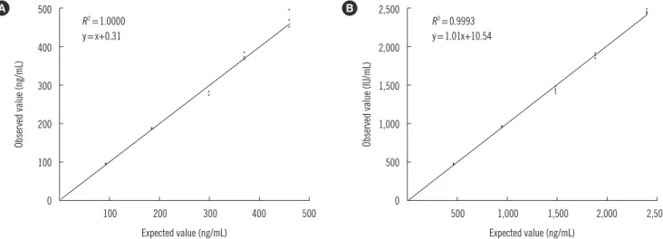
Linearity was observed in both the serum Tg and TgAb at the tested concentrations. R2 was 1.0 for serum Tg and 0.99 for se- rum TgAb (Fig. 1).
Table 1. Precision profile of the DxI 800 assay for serum thyroglobulin and anti-thyroglobulin antibody
Analyte Calibration level Mean SD CV (%)
Within run Between run Between day Total
Tg (ng/mL) Pooled serum 170.6 6.7 2.9 1.7 1.9 3.9
Low level 31.7 1.4 3.9 0 2.2 4.4
Middle level 120.6 5.8 4.2 1.0 2.1 4.8
High level 380.0 21.4 4.0 3.9 1.0 5.6
TgAb (IU/mL) Pooled serum 80.4 4.3 3.5 2.5 3.2 5.3
Low level 4.2 0.3 6.2 3.1 0 6.9
High level 475.5 32.6 4.5 5.1 1.2 6.9
For serum Tg, control materials were manufactured with three concentrations: 32.5 ng/mL (low level calibrator), 250 ng/mL (middle level calibrator), and 400 ng/mL (high level calibrator). For serum TgAb, control materials were manufactured with two concentrations: 4.5 IU/mL (low level calibrator) and 500 IU/mL (high level calibrator).
Abbreviations: Tg, thyroglobulin; TgAb, anti-thyroglobulin antibody.
Fig. 1. Linearity profile of the DxI 800 assay for serum thyroglobulin (Tg) and anti-thyroglobulin antibody (TgAb). For the evaluation of lin- earity, five concentrations of control materials were used for both Tg and TgAb. The intended concentrations for serum Tg were 0, 94, 188, 282, 376, and 470 ng/mL (A), and 0, 480, 960, 1,440, 1,920, and 2,400 IU/mL for serum TgAb (B).
500
400
300
200
100
0
2,500
2,000
1,500
1,000
500
0
100 200 300 400 500 500 1,000 1,500 2,000 2,500
Expected value (ng/mL) Expected value (ng/mL)
Observed value (ng/mL) Observed value (IU/mL)
R2=1.0000 y=x+0.31
R2=0.9993 y=1.01x+10.54
A B
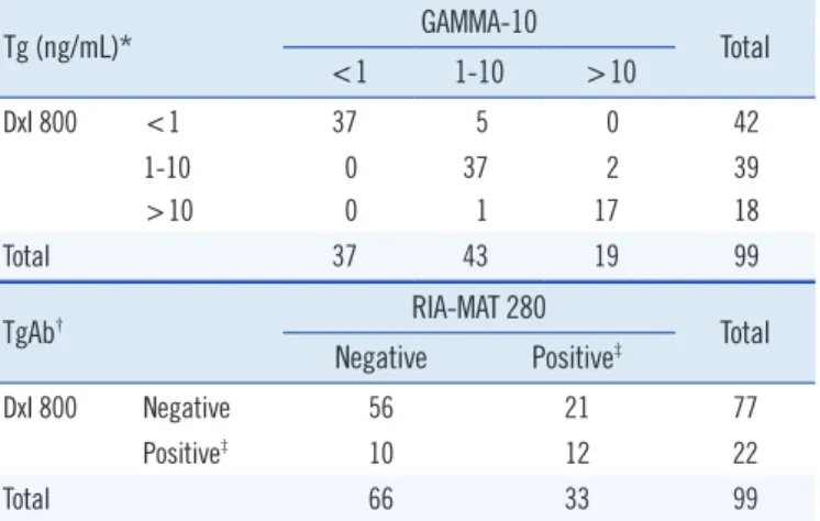
Fig. 2. Correlation of serum thyroglobulin (A) and anti-thyroglobulin antibody (B) concentrations measured by the two assays (results within analytical measurement ranges).
350 300 250 200 150 100 50 0
700 600 500 400 300 200 100 0
50 10 150 200 250 300 350 400 450 500 200 400 600 800 1,000 1,200 1,400 1,600 1,800
GAMMA-10 (ng/mL) RIA MAT-280 (IU/mL)
DxI 800 (ng/mL) DxI 800 (IU/mL)
r=1.9993 y=0.68x+0.89
r=0.8830 y=0.29x+13.93
A B
2. Correlation of serum Tg and TgAb concentrations between the two assays
Some samples exceeded the AMR range of DxI 800. Three samples exceeded 500 ng/mL for Tg, and one sample ex- ceeded 2,500 IU/mL for TgAb. Correlation between each method was evaluated after excluding these results. R was 0.99 and 0.88 for serum Tg and TgAb, respectively. The equation were DxI 800 =0.68 ×GAMMA-10+0.89 (serum Tg) and DxI 800=0.29×RIA-MAT 280+13.93 (serum TgAb) (Fig. 2).
When serum Tg levels were categorized into <1 ng/mL, 1-10 ng/mL, and >10 ng/mL, the weighted Kappa statistic value for Tg was 0.884 (Table 2, top). Serum TgAb levels were compared based on the cut-off value for each assay, resulting in an 84.8%
(56/66) negative agreement and 36.4% (12/33) positive agree- ment, with a total agreement of 68.7% (Cohen’s Kappa statistic, 0.231; Table 2, bottom).
3. Functional sensitivity
In analytes containing low levels of serum Tg (0.2-0.05 ng/mL) manufactured with calibrators, CV values were less than 10% at a
concentration ≤0.075 ng/mL. The CV exceeded 20% (28.5%) in the analyte presenting a 0.05 ng/mL Tg concentration (Table 3).
To assess the functional sensitivity in diluted sera from healthy subjects, CV values ranged from 3.8 to 14.5% at Tg concentra- tions of 0.0625-2.0 ng/mL. When the level was diluted to 0.0313 ng/mL, the CV value exceeded 20% (21.3%). The func- tional sensitivity was estimated to be between 0.0313 and 0.0625 ng/mL (Table 3).
4. A case with discrepant Tg results between the two assays The case with discrepant results between the two assays was a 34-yr-old male patient who underwent total thyroidectomy for classic papillary thyroid cancer (T3N1aM0) and received subse- quent RAIT (50 mCi). The serum Tg level measured using the GAMMA-10 was 1.2 ng/mL, while it was 29.37 ng/mL when measured with the DxI 800. Serum TgAb level was 0.91 IU/mL with the DxI 800 and 69.9 IU/mL with the RIA-MAT 280. One year later, a diagnostic scan (121I) was performed; serum Tg and TgAb levels declined to 0.2 ng/mL with the GAMMA-10 and 16.0 IU/mL with the RIA-MAT 280, respectively. There was no evidence of disease during the 2-yr observation period.
DISCUSSION
We tested the analytic performance of the DxI 800 assay, an ICMA, for serum Tg, which yielded a functional sensitivity be- tween 0.0313 and 0.0625 ng/mL. The results from the DxI 800 for serum Tg and TgAb correlated well with those obtained by using the IRMAs, the GAMMA-10 assay for serum Tg and the RIA-MAT 280 assay for serum TgAb, which are commonly used in Korea.
The advantages of the ICMA method include good functional sensitivity and short processing time. In the present study, the functional sensitivity was expected to be between 0.0313 and 0.0625 ng/mL, which satisfies the criterion suggested by an ex- pert panel, indicating that the Tg assay requires a functional sensitivity of at least 1 ng/mL [17]. Generally, IMAs show a higher sensitivity than RIAs, although some degrees of variation Table 2. Agreement of serum thyroglobulin and anti-thyroglobulin
antibody between the two assays
Tg (ng/mL)* GAMMA-10
Total
<1 1-10 >10
DxI 800 <1 37 5 0 42
1-10 0 37 2 39
>10 0 1 17 18
Total 37 43 19 99
TgAb† RIA-MAT 280
Total Negative Positive‡
DxI 800 Negative 56 21 77
Positive‡ 10 12 22
Total 66 33 99
Values are presented with numbers of subjects.
*Weighted Cohen’s Kappa statistics was 0.884 for serum Tg; †Cohen’s Kap- pa statistics was 0.231 for TgAb, and the agreement rate was 68.7%; ‡The cut-offs for positivity of serum TgAb were 4.0 IU/mL for the DxI 800 and 60.0 IU/mL for the RIA-MAT 280.
Abbreviations: Tg, thyroglobulin; TgAb, anti-thyroglobulin antibody.
Table 3. Functional sensitivity of low levels of serum thyroglobulin manufactured from calibrators and samples from healthy subjects
Target (ng/mL) Calibrators Healthy subject samples
0.2 0.1 0.075 0.05 2 1.5 1 0.75 0.5 0.25 0.125 0.0625 0.0313
Mean (ng/mL) 0.195 0.100 0.072 0.039 2.065 1.489 0.878 0.773 0.504 0.209 0.114 0.062 0.040
SD (ng/mL) 0.013 0.008 0.007 0.011 0.078 0.071 0.041 0.032 0.034 0.012 0.008 0.009 0.008
CV (%) 6.5 7.6 9.3 28.5 3.8 4.7 4.6 4.1 6.7 5.6 6.9 14.5 21.3
in analytic performance exist [5, 6]. Persoon et al [11] reported a superior detection limit of 0.05 ng/mL and functional sensitiv- ity of 0.6 ng/mL with the ICMA, as compared with RIA and IRMA. Recently developed mass spectrometry-based Tg assays have improved functional sensitivity ( ≤0.5 ng/mL). However, false-negative results have been reported for Tg concentrations between 0.1 and 0.5 ng/mL [10].
The correlation of serum Tg between the DxI 800 and the GAMMA-10 methods was excellent. A previous study reported that serum Tg levels measured by using the ICMA, IRMA, and RIA were highly correlated using TgAb-negative samples (R =0.992, R =0.999, respectively) [11]. However, a major con- cern with the ICMA is the TgAb interference, which often results in falsely low or undetectable Tg concentrations [5, 9] in in vitro and clinical studies [8, 9, 19]. Endogenous TgAbs bind to free Tg and conceal the Tg epitopes needed for recognition by signal monoclonal antibody reagents [18]. Nevertheless, we observed a good correlation between the two Tg assays using TgAb-posi- tive and -negative samples, although both assays may not fun- damentally overcome the TgAb interference.
For the ICMA, a 2- or 3-fold difference has been reported be- tween some assays [5, 11]. This might result from differences in specificity for the capture and/or signal monoclonal antibody reagents or standardization, although the ICMA is standardized against the CRM-457 international standard material [5, 11].
Because of high variability and low interchangeability among as- says [5], the current guidelines for differentiated thyroid cancer developed by the ATA recommend serial measurement of se- rum Tg using the same assay in individual patients [20].
Changes in Tg assays, even if it is an ICMA, may result in abrupt serum Tg fluctuation and need to be cautiously considered for adequate decision making.
Eight cases presented discrepant results in term of Tg levels (Table 2). Among them, only subtle variations in Tg concentra- tions were observed in seven cases (i.e. 10.8 ng/mL using the IRMA vs 9.56 ng/mL using the ICMA). These differences were clinically acceptable changes and may not alter the clinical im- plication. However, one case showed a considerable elevation of serum Tg concentration on the DxI 800 when compared with the results obtained by using the GAMMA-10 (29.37 ng/mL vs 1.2 ng/mL). Based on clinical information, there was no evi- dence of remnant or recurrent disease on two instances of thy- roid ultrasonography with a decreasing trend of subsequent stimulated Tg. Thus, the value measured by the DxI 800 might have been falsely elevated. Serum TgAb was positive, even with a slight elevation in this patient. However, the presence of TgAb
always interferes with serum Tg levels, resulting in an underesti- mation in the ICMA [5]. Therefore, overestimation of serum Tg by the DxI 800 might derive from another type of interference.
Human anti-mouse antibody (HAMA) also affects serum Tg re- sults, mainly in terms of false elevation, even though this rarely leads to underestimation of Tg concentrations [21]. This anti- body can bind to animal antigens and form a complex between antibodies, which produces a false elevation of serum Tg results [21]. As the prevalence of HAMA ranges 1.5-3% in automated Tg assays, HAMA interference should be suspected in cases showing Tg results discordant with the clinical situation [21].
This study has several limitations. Neither the ICMA nor IRMA tested in this study was a gold standard, although they are com- monly recommended for Tg measurement. As described above, all IMA methodologies are affected by the interference from TgAbs as well as HAMA. This interference is not as evident as in other assays, including RIA and the LC-MS method. However, they also do not fully overcome it yet. In the present study, we identified one case showing a falsely elevated serum Tg result with the DxI 800, which should be interpreted in the clinical context. The proportion of discordant results (1/99) seems to be acceptable because the prevalence of HAMA has been reported to be up to 3% in IMAs. Even though the IRMA may not serve as the gold standard test, as the most prevalent assay for serum Tg in current laboratories, including South Korea, we sought to validate the excellent discrimination (linear correlation) between the two assays and to identify whether the ICMA would be a via- ble alternative to IRMA. Considering the good correlation be- tween the two assays, irrespective of various titers of TgAb, and the better analytic performance of the ICMA, the ICMA could be considered as an alternative test for the Tg measurement.
The DxI 800 method is a sensitive assay for Tg measurement, and the results were highly correlated with those obtained with the GAMMA-10. Analytic performance and clinical convenience make this assay a good substitute for the IRMA.
Authors’ Disclosures of Potential Conflicts of Interest
No potential conflicts of interest relevant to this article were re- ported.
Acknowledgments
This study was supported by a Samsung Medical Center grant [#PHO 1125465].
REFERENCES
1. Van Herle AJ, Vassart G, Dumont JE. Control of thyroglobulin synthesis and secretion (second of two parts). N Engl J Med 1979;301:307-14.
2. Webb RC, Howard RS, Stojadinovic A, Gaitonde DY, Wallace MK, Ahmed J, et al. The utility of serum thyroglobulin measurement at the time of remnant ablation for predicting disease-free status in patients with differentiated thyroid cancer: a meta-analysis involving 3947 pa- tients. J Clin Endocrinol Metab 2012;97:2754-63.
3. Haugen BR, Alexander EK, Bible KC, Doherty GM, Mandel SJ, Nikiforov YE, et al. 2015 American Thyroid Association Management Guidelines for adult patients with thyroid nodules and differentiated thyroid cancer:
The American Thyroid Association Guidelines Task Force on thyroid nodules and differentiated thyroid cancer. Thyroid 2016;26:1-133.
4. Van Herle AJ, Uller RP, Matthews NI, Brown J. Radioimmunoassay for measurement of thyroglobulin in human serum. J Clin Invest 1973;52:
1320-7.
5. Spencer CA, Bergoglio LM, Kazarosyan M, Fatemi S, LoPresti JS. Clini- cal impact of thyroglobulin (Tg) and Tg autoantibody method differenc- es on the management of patients with differentiated thyroid carcino- mas. J Clin Endocrinol Metab 2005;90:5566-75.
6. Spencer CA, Takeuchi M, Kazarosyan M. Current status and perfor- mance goals for serum thyroglobulin assays. Clin Chem 1996;42:164- 73.
7. Baloch Z, Carayon P, Conte-Devolx B, Demers LM, Feldt-Rasmussen U, Henry JF, et al. Laboratory medicine practice guidelines. Laboratory support for the diagnosis and monitoring of thyroid disease. Thyroid 2003;13:3-126.
8. Görges R, Maniecki M, Jentzen W, Sheu SN, Mann K, Bockisch A, et al. Development and clinical impact of thyroglobulin antibodies in pa- tients with differentiated thyroid carcinoma during the first 3 years after thyroidectomy. Eur J Endocrinol 2005;153:49-55.
9. Spencer CA, Takeuchi M, Kazarosyan M, Wang CC, Guttler RB, Singer PA, et al. Serum thyroglobulin autoantibodies: prevalence, influence on serum thyroglobulin measurement, and prognostic significance in pa- tients with differentiated thyroid carcinoma. J Clin Endocrinol Metab 1998;83:1121-7.
10. Netzel BC, Grebe SK, Carranza Leon BG, Castro MR, Clark PM, Hoof- nagle AN, et al. Thyroglobulin (Tg) testing revisited: Tg assays, TgAb assays and correlation of results with clinical outcomes. J Clin Endocri- nol Metab 2015;100:E1074-83.
11. Persoon AC, Van Den Ouweland JM, Wilde J, Kema IP, Wolffenbuttel BH, Links TP. Clinical utility of an automated immunochemiluminomet-
ric thyroglobulin assay in differentiated thyroid carcinoma. Clin Chem 2006;52:686-91.
12. Spencer C, Petrovic I, Fatemi S, LoPresti J. Serum thyroglobulin (Tg) monitoring of patients with differentiated thyroid cancer using sensitive (second-generation) immunometric assays can be disrupted by false- negative and false-positive serum thyroglobulin autoantibody misclassi- fications. J Clin Endocrinol Metab 2014;99:4589-99.
13. Lee JI, Kim JY, Choi JY, Kim HK, Jang HW, Hur KY, et al. Differences in serum thyroglobulin measurements by 3 commercial immunoradiomet- ric assay kits and laboratory standardization using Certified Reference Material 457 (CRM-457). Head Neck 2010;32:1161-6.
14. Clinical and Laboratory Standards Institute. Evaluation of the linearity of quantitative measurement procedures: a statistical approach; approved guideline. CLSI document EP06-A. Wayne, PA: Clinical and Laboratory Standards Institute, 2003.
15. Clinical and Laboratory Standards Institute. Evaluation of precision per- formance of quantitative measurement methods; approved guideline- second ed. CLSI document EP5-A2. Wayne, PA: Clinical and Laboratory Standards Institute, 2004.
16. Kim TY, Kim WB, Kim ES, Ryu JS, Yeo JS, Kim SC, et al. Serum thyro- globulin levels at the time of 131I remnant ablation just after thyroidec- tomy are useful for early prediction of clinical recurrence in low-risk pa- tients with differentiated thyroid carcinoma. J Clin Endocrinol Metab 2005;90:1440-5.
17. Mazzaferri EL, Robbins RJ, Spencer CA, Braverman LE, Pacini F, Wartofsky L, et al. A consensus report of the role of serum thyroglobulin as a monitoring method for low-risk patients with papillary thyroid carci- noma. J Clin Endocrinol Metab 2003;88:1433-41.
18. Spencer C and Fatemi S. Thyroglobulin antibody (TgAb) methods - Strengths, pitfalls and clinical utility for monitoring TgAb-positive pa- tients with differentiated thyroid cancer. Best Pract Res Clin Endocrinol Metab 2013;27:701-12.
19. Kim WG, Yoon JH, Kim WB, Kim TY, Kim EY, Kim JM, et al. Change of serum antithyroglobulin antibody levels is useful for prediction of clinical recurrence in thyroglobulin-negative patients with differentiated thyroid carcinoma. J Clin Endocrinol Metab 2008;93:4683-9.
20. American Thyroid Association (ATA) Guidelines Taskforce on Thyroid Nodules and Differentiated Thyroid Cancer, Cooper DS, Doherty GM, Haugen BR, Kloos RT, Lee SL, et al. Revised American Thyroid Associ- ation management guidelines for patients with thyroid nodules and dif- ferentiated thyroid cancer. Thyroid 2009;19:1167-214.
21. Preissner CM, O’Kane DJ, Singh RJ, Morris JC, Grebe SK. Phantoms in the assay tube: heterophile antibody interferences in serum thyroglobu- lin assays. J Clin Endocrinol Metab 2003;88:3069-74.