Copyright © 2012, the Korean Surgical Society J Korean Surg Soc 2012;82:370-373
http://dx.doi.org/10.4174/jkss.2012.82.6.370
ORIGINAL ARTICLE
Journal of the Korean Surgical Society
JKSS
pISSN 2233-7903ㆍeISSN 2093-0488
Received February 22, 2012, Revised March 27, 2012, Accepted April 12, 2012 Correspondence to: Sang Jun Park
Department of Surgery, Ulsan University Hospital, University of Ulsan College of Medicine, 877 Bangeojinsunhwan-doro, Dong-gu, Ulsan 682-714, Korea
Tel: +82-52-250-7109, Fax: +82-52-250-7350, E-mail: [email protected]
cc Journal of the Korean Surgical Society is an Open Access Journal. All articles are distributed under the terms of the Creative Commons Attribution Non-Commercial License (http://creativecommons.org/licenses/by-nc/3.0/) which permits unrestricted non-commercial use, distribution, and reproduction in any medium, provided the original work is properly cited.
Extrinsic compression of left iliac vein does not predict the development of post thrombotic syndrome in left side deep venous thrombosis
Sang Jun Park, Ho Jong Park, Eun Kyoung Kwon, Sang Jin Kim, Hong Rae Cho
Department of Surgery, Ulsan University Hospital, University of Ulsan College of Medicine, Ulsan, Korea
Purpose: Left side deep venous thrombosis (DVT) is associated with May-Thurner’s anatomical variation and is often in- stigated by invasive treatment. The aim of this study is to analyze the influence of left iliac vein narrowness on incidence of post thrombotic syndrome (PTS) that developed after left side DVT. Methods: Forty-one left side DVT cases that were fol- lowed up for more than 1 year were enrolled. The iliac vein narrowness was measured by the shortest distance from the right iliac artery to the 5th lumbar vertebra overlying left iliac vein in computed tomography (CT) scan. The incidence of PTS was measured by phone-call history taking for specific symptoms of PTS. The means of the shortest distance were compared by independent t-test. Results: The number of PTS cases was eleven (26.8%). The level of thrombus, demographic data and other risk factors were similar in both PTS and non-PTS groups except the mean risk factor score. The mean of the shortest distance of PTS group and non-PTS group were 5.56 mm and 5.89 mm, respectively. Conclusion: The degree of left iliac vein narrow- ness measured by the shortest distance from the right iliac artery and the 5th lumbar vertebral body was not a predictive fac- tor for PTS.
Key Words: Venous thrombosis, X-ray computed tomography, Postthrombotic syndrome
INTRODUCTION
There are two main goals in treatment of deep venous thrombosis (DVT) of lower extremities: One is to reduce the chance of fatal pulmonary embolism and the other is to prevent post-thrombotic syndrome (PTS) [1]. We try to find out a high risk patient of DVT and to prevent the de- velopment of DVT in a sense that it is not easy to achieve the former once the DVT is established. Prevention of PTS
can be a main focus of managing lower extremity DVT.
Several studies have shown that the frequency of PTS to be up to 30 to 40% and PTS was associated with worsened quality of life [2,3]. Recent studies report the efficacy of early invasive treatment for acute DVT in order to achieve a high venous patency rate and to preserve valve function [4,5]. However, some researchers argue that an invasive treatment such as thrombolytic therapy couldn’t prevent the long-term outcomes, PTS [6]. And proximal DVT of the
Prediction of post thrombotic syndrome
thesurgery.or.kr 371
Fig. 1. Indirect computed tomogram (CT) venography. White ar- rows indicate the thrombus in the left iliac and femoral vein.
Fig. 2. Distances are measured in cross-sectional views. The shortest distance is measured from right iliac artery to 5th vertebral body overlying left iliac vein by picture archive communication system software. RIA, right iliac artery; IVC, inferior vena cava; LV, lumbar vertebra.
left side is closely related to iliac vein compression syn- drome or May-Thurner syndrome [7]. We report that the minor diameter of the left iliac vein between the right iliac artery and the 5th vertebral body impacted the incidence of left side proximal DVT [8].
This study aims to investigate a new non-invasive meth- od to predict PTS after acute DVT. We focused the left iliac vein narrowness measured by the shortest distance from right iliac artery to the 5th vertebral body overlying left iliac vein in computed tomography (CT) scan.
METHODS
One hundred and sixty-six limbs in 113 patients with DVT diagnosed by duplex scan with serum d-dimer test in one institute were reviewed. CT scan was performed to de- tect presence of pulmonary embolism as well as to de- termine the exact location of the thrombus in the vein (Fig.
1). We used a uniform protocol for CT pulmonary arteriog- raphy and indirect venography [9]. We excluded twenty cases that received thrombolysis or other invasive treat- ments. Among the 146 cases, CTs for the diagnosis of pul- monary embolism were checked in 131 cases. We con- tacted 65 cases of the left side DVT over a year after the first diagnosis and ended up with forty one cases for analysis.
All the cases were recommended one year of oral anti- coagulation and anti-gravity stockings. Anticoagulation was performed by treatment with oral warfarin maintain- ing the prothrombin time between 2 to 2.5 international normalized ratio. CT scan and duplex scan were per- formed at a year follow-up of oral anticoagulation. We evaluated the recanalization of thrombosed vein by CT scan and duplex scan at a year follow-up. According to our definitions, recanalization was the partial or complete re- solved thrombus while non-recanalization was complete occlusion of a whole venous lumen in any segment of thrombosed vein.
The shortest distance from right iliac artery to 5th verte- bral body was measured in picture archiving and commu- nicating system program using π-ViewSTAR (INFINITT Healthcare Co., Seoul, Korea). The distance was measured at the shortest distance between the right iliac artery and the 5th vertebral body overlying left iliac vein on cross sec- tional view (Fig. 2).
Diagnosis of PTS was checked by phone call with questionnaire. We used the Ginsberg measure of PTS diag- nosis [10]. We asked the following questions: 1) Do you have newly onset chronic venous insufficiency signs, such as varicose veins, swelling of foot or calf, skin pigmenta- tion or discoloration, and venous ulcers? 2) Is there any
Sang Jun Park, et al.
372 thesurgery.or.kr
Table 2. The mean of the shortest distance from right iliac artery to 5th vertebral body
PTS group (n = 11)
Non-PTS group
(n = 29) P-value Mean ± SD (mm) 5.56 ± 3.04 5.89 ± 4.23 0.882 PTS, post-thrombotic syndrome.
Fig. 3. Patient selection diagram for evaluation of left side post throm- botic syndrome after deep venous thrombo- sis (DVT).
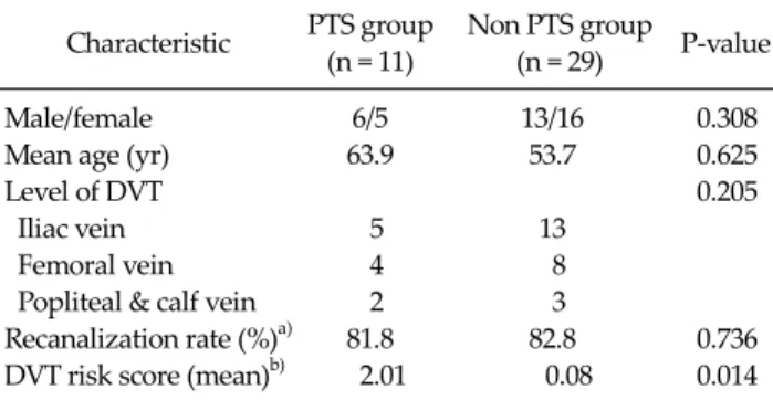
Table 1. Clinical characteristics of left side DVT
Characteristic PTS group (n = 11)
Non PTS group (n = 29) P-value
Male/female 6/5 13/16 0.308
Mean age (yr) 63.9 53.7 0.625
Level of DVT 0.205
Iliac vein 5 13
Femoral vein 4 8
Popliteal & calf vein 2 3
Recanalization rate (%)a) 81.8 82.8 0.736 DVT risk score (mean)b) 2.01 0.08 0.014 PTS, post-thrombotic syndrome; DVT, deep venous thrombosis.
a)Complete or partial resolution of venous lumen/total number.
b)DVT risk scores were followed by New York Heart Association protocols.
newly onset spontaneous calf pain when walking or standing? 3) Are you experiencing newly onset heaviness of leg? We sorted the cases to the PTS group if he or she an- swered ‘yes’ to least one of the above questions.
New York Heart Association DVT risk score system was used for DVT risk assessment [10].
The mean of the shortest distance from the right iliac ar- tery to the 5th vertebral body was compared between PTS group and non PTS group by independent t-test. The stat- istical analysis was performed with SPSS ver. 11.0 (SPSS Inc., Chicago, IL, USA).
RESULTS
Forty-one patients were finally enrolled in the study.
Median follow-up period was 16 months (Fig. 3). Eleven of
forty-one cases (26.8%) had PTS symptoms or signs. Male and female ratio of PTS group was 6:5 and 13:16 for non-PTS group. Mean ages were 63.9 for PTS group and 54.9 for non PTS group. The levels of thrombus were 5 iliac veins, 4 femoral veins and 2 popliteal veins in PTS group and 13 iliac veins, 8 femoral veins and 3 femoral veins in non-PTS group. The recanalization rate was achieved in 81.8% (9/11) of PTS group and 82.8% (24/29) of non-PTS group. Mean of DVT risk score for PTS group was 2.00 and 0.08 for non-PTS group. Most clinical features were similar except the DVT risk score by means of Fisher exact test or t-test (Table 1). Mean of the shortest distance from right iliac artery to 5th vertebral body of PTS group was 5.56 mm and 5.89 mm in non-PTS group (Table 2).
DISCUSSION
About twenty-five percent of all the patients with DVT in their lower extremities developed PTS within a year, and PTS is one of the most important complications of DVT [11]. In our study, a figure of 26.8% incidence of PTS is similar to that of other study that reported on patients
Prediction of post thrombotic syndrome
thesurgery.or.kr 373
who used elastic stockings [12]. However, there was a var- iation in the incidence of PTS because of a difference of di- agnostic criteria and follow-up duration.
While several studies reported that invasive treatment such as catheter directed thrombolysis could reduce the risk of PTS in short-term results [6], long-term results were not established and the complication of invasive treatment was not possible to overcome.
We reported that the shortest distance between the right iliac artery and the 5th vertebral body overlying the left iliac vein could be a risk factor for developing DVT in the left proximal vein [9]. The main mechanism in the devel- opment of DVT might be similar to that of iliac com- pression syndrome or May-Thurner syndrome. Accord- ing to our hypothesis, this short distance representing the narrowness of the iliac vein would be also related to the in- cidence of PTS after left-side DVT. If this method could predict PTS development, we could easily select patients who are in need of invasive treatment. However, no result could be found to show any relationship between them.
Rather, the result concluded that the higher the risk factor scores are, the higher the PTS incidence is (Table 1). It is ac- knowledged that DVT risk factors such as the previous documented DVT are shared with PTS risk factors [3].
There are some other risk factors such as development of PTS after DVT. The recanalization rate of thrombosed vein is considered one of the risk factors for PTS [13]. It is found in this study that the recanalization rates were similarly achieved in both groups.
There were some limitations in this study: small num- ber of cases, short follow-up period, and inexactitude of diagnostic criteria for PTS. The thrombolysis cases were excluded in that they were suspected as severe iliac vein compression.
We aimed for a non-invasive and simple method to pre- dict the necessity of invasive intervention for DVT.
However, it doesn’t seem that this study provides strong effects on external iliac vein compression in the develop- ment of PTS after left-side DVT or less effect than DVT risk factor scores.
CONFLICTS OF INTEREST
No potential conflict of interest relevant to this article was reported.
REFERENCES
1. Lee JS, Kim YS. Deep vein thrombosis in lower extremity. J Korean Surg Soc 1990;38:110-8.
2. Kahn SR, Shrier I, Julian JA, Ducruet T, Arsenault L, Miron MJ, et al. Determinants and time course of the post- thrombotic syndrome after acute deep venous thrombosis.
Ann Intern Med 2008;149:698-707.
3. Prandoni P, Lensing AW, Cogo A, Cuppini S, Villalta S, Carta M, et al. The long-term clinical course of acute deep venous thrombosis. Ann Intern Med 1996;125:1-7.
4. Couturaud F, Kearon C. Treatment of deep vein throm- bosis. Semin Vasc Med 2001;1:43-54.
5. Wells PS, Forster AJ. Thrombolysis in deep vein thrombo- sis: is there still an indication? Thromb Haemost 2001;86:
499-508.
6. Schweizer J, Kirch W, Koch R, Elix H, Hellner G, Forkmann L, et al. Short- and long-term results after thrombolytic treatment of deep venous thrombosis. J Am Coll Cardiol 2000;36:1336-43.
7. Ouriel K, Green RM, Greenberg RK, Clair DG. The anat- omy of deep venous thrombosis of the lower extremity. J Vasc Surg 2000;31:895-900.
8. Kim SJ, Park SJ, Kwon EK, Park HJ, Rho SY, Hwang JC, et al. Impact of distance from the right iliac artery to the lum- bar vertebra in left side deep venous thrombosis. Korean J Vasc Endovasc Surg 2011;27:71-5.
9. Yi JH, Park SJ, Kwon EK, Oh YJ, Kang TW, Hwang JC, et al.
The usefulness of the computed tomography for diagnos- ing deep venous thrombosis of the lower extremities. J Korean Soc Vasc Surg 2009;25:12-6.
10. Gloviczki P. Handbook of venous disorders: guidelines of American venous forum. 3rd ed. London: Hodder Anold;
2009.
11. Scarvelis D, Wells PS. Diagnosis and treatment of deep- vein thrombosis. CMAJ 2006;175:1087-92.
12. Tick LW, Kramer MH, Rosendaal FR, Faber WR, Doggen CJ. Risk factors for post-thrombotic syndrome in patients with a first deep venous thrombosis. J Thromb Haemost 2008;6:2075-81.
13. Byun JW, Kwun WH, Suh BY. Predictor of recanalization in lower extremity deep vein thrombosis. Korean J Vasc Endovasc Surg 2012;28:43-7.