J Korean Soc Surg Hand 2015;20(4):198-203.
http://dx.doi.org/10.12790/jkssh.2015.20.4.198
THE HAND
INTRODUCTION
Even though fractures of the distal radius are the most com- mon fracture of the upper extremity, complications con- cerning the injury are relatively poorly reported. It is thought that flexor tendon ruptures associated with distal radius fracture are caused by attrition by rough surfaces of
malunited fracture and irritation by fixation implants. The majority of reported cases usually presents with the compli- cation four or more weeks after the injury1. Extensor tendon damage is thought to be more common than flexor tendon damage in patients with distal radius fractures2,3. Concurrent flexor digitorum profundus tendon rupture with distal radius fracture is very rare. Therefore, we are
Acute Rupture of Flexor Digitorum Profundus Tendon Associated with Distal Radius Fracture:
A Case Report
Dong-Yeong Lee, Sun-Chul Hwang, Dae-Cheol Nam, Jin-Sung Park
Department of Orthopaedic Surgery, Gyeongsang National University Hospital, Gyeongsang National University School of Medicine, Jinju, Korea
Received:August 1, 2015 Revised:[1] August 18, 2015
[2] August 29, 2015 Accepted:August 31, 2015 Correspondence to:Jin-Sung Park Department of Orthopaedic Surgery, Gyeongsang National University Hospital, Gyeongsang National University School of Medicine, 79 Gangnam-ro, Jinju 52727, Korea TEL:+82-55-750-8858
FAX:+82-55-754-0477 E-mail:[email protected]
Acute ruptures of flexor tendons in patients with distal radius fractures are very rare complications. The majority of reported cases, flexor tendon rupture asso- ciated with distal radius fracture, is chronic flexor tendon ruptures, which are caused by implants for fixation or rough surfaces of malunited distal radius. We experienced an unusual case of an acute rupture of the flexor digitorum pro- fundus tendon in a patient with a distal radius fracture, in addition to provid- ing an auxiliary review of the literature.
Keywords:Distal radius fracture, Flexor tendon rupture, Complication
This is an Open Access article distributed under the terms of the Creative Commons Attribution Non-Commercial License (http://creativecommons.org/licenses/by- nc/3.0/) which permits unrestricted noncommercial use, distribution, and reproduction in any medium, provided the original work is properly cited.
reporting this case in this article with an auxiliary review of the relevant literature.
CASE REPORT
A 59-year-old, right-handed female presented to the emer- gency department after a pedestrian traffic accident injury to her right wrist. On physical examination, the range of motion of all fingers was mildly limited due to pain, and the distal sensation and circulation were intact. There was severe swelling with crepitus, and “distal radius fracture”
was observed at wrist. Also, there was the 0.4 cm sized lacer- ation located on the anteromedial aspect of her wrist.
Radiographs showed a displaced, comminuted, and intra- articular distal radius fracture with an ulnar styloid process fracture. A three-dimensional computed tomography (CT) was performed for the structural evaluation of the intra- articular fracture. On the basis of radiographs and CT images, according to the AO/OTA classification of distal radius fracture, our case was classified as an AO/OTA classi- fication C3 distal radius fracture (Fig. 1). Closed reduction was performed, and the sugar tong splint was applied.
However, the fracture site was reduced unsatisfactorily, because dorsal displacement of the distal fragment was sig- nificant. We recommended her a surgery for anatomic reduction and stable fixation of radius, after improvement of swelling.
Eight days after the injury, the patient was taken to the operating room. Under general anesthesia, we were able to observe a disrupted finger cascade of the distal interpha- langeal joint of the third finger. So, we have supposed that a flexor digitorum profundus tendon of the third finger was ruptured. To treat the distal radius fracture, we used volar approach. The flexor carpi radialis was retracted ulnarly, then flexor pollicis longus was exposed. After ulnar retrac- tion of flexor pollicis longus, we were able to find ruptured pronator quadratus by proximal fragment of the radius.
After dividing pronator quadratus, fracture site was exposed. The torn pronator quadratus muscle was inter- posed between proximal and distal fragment of radius. We performed reduction distal fragments using freer as a lever- age, then fixation of volar locking plate and screws (2.4-mm
LCP Distal Radius Plates, Synthes, Salzburg, Austira). After fixation of plate, we repaired pronator quadratus muscle.
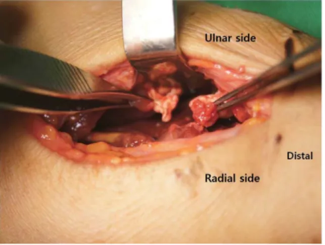
We explored flexor digitorum profundus and were able to find distal stump of the third tendon of flexor digitorum profundus. It was located in flexor tendon zone V and it ruptured at the tendinous portion (Fig. 2). We did not find other injured structures.
We reparied it using a two-strand modified Kessler method with 3-0 nylon and circumferential epitendinous suture with 5-0 nylon (Fig. 3). After the ruptured tendon was
Fig. 1. (A)Initial radiographs of the wrist showing a comminuted and displaced distal radius fracture with dorsal angulation and ulnar styloid process fracture. Note the bony beak at the proximal fractured end and the obvious deformity of the wrist.(B) Computed tomography scan showing simple intra-articular and simple metaphysic fractures of the distal radius. The bony beak at the proximal fractured end is prominently shown (white arrow).
repaired, we were able to observe a disrupted finger cascade of the distal interphalangeal joint of the third finger, which was visible before the repair (Fig. 4). Our patient’s laboratory
results, including white blood cell, erythrocyte sedimenta- tion rate, C-reactive protein, and rheumatoid factor were within the normal range. On simple radiographs and com- puted tomography scan, there was no evidence of inter- carpal arthritis or radiocarpal joint arthritis. We were unaware of any risk factors of tendon rupture, such as steroid medication or injection, or diagnosis or family histo- ry of rheumatoid arthritis. Finally, we concluded the patient suffered from a concurrent distal radius fracture and acute third flexor digitorum profundus tendon rupture.
After surgery, the patient’s wrist and fingers were immo- bilized in dorsal block splint for 4 weeks. Postoperative radiographs of the left wrist showed a normal alignment of the carpals. Early controlled mobilization of the fingers was initiated on the first day after surgery in accordance with the Duran protocol including active finger extension with patient-assisted passive finger flexion. At six months after surgery, the patient demonstrated excellent active distal interphalangeal joint range-of-motion (0°-70°) and proxi- mal interphalangeal joint range-of-motion (0°-120°) of the third finger. Wrist motion was 60° in flexion and 60° in extension. There were no clinical signs of carpal instability.
DISCUSSION
Distal radius fractures are the most common fracture of the upper extremity. Despite its prevalence, acute and delayed ruptures of tendons at the wrist level in patients with closed distal radius fractures are a rare complication. The inci- dence of tendon rupture in nonoperative management of Fig. 2.Intraoperative gross picture showing ruptured third flex-
or digitorum profundus tendon at the musculotendinous junc- tion in flexor tendon zone V.
Fig. 3. Intraoperative gross picture showing repaired tendon (black arrow) using the modified Kessler technique and a run- ning epitendinous suture for reinforcement.
Fig. 4. (A) Preoperative gross picture showing a disruption of finger cascade at the third distal interphalangeal joint.(B) Postoperative gross picture showing restored finger cascade at the third distal interphalangeal joint.
distal radius fractures has been reported to be as high as 3%, and tendon rupture after volar locking plate fixation has been as high as 12%4,5. In addition, most of these reports revealed that tendon ruptures accompanying distal radius fracture were associated with a delayed complication. In our literature review, we could not find a report concerning the incidence of concurrent acute tendon rupture and distal radius fracture due to its rarity.
The most common etiology of the tendon ruptures is attrition between the sharp bony spurs on the volar aspect of the distal radius and the involved tendons. The majority of cases involve the extensor tendons of the digit, most commonly the extensor pollicis longus tendon2. Extensor tendons are particularly vulnerable due to their flattened structure, their close association to the distal radius, and the unyielding, restraining force of the extensor retinaculum.
Furthermore, the mechanical factors that disrupt the anteri- or interosseous artery at the time of injury has been postu- lated to lead to ischemic degeneration of damaged extensor tendons, predisposing them to attritional and ischemic rup- ture6. For these reasons, extensor tendon ruptures have been more frequently associated with distal radius frac- tures. In contrast, flexor tendon ruptures caused by denud- ed carpal bones in patients with rheumatoid arthritis have been described by Vaughan-Jackson7. However, flexor ten- don complications after distal radius fractures are much less common3,5,8, and may occur early with fracture displace- ment, later after malunion with bony prominence causing mechanical irritation or in physiologically abnormal ten-
dons2,6,8. McMaster3, in 1932, was the first to report a case of
flexor tendon rupture after a Colles’ fracture. He postulated that the rupture was either due to partial tendon division by a sharp bony spur at the time of fracture, followed by insuf- ficient healing and subsequent rupture, or that the blood supply to the flexor tendons was compromised by pro- longed pressure over the malunion, eventually leading to weakening and subsequent rupture3.
Previous studies report that flexor and extensor tendon injuries may result from improper plate design and posi- tion, prominent screw protrusion, steroid use, loss of reduc- tion or fracture collapse, and various bone abnormalities around the wrist1,8. In most cases, the ruptures occurred sev-
eral weeks or even months after the fracture. In the cases reported throughout the literature, the timing of the rupture ranges between 0 and 300 months. Unlike previous cases, the case we report is the rare one with concurrent distal radius fracture and third flexor digitorum profundus tendon rupture. In contrast to extensor tendons, the flexor tendons are not closely associated with bone at the metadiaphyseal level of the distal radius and are less likely to be damaged after a fracture at this level. Three factors contribute to this.
First, the high tensile strength of the tendon and its flexibili- ty protect it from rupture. Second, the pronator quadratus muscle overlies the volar surface of the distal radius and such position provides cushion effect for its superficial structures. When a fracture occurs or is treated, the cushion effect of the pronator quadratus muscle protects the flexor tendons from deeper structures, such as prominent bony fragments or hardware8. Third, the normal prominence of the volar lip in combination with the bony anatomy of the pronator fossa creates a slight bowstring over this area, thus causing the resting position of the flexor tendons to lie away from the volar cortex9. For these reasons, acute and delayed flexor tendon ruptures associated with distal radius frac- tures are a much less likely complication compared to those of the extensor tendons. Our case represents a flexor tendon rupture accompanying distal radius fracture with partial tear of the pronator quadratus muscle. Accordingly, the flexor tendon rupture was caused by a direct injury from one of the fracture fragments. The proximal fragment of radius pierced pronator quadratus muscle, and then sev- ered the third flexor digitorum profundus tendon.
The optimal timing of flexor tendon repair has not been established. Generally, primary tendon repair results in a better functional outcome compared with secondary ten- don repair or tendon graft surgery in patients with flexor tendon injuries. Recent studies revealed that the timing of the repair has changed from emergency repair to delayed primary repair, usually within one week of the injury10. Early tendon exploration would allow primary repair and the fracture would simultaneously be reduced and stabilized.
When either diagnosis or operation is delayed, the short- ened and retracted tendons are no longer available for a direct repair, and then methods such as tendon transfer or
tendon graft must be considered in the repair of a deficient tendon. As a result, concurrent distal radius fractures and flexor tendon ruptures do not require emergency surgery, but early recognition of the tendon rupture is crucial for the timely treatment and functional recovery of the patient.
The pain associated with the fracture makes easy to ovelook the concurrent tendon rupture. Physicians mistake that the cause of limitation of the active finger motion is pain rather than tendon injury. We did not consider per- forming a careful physical examination or further radiologic evaluation for diagnosing a tendon rupture because of its rarity, in the present case. Ultrasound is very useful in diag- nosing pathologic conditions of the hand and wrist, as it can effectively diagnose ruptured tendons by demonstrating tendon nonvisualization, blunt torn ends, refractive shad- owing, and adjacent fluid. It also provides a cost-effective and expedient alternative and/or adjunct to MRI. Therefore, by using radiologic modalities, we can prevent misdiagnosis and malpractice like occult flexor tendon ruptures.
In summary, acute rupture of flexor tendon associated with distal radius fracture is very rare, but it is possible.
Therefore, suspicion and meticulous physical examination is required to avoid misdiagnosis.
REFERENCES
1. Soong M, van Leerdam R, Guitton TG, Got C, Katarincic J, Ring D. Fracture of the distal radius: risk factors for
complications after locked volar plate fixation. J Hand Surg Am. 2011;36:3-9.
2. Kato N, Nemoto K, Arino H, Ichikawa T, Fujikawa K.
Ruptures of flexor tendons at the wrist as a complica- tion of fracture of the distal radius. Scand J Plast Reconstr Surg Hand Surg. 2002;36:245-8.
3. McMaster PE. Late ruptures of extensor and flexor pol- licis longus tendons following Colles' fracture. J Bone Joint Surg Am. 1932;14:93-101.
4. Bonatz E, Kramer TD, Masear VR. Rupture of the exten- sor pollicis longus tendon. Am J Orthop (Belle Mead NJ). 1996;25:118-22.
5. Drobetz H, Kutscha-Lissberg E. Osteosynthesis of distal radial fractures with a volar locking screw plate system.
Int Orthop. 2003;27:1-6.
6. Roberts JO, Regan PJ, Roberts AH. Rupture of flexor pollicis longus as a complication of Colles' fracture: a case report. J Hand Surg Br. 1990;15:370-2.
7. Vaughan-Jackson OJ. Rheumatoid hand deformities considered in the light of tendon imbalance. J Bone Joint Surg Br. 1962;44:764-75.
8. Bell JS, Wollstein R, Citron ND. Rupture of flexor polli- cis longus tendon: a complication of volar plating of the distal radius. J Bone Joint Surg Br. 1998;80:225-6.
9. Orbay JL, Touhami A. Current concepts in volar fixed- angle fixation of unstable distal radius fractures. Clin Orthop Relat Res. 2006;445:58-67.
10. Lalonde DH. An evidence-based approach to flexor ten- don laceration repair. Plast Reconstr Surg. 2011;127:
885-90.
원위 요골 골절에 동반된 급성 심 수지 굴곡건 파열:
증례 보고
이동영∙황선철∙남대철∙박진성
경상대학교 의학전문대학원 정형외과학교실
원위 요골 골절 환자에 있어 급성 굴곡건 파열은 매우 드문 합병증 중 하나이다. 많은 저자들이 보고한 연구에서 원위 요 골 골절에 동반된 건 파열은 원위 요골 골절의 부정 유합 및 내고정 장치의 금속 자극에 의한 지연형 파열이다. 본 교실에 서는 원위 요골 골절에 동반된 급성 심 수지 굴곡건 파열이 일어난 증례를 경험하여 문헌 고찰과 함께 보고하고자 한다.
색인단어:원위 요골 골절, 굴곡건 파열, 합병증
접수일2015년 8월 1일수정일1차: 2015년 8월 18일, 2차: 2015년 8월 29일 게재확정일2015년 8월 31일
교신저자박진성
경상남도 진주시 강남로 79
경상대학교 의학전문대학원 정형외과학교실 TEL055-750-8858 FAX055-754-0477 [email protected]