Letter to the Editor
256 Ann Dermatol
Received March 7, 2013, Revised April 1, 2013, Accepted for publication April 15, 2013
Corresponding author: Seong Jin Kim, Department of Dermatology, Chonnam National University Medical School, 160 Baekseo-ro, Dong- gu, Gwangju 501-746, Korea. Tel: 82-62-220-6683, Fax: 82-62-222- 4058, E-mail: [email protected]
This is an Open Access article distributed under the terms of the Creative Commons Attribution Non-Commercial License (http://
creativecommons.org/licenses/by-nc/3.0) which permits unrestricted non-commercial use, distribution, and reproduction in any medium, provided the original work is properly cited.
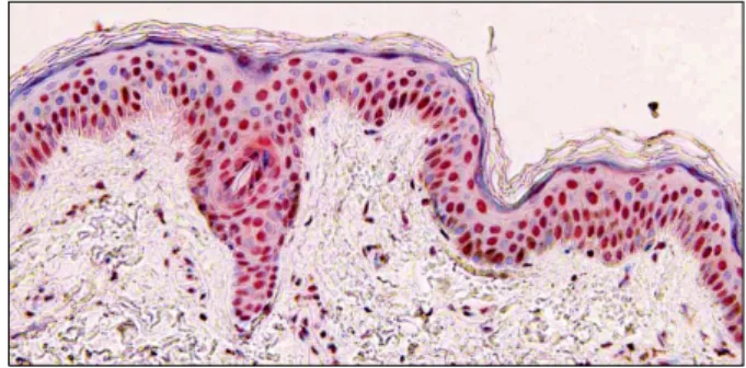
Fig. 1. Cold-inducible RNA-binding protein (CIRP) expression is evident in the nuclei of epidermal keratinocytes in normal skin from back (CIRP immunohistochemical stain, ×200).
http://dx.doi.org/10.5021/ad.2014.26.2.256
Expression of Cold-Inducible RNA-Binding Protein in Normal Skin, Actinic Keratosis and Squamous Cell Carcinoma
Bo Mi Park, Jae Hyuk Lee
1, Seong Jin Kim
Departments of Dermatology and 1Pathology, Chonnam National University Medical School, Gwangju, Korea
Dear Editor:
Cold-inducible RNA-binding proteins (CIRP) are a kind of RNA binding protein associated with diverse cellular res- ponses including cell growth, proliferation, and apop- tosis1. CIRP are induced by various cellular stresses in ad- dition to cold exposure such as ultraviolet (UV) radiation and hypoxia1,2. Recently, their proto-oncogenic functions have been suggested, but a detailed expression pattern in skin tumor has not been reported3-5. Therefore, we aimed to simply observe the expression pattern of CIRP in photo- damaged epidermis of actinic keratosis (AK), squamous cell carcinoma (SCC) and normal skin by immunohisto- chemical staining.
Five samples of normal skin were obtained from 5 healthy individuals undergoing cosmetic surgery. Among them, 3 were taken from the back and 2 from the face. Specimens of AK and SCC were obtained from the faces of 5 patients who underwent excisional surgery. SCC samples were confined to that which developed after long-standing AK.
Clinically and pathologically active lesions were taken as the samples. In all cases, informed consent was obtained from patients according to the ethics committee of the Chonnam National University Hospital. Serial paraffin sec- tions of each specimen were stained with monoclonal
antibodies specific for CIRP (Proteintech Group, Chicago, IL, USA) at a dilution of 1 : 100 according to the manu- facturers’ protocols. The expression of CIRP in epidermal keratinocytes was scored semi-quantitatively by two der- matologists. Nuclear and cytoplasmic staining were asse- ssed separately. We considered both staining intensity and the ratio of positively stained cells in comparison with adjacent stromal cells, lymphocytes, and sebaceous and eccrine glands. No staining was cited as 0, weaker staining than stromal cells as 1, similar to stromal cells as 2, and stronger as 3. Statistical analysis was performed using the chi-square test and Wilcoxon’s rank sum test to compare the expression pattern (SPSS ver. 17.0; SPSS Inc., Chicago, IL, USA).
In normal skin specimens, CIRP expression was more evident in nuclei than in cytoplasm throughout the epi- dermal keratinocytes (Fig. 1). However, in specimens from the face, the most sun exposed area, cytoplasmic CIRP staining intensity was increased compared to that in spec- imens from the back, a less-sun exposed area. In the case of AK, nuclear CIRP expression was decreased while cyto-
Letter to the Editor
Vol. 26 No. 2, 2014 257 Fig. 2. Expression patterns of Cold-inducible RNA-binding protein (CIRP). Representative pictures of five cases in each normal skin from face (A), actinic keratosis (AK) (B), and squamous cell carcinoma (SCC) (C) (A∼C: CIRP immunohistochemical stain, ×200).
Inset: higher magnification of the hot spot of the specimen (CIRP immunohistochemical stain, ×400). (D) Statistical analysis of staining intensity shows significantly decreased nuclear CIRP expression in actinic keratosis and squamous cell carcinoma compared with normal skin specimens. Open circles: tissue sample from back. Closed circle: tissue sample from face. Open circle indicates a normal skin specimen from the back. *p<0.05, **p<0.01, Wilcoxon’s rank sum test.
plasmic expression was maintained or rather increased.
And in the most pathologic spots of SCC, CIRP expression was significantly decreased both in the nuclei and cyto- plasm. Statistical analysis revealed significantly decreased expression of CIRP in nuclei of AK and consequent SCC compared with normal skin. There was no statistically significant difference in cytoplasmic staining intensity among them (Fig. 2).
In previous studies of human cancer, the majority of endo- metrial carcinoma showed decreased staining intensity4. However, staining intensity was increased in several other human tumors, such as colon and prostate cancer5. These conflicting results may come from the early inducing me- chanism of CIRP. CIRP regulates gene expression at the level of translation1,3. Therefore, the exact cellular func-
tion of CIRP remains unknown at the moment and the expression pattern in cancer cells could vary according to the state of the tumor. The increased cytoplasmic CIRP expression we observed in sun-exposed areas might be explained by relocalization of CIRP that was triggered by UV exposure2.
We observed a significant decrease of nuclear CIRP ex- pression in AK and SCC compared with normal skin.
Further studies are needed to elucidate the relationship between CIRP and UV radiation and consequent tumo- rigenesis in the skin.
ACKNOWLEDGMENT
This work was supported by Chonnam National Univer-
Letter to the Editor
258 Ann Dermatol
Received December 6, 2012, Revised March 23, 2013, Accepted for publication April 16, 2013
Corresponding author: Seung Phil Hong, Department of Dermatology, Dankook University Medical College, 119 Dandae-ro, Dongnam-gu, Cheonan 330-714, Korea. Tel: 82-41-550-6485, Fax: 82-41-552-7541, E-mail: [email protected]
This is an Open Access article distributed under the terms of the Creative Commons Attribution Non-Commercial License (http://
creativecommons.org/licenses/by-nc/3.0) which permits unrestricted non-commercial use, distribution, and reproduction in any medium, provided the original work is properly cited.
sity Medical School (BK+21) and Chonnam National University Hospital Biomedical Research Institute (CRE- 13118-7).
REFERENCES
1. Nishiyama H, Itoh K, Kaneko Y, Kishishita M, Yoshida O, Fujita J. A glycine-rich RNA-binding protein mediating cold- inducible suppression of mammalian cell growth. J Cell Biol 1997;137:899-908.
2. Yang C, Carrier F. The UV-inducible RNA-binding protein A18 (A18 hnRNP) plays a protective role in the genotoxic
stress response. J Biol Chem 2001;276:47277-47284.
3. Lleonart ME. A new generation of proto-oncogenes: cold- in- ducible RNA binding proteins. Biochim Biophys Acta 2010;
1805:43-52.
4. Hamid AA, Mandai M, Fujita J, Nanbu K, Kariya M, Kusakari T, et al. Expression of cold-inducible RNA-binding protein in the normal endometrium, endometrial hyperplasia, and en- dometrial carcinoma. Int J Gynecol Pathol 2003;22:240-247.
5. Artero-Castro A, Callejas FB, Castellvi J, Kondoh H, Carnero A, Fernández-Marcos PJ, et al. Cold-inducible RNA-binding protein bypasses replicative senescence in primary cells through extracellular signal-regulated kinase 1 and 2 activa- tion. Mol Cell Biol 2009;29:1855-1868.
http://dx.doi.org/10.5021/ad.2014.26.2.258
Pemphigus Vulgaris in Pregnancy Associated with Herpes Virus Type 1 Infection
Jiwon Gye, Chan Hee Nam, Ji Seok Kim, Jee Young Kim, Byung Cheol Park, Myung Hwa Kim, Seung Phil Hong
Department of Dermatology, Dankook University Hospital, Cheonan, Korea
Dear Editor:
Pemphigus vulgaris (PV) rarely occurs during pregnancy.
We report a case of PV associated with herpes simplex type 1 virus (HSV-1) which occurred in the third trimester of pregnancy.
A 32-year-old second gravida at 37 weeks’ gestation was admitted for multiple bullous skin lesions that persisted for over a month.
These vesicular and erosive lesions initiated from the periumbilical region and spread to the oral mucosa and the skin of the back (Fig. 1A, B). The diagnosis of PV was
confirmed by biopsy (Fig. 1C), and direct immufluore- scence detected anti-immunoglobulin G and C3 antibodies.
Anti-desmoglein 1 and anti-desmoglein 3 antibodies were elevated at 82.1 U/ml (normal, <14 U/ml) and 184.9 U/ml (normal, <7 U/ml) in peripheral blood. Tzanck smear and viral polymerase chain reaction (Seeplex STD B41 Detedtion; Seegene, Seoul, Korea) were done on the base of a vesicular lesion on the trunk. Tzanck smear was negative, but, viral polymerase chain reaction (PCR) was positive for HSV-1 (Fig. 2).
Prednisolone at a dose of 20 mg/d was initiated. Foll-