Preoperative Predictive Factors for Parathyroid Carcinoma in Patients with Primary Hyperparathyroidism
This study was conducted to review the clinical characteristics of parathyroid carcinoma (PC) and to evaluate potential preoperative predictive factors for PC in patients with primary hyperparathyroidism (PHPT). We performed a retrospective review of electronic medical records of 194 patients with pathologically confirmed PHPT in affiliated teaching hospitals of Seoul National University from January 2000 to March 2011. Adenoma was diagnosed in 171 patients, hyperplasia in 12, and carcinoma in 11. Several biochemical measurements were higher in patients with PC than in patients with benign disease, including serum total calcium (P < 0.001), intact parathyroid hormone (P = 0.003), and alkaline phosphatase (ALP) (P < 0.001). Tumors were larger in PC than in benign disease (P < 0.001). Multivariate analysis revealed that serum ALP level (P < 0.001) and tumor size were associated with PC (P = 0.03). Tumor size and serum ALP level were evaluated as preoperative predictive factors for PC using ROC analyses: a tumor size of 3.0 cm
(sensitivity 90.9%, specificity 92.1%) and serum ALP level of 285 IU/L (83.3%, 97.0%) had predictive value for the diagnosis of PC in patients with PHPT. In conclusion, elevated serum ALP and a large parathyroid mass at the time of diagnosis can be helpful to predict PC in patients with PHPT.
Key Words: Hyperparathyroidism; Parathyroid Neoplasms; Alkaline Phosphatase Jae Hyun Bae1, Hyung Jin Choi1,
Yenna Lee2, Min Kyong Moon3, Young Joo Park1, Chan Soo Shin1, Do Jun Park1, Hak Chul Jang2, Seong Yeon Kim1, and Sang Wan Kim3
1Division of Endocrinology and Metabolism, Department of Internal Medicine, Seoul National University Hospital, Seoul National University College of Medicine, Seoul; 2Division of Endocrinology and Metabolism, Department of Internal Medicine, Seoul National University Bundang Hospital, Seoul National University College of Medicine, Seongnam; 3Division of Endocrinology and Metabolism, Department of Internal Medicine, Seoul Metropolitan Government Borame Medical Center, Seoul National University College of Medicine, Seoul, Korea
Received: 26 February 2012 Accepted: 21 May 2012 Address for Correspondence:
Sang Wan Kim, MD
Division of Endocrinology and Metabolism, Department of Internal Medicine, Seoul Metropolitan Government Borame Medical Center, Seoul National University College of Medicine, 5 Boramae-ro, Dongjak-gu, Seoul 156-707, Korea Tel: +82.2-870-2223, 3861, Fax: +82.2-870-3863 E-mail: swkimmd@snu.ac.kr
This study was supported by a grant from Seoul National University Hospital (No.04-2005-0200).
http://dx.doi.org/10.3346/jkms.2012.27.8.890 • J Korean Med Sci 2012; 27: 890-895
INTRODUCTION
Primary hyperparathyroidism (PHPT) is a common endocrine disorder that is being diagnosed more frequently due to the in- creasing use of routine serum total calcium tests as part of medi- cal examinations (1). The clinical presentation of PHPT is chang- ing from a severe disease with renal stones and metabolic bone disease to mild asymptomatic disease (2). Parathyroid carcino- ma (PC) comprises a very small proportion of PHPT cases. A meta-analysis of 20,225 cases of PHPT in the USA reported that PC occurred only in 0.74% of cases (3). Nevertheless, PC requires early recognition and complete resection because its prognosis is quite variable (4-6). The average time to the first recurrence is approximately 3-yr and intervals of up to 20-yr have been re- ported (7).
The current definitive treatment of PC is en-bloc resection, which avoids rupture and spillage of the tumor. Once the tumor
has recurred, curative treatment is unlikely and the surgical com- plication rate is high, although PC patients typically have a pro- longed survival (8). Therefore, it is desirable to distinguish be- nign disease such as parathyroid adenoma and hyperplasia from PC prior to surgery. Unfortunately, PC is frequently only diag- nosed after surgery for hyperparathyroidism because the clini- cal presentation of PC and benign disease are strikingly similar.
In this retrospective study, we compared the clinical presen- tation of PC with benign disease and reevaluated the biochemi- cal parameters that have been used to differentiate PC from be- nign disease at the time of the first operation.
MATERIALS AND METHODS
We performed a retrospective review of electronic medical re- cords in affiliated teaching hospitals of Seoul National Univer- sity (Seoul National University Hospital, Seoul National Univer-
sity Bundang Hospital, and Seoul Metropolitan Government Borame Medical Center). The records were reviewed, over a 10- yr period from January 2000 to March 2011, of patients who had been diagnosed with hyperparathyroidism, parathyroid adeno- ma, parathyroid hyperplasia, PC, parathyroid tumor, or hyper- calcemia. Patients with secondary or tertiary hyperparathyroid- ism, or other causes of hypercalcemia such as malignancy, vita- min D intoxication, immobilization, medications, hyperthyroid- ism, and other endocrine diseases, were excluded. Data collect- ed from the records included the clinical features (hypercalce- mia-related symptoms at the time of diagnosis, previous history of osteoporosis, fracture, and renal or urinary stone), laboratory data (serum levels of total calcium, phosphorous, intact para- thyroid hormone [iPTH], creatinine, alkaline phosphatase [ALP], and 25-hydroxy vitamin D; 24 hr urinary calcium and phospho- rus; and dual energy X-ray absorptiometry [DEXA]), and surgi- cally proven pathology (adenoma, hyperplasia, and carcinoma).
Serum iPTH was measured preoperatively by means of immu- noradiometric assay (Cis Bio International, Gif-Sur-Yvette, Ce- dex, France). Follow-up duration, interval change of clinical features, and laboratory data after surgery were also included.
Descriptive statistics are reported as mean ± standard devia- tion, and confidence intervals were computed as two-tailed us- ing 95% coverage. Categorical variables are reported as frequen- cies and proportions. Comparisons between continuous vari- ables were performed using repeated measures ANOVA. Com- parisons between categorical variables were performed using a chi-squared test. Univariate and multivariate analyses of vari- ables were performed. Statistical analyses were done with SPSS version 18.0 (SPSS Inc., Chicago, IL, USA). P values < 0.05 were accepted as significant.
Ethics statement
The study protocol was approved by the institutional review board (IRB) of Seoul National University Hospital (IRB No. H- 1101-070-348). An informed consent was waived by the IRB.
RESULTS
Subjects and clinical characteristics of patients with PHPT according to the etiology
Two hundred and ninety-one patients with a diagnosis of PHPT were eligible for the study, and 194 of these patients with surgi-
cally proven parathyroid adenoma, hyperplasia, or carcinoma were included in the analysis. Adenoma was diagnosed in 171 patients, hyperplasia in 12 patients, and carcinoma in 11 patients.
PC was diagnosed based on several characteristic pathologic findings (capsular penetration, vascular invasion, perineural space invasion, trabecular growth pattern, and mitotic figures) or definite evidence of metastasis (9, 10). Five patients revealed capsular penetration. Two patients revealed capsular invasion with extension into soft tissue, trabecular growth pattern, and partly cystic change. Two patients revealed capsular invasion with extension into soft tissue, mitotic figures, and partly cystic or hemorrhagic change. One patient revealed capsular penetra- tion and vascular invasion. And the last one patient revealed vascular invasion with solid and spindle cell change.
The mean age at diagnosis in the adenoma, hyperplasia and carcinoma groups were 60 ± 6 (range, 46-74), 48 ± 13 (35-61), and 54 ± 14 (40-68) yr old, respectively. The mean follow-up duration for each of the three groups was 46.5, 40.4, and 56.3 months, respectively. Female predominance was observed in the adenoma and hyperplasia groups, as is generally observed in patients with PHPT. In contrast, carcinoma predominantly occurred in males (7 males, 4 females). However, there were no significant differences in gender distribution in each of the three different etiological groups (Table 1).
Patients with PHPT generally presented without any symp-
Table 1. Demographics of patients with pathologically confirmed primary hyperpara- thyroidism
Parameters Adenoma
(n = 171) Hyperplasia
(n = 12) Carcinoma (n = 11)
Age (yr) 60 ± 6 48 ± 13 54 ± 14
Sex Male
Female 55 (32%)
116 (68%) 4 (33%)
8 (67%) 7 (64%)
4 (36%)
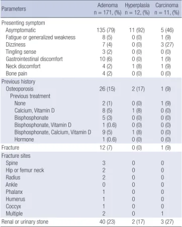
Table 2. Clinical characteristics of patients with pathologically confirmed hyperpara- thyroidism
Parameters Adenoma
n = 171, (%) Hyperplasia
n = 12, (%) Carcinoma n = 11, (%) Presenting symptom
Asymptomatic
Fatigue or generalized weakness Dizziness
Tingling sense
Gastrointestinal discomfort Neck discomfort Bone pain
135 (79) 8 (5) 7 (4) 3 (2) 10 (6) 4 (2) 4 (2)
11 (92) 0 (0) 0 (0) 0 (0) 0 (0) 1 (8) 0 (0)
5 (46) 1 (9) 3 (27) 0 (0) 1 (9) 1 (9) 0 (0) Previous history
Osteoporosis Previous treatment None
Calcium, Vitamin D Bisphosphonate Bisphosphonate, Vitamin D Bisphosphonate, Calcium, Vitamin D Hormone
26 (15) 2 (1) 8 (5) 5 (3) 1 (0.6) 9 (5) 1 (0.6)
2 (17) 0 (0) 1 (8) 0 (0) 0 (0) 1 (8) 0 (0)
1 (9) 1 (9) 0 (0) 0 (0) 0 (0) 0 (0) 0 (0)
Fracture 12 (7) 0 (0) 1 (9)
Fracture sites Spine
Hip or femur neck Radius Ankle Phalanx Humerus Coccyx Multiple
3 2 2 0 1 1 1 2
0 0 0 0 0 0 0 0
0 0 0 0 0 0 0 1
Renal or urinary stone 40 (23) 2 (17) 3 (27)
tom or sign regardless of the etiology. Asymptomatic presenta- tion was seen in 135 adenoma patients (79% of the PHPT patients with adenoma), 11 patients with hyperplasia (92%), and 5 pa- tients with carcinoma (46%). In the remaining patients, present- ing symptoms and signs at the time of diagnosis included fatigue or generalized weakness, dizziness, tingling sensation in the ex- tremities, gastrointestinal discomfort, neck discomfort, and bone pain. Dizziness was observed in 3 patients with carcinoma (27%), but the number of patients was too small and this was not sta- tistically significant compared to benign disease. There was no difference between the three etiological groups in the occurrence of other symptoms and signs. There was no significant differ- ence in the frequency of a history of osteoporosis, fracture, or renal or urinary stone between the different etiological groups (Table 2).
Comparison of biochemical parameters in PHPT patients according to etiology
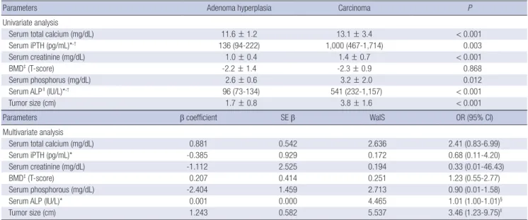
Measurements were obtained for serum total calcium, iPTH, creatinine, phosphorus, ALP, 25-hydroxy vitamin D, and 24 hr urine calcium and phosphorus. In the patients with PC, serum levels of total calcium, iPTH, creatinine, phosphorus and ALP were significantly higher than in patients with adenoma and hyperplasia. There were no significant differences between the three etiological groups in serum 25-hydroxy vitamin D, 24 hr urine calcium and phosphorous, and bone mineral density (BMD) measured by DEXA (Table 3).
Univariate analysis was performed to compare carcinoma and benign disease (adenoma and hyperplasia). Serum total calcium, iPTH, creatinine, phosphorous, and ALP were includ- ed in the analysis. All biochemical parameters in the univariate
analysis were significantly higher in carcinoma than in benign disease (Table 3).
Multivariate analysis was done by logistic regression analysis.
The same biochemical parameters were included in the analy- sis as independent variables comparing carcinoma and benign disease. When all variables were adjusted, only serum ALP level was significantly higher in carcinoma than in benign disease (P < 0.001) (Table 3).
Tumor size
The mean tumor (or hyperplasic gland) size was 3.8 ± 1.6 cm for carcinoma and 1.7 ± 0.8 cm for benign disease. The carcino- mas were significantly larger in univariate (P < 0.001) and mul- tivariate analysis (P = 0.03). The locations of the carcinomas were intrathyroid (5 patients), right lower (2 patients), ectopic (2 pa- tients), right upper (1 patient), and left upper (1 patient). The number of carcinomas in the parathyroid gland was only one for all patients. The location and number of involved parathy- roid glands was not statistically significant between carcinoma and benign diseases.
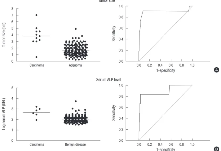
Receiver operating characteristic (ROC) curve analysis of serum ALP level and tumor size for prediction of PC To evaluate the diagnostic values of serum ALP level and tumor size for PC, ROC were calculated for each parameters. ROC curve compared true positive rate, or sensitivity with false positive rate, or 1-specificity. Cut-off value was determined at the highest points of sensitivity and specificity. Confidential interval was 95%. When the cut-off value of tumor size was 3.0 cm, sensitivity and speci- ficity for prediction of carcinoma were 90.9% and 92.1%, respec- tively. Serum ALP level was 540.5 (232.3-1,156.8) (median, [in-
Table 3. Biochemi cal parameters of patients with pathologically confirmed hyperparathyroidism
Parameters Adenoma hyperplasia Carcinoma P
Univariate analysis
Serum total calcium (mg/dL) 11.6 ± 1.2 13.1 ± 3.4 < 0.001
Serum iPTH (pg/mL)*,† 136 (94-222) 1,000 (467-1,714) 0.003
Serum creatinine (mg/dL) 1.0 ± 0.4 1.4 ± 0.7 < 0.001
BMD‡ (T-score) -2.2 ± 1.4 -2.3 ± 0.9 0.868
Serum phosphorus (mg/dL) 2.6 ± 0.6 3.2 ± 2.0 0.012
Serum ALPll (IU/L)*,† 96 (73-134) 541 (232-1,157) < 0.001
Tumor size (cm) 1.7 ± 0.8 3.8 ± 1.6 < 0.001
Parameters β coefficient SE β WalS OR (95% CI)
Multivariate analysis
Serum total calcium (mg/dL) 0.881 0.542 2.636 2.41 (0.83-6.99)
Serum iPTH (pg/mL)* -0.385 0.929 0.172 0.68 (0.11-4.20)
Serum creatinine (mg/dL) -1.112 2.525 0.194 0.33 (0.01-46.43)
BMD‡ (T-score) 0.207 0.414 0.251 1.23 (0.55-2.77)
Serum phosphorous (mg/dL) -2.404 1.459 2.713 0.90 (0.01-1.58)
Serum ALP (IU/L)* 0.001 0.000 4.465 1.01 (1.00-1.01)§
Tumor size (cm) 1.243 0.582 5.537 3.46 (1.23-9.75)ll
*Log-transformed values were used for analysis. †Median (interquartile range) values were shown. ‡Bone mineral density. §P < 0.001, llP = 0.03. SE β, standard error of β co- efficient; WalS, Wald statistic; OR, odds ratio; CI, confidence interval.
terquartile range]) IU/L in carcinoma and 96.0 (73.0-133.8) IU/
L in benign disease (P < 0.001). When the cut-off value of serum ALP was 285 IU/L, sensitivity and specificity for prediction of carcinoma were 83.3% and 97.0%, respectively (Fig. 1).
Correlation of tumor size with biochemical parameters in patients with PHPT
The tumor size was correlated with serum ALP and iPTH levels in patients with parathyroid adenoma (P = 0.009, and P < 0.001).
However, correlation between tumor size and biochemical pa- rameters was not significant in PC patients (Table 4).
DISCUSSION
In this retrospective study of 194 patients with pathologically confirmed PHPT, the tumor size and serum ALP level were pre- dictors of PC before the first operation. PC is an extremely rare tumor. In this study, only 11 patients were diagnosed with PC in Seoul National University’s three affiliated teaching hospitals during a 10-yr period. The proportion of PC in patients with PHPT was relatively high in our study, 5.7% considering that previous studies reported that PC accounts for only 0.5%-5% of all patients with PHPT (3, 5, 11). This discrepancy could result from referral bias since the hospitals involved in the present study are refer- Fig. 1. Receiver operating curve analyses of tumor size and serum ALP level. Tumor size and serum ALP level were evaluated for preoperative predictive factors for PC. Tumor size (A) 3.0 cm had 90.9% of sensitivity, 92.1% of specificity and 43.4% of positive predictive values (PPV), 99.9% of negative predictive value (NPV). Serum ALP level (B) 285 IU/L had 83.3% of sensitivity, 97.0% of specificity and 33.3% of PPV, 99.9% of NPV.
Tumor size (cm) Sensitivity
Carcinoma Adenoma 0.0 0.2 0.4 0.6 0.8 1.0
8 7 6 5 4 3 2 1 0
1.0
0.8
0.6
0.4
0.2
0.0 Tumor size
1-specificity A
Log serum ALP (IU/L) Sensitivity
Carcinoma Benign disease 0.0 0.2 0.4 0.6 0.8 1.0
5 4 3 2 1 0
1.0
0.8
0.6
0.4
0.2
0.0 Serum ALP level
1-specificity B
Table 4. Correlation of tumor size with biochemical parameters in patients with primary hyperparathyroidism
Parameters in serum All patients Parathyroid adenoma Parathyroid carcinoma
r P r P r P
Calcium 0.109 0.145 0.122 0.127 0.133 0.715
Phosphorus 0.217 0.004* 0.011 0.895 0.458 0.183
iPTH 0.256 0.001* 0.209 0.009* 0.028 0.947
ALP 0.401 0.001* 0.379 0.000* 0.129 0.807
Creatinine 0.215 0.005* 0.101 0.218 0.133 0.715
*P < 0.01.
ral centers.
There were no significant differences between PC and the benign disease in terms of clinical symptoms, bone loss or frac- ture, or nephrolithiasis. PC affects the classical target organs, such as bone and kidney, more severely than benign PHPT, since patients with PC commonly have profound hypercalcemia (8, 12-14). Although most PHPT patients in contemporary clinical practice are asymptomatic, some patients with benign PHPT present with more severe manifestations (2, 15, 16). In such pa- tients, it may be extremely difficult to differentiate the clinical manifestations of malignancy from those of benign diseases. As the number of benign, symptomatic PHPT patients is progres- sively decreasing, the sample size of 194 patients in the present study would be unlikely to find a statistical difference between the clinical manifestations of malignant and benign PHPT.
Serum ALP level was higher in patients with PC than in those with benign disease (17), and our results suggest that a patient with serum ALP level less than 300 IU/L is unlikely to have car- cinoma. Elevated serum ALP level in patients with PHPT may reflect the longstanding effect of PTH on the bone. This result is supported by the previous study that serum levels of PTH and ALP are useful for the prediction of PC (18). In this study, serum iPTH levels in patients with benign PHPT showed relatively wide distribution compared to serum ALP levels. This result suggests that serum ALP rather than serum iPTH could be predictive fac- tors of PC. Persistent excess of PTH in the circulation causes an increase in the RANKL/OPG ratio, which is the main mechanism by which PTH stimulates osteoclastogenesis and bone resorp- tion (19, 20). Histomorphometric studies have shown that PHPT commonly causes preferential cortical bone loss with preserva- tion of cancellous bone (21, 22). Therefore, serum ALP might reflect the effects of PTH on the bone earlier than skeletal phe- notypes such as bone mineral density or fractures.
The size of a parathyroid tumor may be an important factor in predicting PC in a patient with PHPT. In this study, 10 of 11 patients with PC had a parathyroid mass larger than 3.0 cm, and ROC analysis showed that the sensitivity and specificity for PC was 90.9% and 92.1%, respectively. This result is in agreement with previous findings that PC are usually larger than 3 cm and may be palpable (23). Interestingly, the median tumor size at the time of diagnosis in 286 cases of PC treated in the USA from 1985 to 1995 was approximately 3.3 cm (8). A palpable mass in the neck has been suggested as a strong predictive factor for PC (7, 11, 24), and particular attention should be paid to a hyper- parathyroid patient with a parathyroid mass larger than 3.0 cm.
We also examined PTH as a potential factor to distinguish PC from benign causes of PHPT. In multivariate analysis, patients with PC did not show a statistically significant difference in se- rum levels of iPTH compared to those with benign PHPT. The relationship between preoperative serum PTH levels and the size of a parathyroid adenoma or carcinoma is controversial (25,
26). In the present study, serum iPTH levels were correlated with tumor size in patients with parathyroid adenoma but not in pa- tients with PC. This difference may be resulted from the small number of patients with carcinoma. A recent report suggested that an N-terminal PTH distinct from iPTH can be recognized in a 3rd generation assay of whole PTH and that this form is over- produced in some patients with PC (27). Oncogenes and tumor suppressor genes involved in the control of the cell cycle have been linked to the pathogenesis of PC, including retinoblastoma (Rb), p53, breast carcinoma susceptibility (BRCA2), and cyclin D l/parathyroid adenomatosis gene 1 (CCND1/PRAD1) genes (28, 29). The excessive expression of CCND1 by the highly active PTH gene promoter in the parathyroid cell may contribute to excess cellular proliferation and development of PC. This result indicates that PC is frequently functional. Although CCND1 is overexpressed in more than 90% of cases of PC, its role in patho- genesis remains to be determined (30).
In conclusion, the results of this study suggest that the size of tumor and serum level of ALP may be helpful to predict PC in patients with PHPT before the primary operation.
REFERENCES
1. Wermers RA, Khosla S, Atkinson EJ, Achenbach SJ, Oberg AL, Grant CS, Melton LJ 3rd. Incidence of primary hyperparathyroidism in Rochester, Minnesota, 1993-2001: an update on the changing epidemiology of the disease. J Bone Miner Res 2006; 21: 171-7.
2. Fraser WD. Hyperparathyroidism. Lancet 2009; 374: 145-58.
3. Ruda JM, Hollenbeak CS, Stack BC Jr. A systematic review of the diagno- sis and treatment of primary hyperparathyroidism from 1995 to 2003.
Otolaryngol Head Neck Surg 2005; 132: 359-72.
4. Iacobone M, Lumachi F, Favia G. Up-to-date on parathyroid carcino- ma: analysis of an experience of 19 cases. J Surg Oncol 2004; 88: 223-8.
5. Iihara M, Okamoto T, Suzuki R, Kawamata A, Nishikawa T, Kobayashi M, Obara T. Functional parathyroid carcinoma: long-term treatment outcome and risk factor analysis. Surgery 2007; 142: 936-43.
6. Kleinpeter KP, Lovato JF, Clark PB, Wooldridge T, Norman ES, Bergman S, Perrier ND. Is parathyroid carcinoma indeed a lethal disease? Ann Surg Oncol 2005; 12: 260-6.
7. Shane E. Clinical review 122: Parathyroid carcinoma. J Clin Endocrinol Metab 2001; 86: 485-93.
8. Hundahl SA, Fleming ID, Fremgen AM, Menck HR. Two hundred eighty-six cases of parathyroid carcinoma treated in the U.S. between 1985-1995: a National Cancer Data Base Report. The American College of Surgeons Commission on Cancer and the American Cancer Society.
Cancer 1999; 86: 538-44.
9. Okamoto T, Iihara M, Obara T, Tsukada T. Parathyroid carcinoma: eti- ology, diagnosis, and treatment. World J Surg 2009; 33: 2343-54.
10. Owen RP, Silver CE, Pellitteri PK, Shaha AR, Devaney KO, Werner JA, Rinaldo A, Ferlito A. Parathyroid carcinoma: a review. Head Neck 2011;
33: 429-36.
11. Schaapveld M, Jorna FH, Aben KK, Haak HR, Plukker JT, Links TP. Inci- dence and prognosis of parathyroid gland carcinoma: a population-
based study in The Netherlands estimating the preoperative diagnosis.
Am J Surg 2011; 202: 590-7.
12. Obara T, Okamoto T, Kanbe M, Iihara M. Functioning parathyroid car- cinoma: clinicopathologic features and rational treatment. Semin Surg Oncol 1997; 13: 134-41.
13. Wang CA, Gaz RD. Natural history of parathyroid carcinoma. Diagno- sis, treatment, and results. Am J Surg 1985; 149: 522-7.
14. Wynne AG, van Heerden J, Carney JA, Fitzpatrick LA. Parathyroid car- cinoma: clinical and pathologic features in 43 patients. Medicine (Balti- more) 1992; 71: 197-205.
15. Chon S, Kim YH, Park JY, Ko KP, Park CY, Kim DY, Woo JT, Kim SW, Kim JW, Kim YS, et al. A case of cystic parathyroid adenoma presenting as se- vere bony lesion. J Korean Soc Endocrinol 2003; 18: 214-20.
16. Chung JO, Jeong GH, Hong SE, Cho DH, Chung DJ, Chung MY. A case of primary hyperparathyroidism due to cystic parathyroid adenoma presenting as hypercalcemic crisis associated with intracranial hemor- rhage. J Korean Endocr Soc 2007; 22: 292-8.
17. Silverberg SJ, Shane E, Jacobs TP, Siris ES, Gartenberg F, Seldin D, Cle- mens TL, Bilezikian JP. Nephrolithiasis and bone involvement in prima- ry hyperparathyroidism. Am J Med 1990; 89: 327-34.
18. Chen Q, Kaji H, Nomura R, Sowa H, Yamauchi M, Tsukamoto T, Yama- guchi T, Kobayashi A, Sugimoto T, Chihara K. Trial to predict malig- nancy of affected parathyroid glands in primary hyperparathyroidism.
Endocr J 2003; 50: 527-34.
19. Huang JC, Sakata T, Pfleger LL, Bencsik M, Halloran BP, Bikle DD, Nis- senson RA. PTH differentially regulates expression of RANKL and OPG.
J Bone Miner Res 2004; 19: 235-44.
20. Ma YL, Cain RL, Halladay DL, Yang X, Zeng Q, Miles RR, Chandrasekhar S, Martin TJ, Onyia JE. Catabolic effects of continuous human PTH (1-- 38) in vivo is associated with sustained stimulation of RANKL and inhi- bition of osteoprotegerin and gene-associated bone formation. Endocri- nology 2001; 142: 4047-54.
21. Parisien M, Mellish RW, Silverberg SJ, Shane E, Lindsay R, Bilezikian JP,
Dempster DW. Maintenance of cancellous bone connectivity in primary hyperparathyroidism: trabecular strut analysis. J Bone Miner Res 1992;
7: 913-9.
22. Parisien M, Silverberg SJ, Shane E, de la Cruz L, Lindsay R, Bilezikian JP, Dempster DW. The histomorphometry of bone in primary hyper- parathyroidism: preservation of cancellous bone structure. J Clin Endo- crinol Metab 1990; 70: 930-8.
23. Marcocci C, Cetani F, Rubin MR, Silverberg SJ, Pinchera A, Bilezikian JP. Parathyroid carcinoma. J Bone Miner Res 2008; 23: 1869-80.
24. Robert JH, Trombetti A, Garcia A, Pache JC, Herrmann F, Spiliopoulos A, Rizzoli R. Primary hyperparathyroidism: can parathyroid carcinoma be anticipated on clinical and biochemical grounds? Report of nine cas- es and review of the literature. Ann Surg Oncol 2005; 12: 526-32.
25. Randhawa PS, Mace AD, Nouraei SA, Stearns MP. Primary hyperpara- thyroidism: do perioperative biochemical variables correlate with para- thyroid adenoma weight or volume? Clin Otolaryngol 2007; 32: 179-84.
26. Williams JG, Wheeler MH, Aston JP, Brown RC, Woodhead JS. The rela- tionship between adenoma weight and intact (1-84) parathyroid hor- mone level in primary hyperparathyroidism. Am J Surg 1992; 163: 301-4.
27. Rubin MR, Silverberg SJ, D’Amour P, Brossard JH, Rousseau L, Sliney J Jr, Cantor T, Bilezikian JP. An N-terminal molecular form of parathyroid hormone (PTH) distinct from hPTH(1 84) is overproduced in parathy- roid carcinoma. Clin Chem 2007; 53: 1470-6.
28. Arnold A, Shattuck TM, Mallya SM, Krebs LJ, Costa J, Gallagher J, Wild Y, Saucier K. Molecular pathogenesis of primary hyperparathyroidism. J Bone Miner Res 2002; 17: N30-6.
29. Cryns VL, Rubio MP, Thor AD, Louis DN, Arnold A. p53 abnormalities in human parathyroid carcinoma. J Clin Endocrinol Metab 1994; 78:
1320-4.
30. Vasef MA, Brynes RK, Sturm M, Bromley C, Robinson RA. Expression of cyclin D1 in parathyroid carcinomas, adenomas, and hyperplasias: a paraffin immunohistochemical study. Mod Pathol 1999; 12: 412-6.