Relative Prevalence and Antimicrobial
Susceptibility of Clinical Isolates of
Elizabethkingia Species Based on 16S
rRNA Gene Sequencing
Mi-Soon Han,a,dHyunsoo Kim,aYangsoon Lee,cMyungsook Kim,aNam Su Ku,b
Jun Yong Choi,bDongeun Yong,aSeok Hoon Jeong,aKyungwon Lee,a
Yunsop Chonga
Department of Laboratory Medicine and Research Institute of Bacterial Resistance, Yonsei University College of Medicine, Seoul, Republic of Koreaa; Department of Internal Medicine and AIDS Research Institute, Yonsei University College of Medicine, Seoul, Republic of Koreab; Department of Laboratory Medicine, Hanyang University College of Medicine, Seoul, Republic of Koreac; Medical Clinic Laboratory Department of U
2Bio Co. Ltd., Seoul, Republic of Koread
ABSTRACT Some of the previously reported clinical isolates of Elizabethkingia me-ningoseptica may be later named species of Elizabethkingia. We determined the ac-curacy of species identification (with two matrix-assisted laser desorption ionization– time of flight mass spectrometry [MALDI-TOF MS] systems and the Vitek 2 GN card), relative prevalence of three Elizabethkingia spp. in clinical specimens, and antimicro-bial susceptibility of the species identified by 16S rRNA gene sequencing. Specimens for culture were collected from patients in a university hospital in Seoul, South Ko-rea, between 2009 and 2015. All 3 Elizabethkingia spp. were detected in patients; among the 86 isolates identified by 16S rRNA gene sequencing, 17 (19.8%) were E. meningoseptica, 18 (20.9%) were Elizabethkingia miricola, and 51 (59.3%) were Eliza-bethkingia anophelis. Only the MALDI-TOF Vitek MS system with an amended data-base correctly identified all of the isolates. The majority (76.7%) of the isolates were from the lower respiratory tract, and 8 (9.3%) were from blood. Over 90% of E. me-ningoseptica and E. anophelis isolates were susceptible to piperacillin-tazobactam and rifampin. In contrast, all E. miricola isolates were susceptible to fluoroquinolones except ciprofloxacin. Further studies are urgently needed to determine the optimal antimicrobial agents for the treatment of infections due to each individual Elizabeth-kingia species.
KEYWORDS Elizabethkingia meningoseptica, Elizabethkingia miricola, Elizabethkingia anophelis, antimicrobial susceptibility, 16S rRNA gene sequencing
E
lizabethkingia species are aerobic, nonmotile, oxidase-positive, indole-positive, Gram-negative bacilli that do not ferment glucose. Elizabethkingia spp. can be found frequently in soil, freshwater, salt water, and in hospital environments (1). However, they do not normally exist in the human body. Elizabethkingia meningoseptica (formerly Chryseobacterium meningosepticum) has been a well-known human pathogen since its first description in a case of neonatal meningitis by Elizabeth O. King in 1959 (2). This organism was reported to cause various invasive infections in immunocom-promised hosts and to be associated with nosocomial infections and outbreaks in intensive care units (ICUs) (3–5). It has been considered that the incidence of E. meningoseptica bacteremia has increased over the last decade (6). Two new species of Elizabethkingia, Elizabethkingia miricola and Elizabethkingia anophelis, were proposed in 2003 and 2011, respectively (7–9). Therefore, some of the previously reported clinical isolates of E. meningoseptica may be later named species of Elizabethkingia. The firstReceived 1 August 2016 Returned for modification 17 August 2016 Accepted 31
October 2016
Accepted manuscript posted online 9
November 2016
Citation Han M-S, Kim H, Lee Y, Kim M, Ku NS,
Choi JY, Yong D, Jeong SH, Lee K, Chong Y. 2017. Relative prevalence and antimicrobial susceptibility of clinical isolates of
Elizabethkingia species based on 16S rRNA
gene sequencing. J Clin Microbiol 55:274 –280. https://doi.org/10.1128/JCM.01637-16.
Editor Karen C. Carroll, The Johns Hopkins
University School of Medicine
Copyright © 2016 American Society for
Microbiology. All Rights Reserved. Address correspondence to Kyungwon Lee, [email protected].
M.-S.H. and H.K. contributed equally to this article.
case of E. miricola sepsis was reported in 2008 (10), and a case of E. anophelis neonatal meningitis was reported in 2013 (11).
E. meningoseptica isolates are often resistant (R) to multiple-lactam antibiotics due to intrinsic class A extended-spectrum -lactamases (ESBLs) and inherent class B metallo--lactamases (MBLs) (12). The antimicrobial susceptibility of Elizabethkingia may vary depending on the species. However, there are scanty data on the suscepti-bility of the new species. The aims of this study were to determine the accuracy of species identification systems and the relative prevalence of three Elizabethkingia spp. in clinical specimens and to compare the antimicrobial susceptibility of the species identified by 16S rRNA gene sequencing.
RESULTS
Elizabethkingia spp. identified. Among the total 86 Elizabethkingia isolates, the species identified using 16S rRNA gene sequencing were 17 isolates (19.8%) of E. meningoseptica (99.5% to 99.9% nucleotide identity to E. meningoseptica type strain ATCC 13253), 18 isolates (20.9%) of E. miricola (98.9% to 99.8% nucleotide identity to E. miricola type strain GTC862), and 51 isolates (59.3%) of E. anophelis (99.1% to 100.0% nucleotide identity to E. anophelis type strain FMS-007).
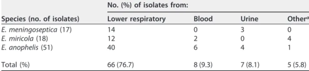
The matrix-assisted laser desorption ionization–time of flight (MALDI-TOF) Vitek MS system with an amended database correctly identified all of the 17, 18, and 51 isolates of E. meningoseptica, E. miricola, and E. anophelis, respectively. However, the Bruker Biotyper correctly identified 16 of 17 E. meningoseptica isolates and 17 of 18 E. miricola isolates but none of the E. anophelis isolates (Table 1). The Vitek 2 GN card system correctly identified 16 of 17 E. meningoseptica isolates but none of the other species. Among the 86 isolates of Elizabethkingia spp., 66 (76.7%) were recovered from the lower respiratory tract, 8 (9.3%) from blood, 7 (8.1%) from urine, and 5 (5.8%) from other specimens (Table 2). Among the 8 isolates from blood, 2 isolates were identified as E. miricola and 6 isolates were identified as E. anophelis. There were no E. meningoseptica isolates from blood. Careful clinical evaluation suggested that the positive blood
TABLE 1 Comparison of species identified by 16S rRNA gene sequencing with those by
the two MALDI-TOF systems and the Vitek 2 GN card system
16S rRNA gene sequencing (no. of isolates) MALDI-TOF Vitek MSa (no. of isolates) MALDI-TOF Bruker Biotyper (no. of isolates)
Vitek 2 with GN card (no. of isolates) E. meningoseptica (17) E. meningoseptica (17) E. meningoseptica (16) E. meningoseptica (16)
Chryseobacterium indologenes (1)
C. indologenes (1) E. miricola (18) E. miricola (18) E. miricola (17) E. meningoseptica (16)
C. indologenes (1) C. indologenes (2) E. anophelis (51) E. anophelis (51) E. meningoseptica (49) E. meningoseptica (48)
C. indologenes (1) E. meningoseptica/
E. miricola (1)
E. meningoseptica (2) E. miricola (1)
aIdentification was based on a SARAMIS database amended with Elizabethkingia spp. spectra provided to
bioMérieux.
TABLE 2 Source of detection of 86 isolates of Elizabethkingia spp. at a tertiary care
hospital from 2009 to 2015
Species (no. of isolates)
No. (%) of isolates from:
Lower respiratory Blood Urine Othera
E. meningoseptica (17) 14 0 3 0
E. miricola (18) 12 2 0 4
E. anophelis (51) 40 6 4 1
Total (%) 66 (76.7) 8 (9.3) 7 (8.1) 5 (5.8)
cultures in 4 of the 8 patients were not significant. However, they may have been transient invaders from central arterial or venous lines or endotracheal tubes. Therefore, no antimicrobial agents were administered for Elizabethkingia infection (Table 3). All of the 8 patients had various underlying diseases and had indwelling catheters or endo-tracheal tubes. Four patients were admitted to the ICU. All of the patients, except for one, were treated with various antimicrobial agents during the 7 days prior to blood culture. Among the four patients, two were cured with tigecycline and trimethoprim-sulfamethoxazole, to which the isolates were susceptible (S). The remaining two patients were cured with trimethoprim-sulfamethoxazole, although the MICs for the isolates were 4 and 76g/ml (resistance breakpoints, ⱖ4 and 76 g/ml), respectively. In pulsed-field gel electrophoresis (PFGE) analysis, isolates of each Elizabethkingia species belonged to 5 to 10 different PFGE groups, while identical pulsotypes were found in 8 of 17 E. meningoseptica isolates, 6 of 18 E. miricola isolates, and 17 of 51 E. anophelis isolates (see Fig. S1 in the supplemental material).
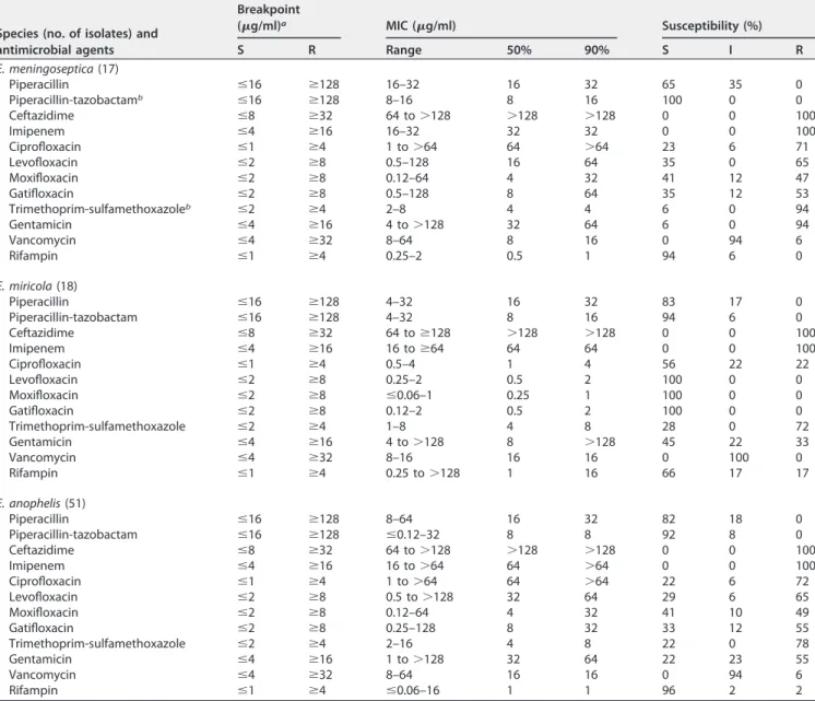
Antimicrobial susceptibilities. The MICs of the antimicrobial agents and the susceptibilities of the isolates are shown in Table 4 (see Data Set S1 in the supplemental material). Among the E. meningoseptica isolates, 100% and 94% were susceptible to piperacillin-tazobactam and rifampin, respectively, but only 23% to 41% were tible to fluoroquinolones. Unlike E. meningoseptica, all E. miricola isolates were suscep-tible to fluoroquinolones, except for ciprofloxacin. Over 90% of E. meningoseptica and E. anophelis isolates were susceptible to piperacillin-tazobactam and rifampin. Although none of the species were susceptible to vancomycin, all three species exhibited at least 94% intermediate (I) reaction to this agent.
DISCUSSION
The accuracy of species identification was low with the Vitek 2 GN card system. Although our Bruker Biotyper without an amended database failed to identify E. anophelis isolates, the addition of a database for E. anophelis was recently reported (13). This result indicates that a MALDI-TOF mass spectrometry (MS) system can be a reliable species identification system for the genus Elizabethkingia.
Although E. meningoseptica infections are well known, E. miricola sepsis in a lym-phoma patient was reported in the United States after the proposal of new species (10). Several E. anophelis infections have been reported from tropical or subtropical regions, the Central African Republic (11), Singapore (14), and Hong Kong (15). A recent study in Hong Kong urged researchers to consider the clinically significant morbidity and mortality of patients with E. anophelis bacteremia (13). An E. anophelis outbreak in Wisconsin in 2016 resulted in the deaths of at least 18 patients (16).
In our study, which took place in the temperate country of South Korea, all three species of Elizabethkingia were detected in patients, and E. anophelis was the most prevalent. It is interesting that E. meningoseptica was not present while E. anophelis was the most common among blood isolates (Table 2). These findings suggest that E. anophelis plays a significant role as a human pathogen. In general, the majority of blood isolates are clinically significant (5, 13, 16). However, in our study, only 4 of the 8 patients with positive Elizabethkingia blood cultures were clinically significant, although all of the patients had risk factors for infection (Table 3). The majority of the Elizabeth-kingia isolates were detected in lower respiratory tract specimens, but it was difficult to distinguish infection from colonization as reported in other studies (4, 17).
Our PFGE analysis of each species showed that certain pulsotypes were more prevalent than others, suggesting that these types are either more prevalent in the hospital environment or that they have a higher capability to infect or colonize.
E. meningoseptica has been known to be resistant to multiple antimicrobial agents (18, 19). However, as mentioned above, the susceptibility of E. meningoseptica in the previous study may include those of the other 2 species. To the best of our knowledge, our study is the first one to compare the susceptibilities of all Elizabethkingia species (Table 4). The organisms are typically resistant to-lactams (15, 18). In a previous study from our group (19), all 31 isolates of E. meningoseptica (which may include other Elizabethkingia species)
TABLE 3 Case summary of eight patients with Elizabethkingia spp. isolation from blood No. Age/sex a Initial identification b Final identification c Underlying disease d Indwelling device Ward e No. positive/total pairs, bottles (culture date) Infection sign f Previous antibiotic therapy (1 week before culture) Antimicrobial therapy for Elizabethkingia infection Outcome BT (°C) WBC (per l) Microbiologic eradication g Clinical response h 1 79/M E. meningoseptica E. miricola Bladder cancer, hypoxic brain damage Endotracheal tube, central venous line ICU 1/3 (Aug. 8, 2011) 38.0 23,990 None None NA i NA 1/1 (Aug. 9, 2011) 2 69/M E. meningoseptica E. miricola COPD, CRF, cerebral infarction, fungal pneumonia Endotracheal tube GW 1/4 (Aug. 26, 2011) 35.9 9,990 Meropenem, teicoplanin, levofloxacin None NA NA 3 69/M E. meningoseptica E. anophelis Rectal cancer Endotracheal tube, arterial line, central venous line ICU 1/3 (Sep. 25, 2010) 38.0 8,320 Imipenem, metronidazole None NA NA 4 72/F E. meningoseptica E. anophelis Klatskin tumor, DM Endotracheal tube, arterial line, central venous line GW 2/3 (Oct. 30, 2010) 36.2 7,420 Tigecycline Tigecycline, ciprofloxacin Cured Failed 5 72/M E. meningoseptica E. anophelis Alcoholic LC Endotracheal tube, central venous line GW 3/3 (Nov. 13, 2012) 37.8 2,400 Imipenem, colistin, teicoplanin, minocycline Trimethoprim-sulfamethoxazole Cured Cured 2/2 (Nov. 15, 2012) 6 67/F E. meningoseptica E. anophelis Myelofibrosis, splenomegaly, thrombotic endocarditis Central venous line GW 4/4 (Nov. 22, 2012) 37.5 11,180 Ciprofloxacin, cefazolin Trimethoprim-sulfamethoxazole Cured Cured 1/3 (Nov. 23, 2012 1/1 (Nov. 24, 2012) 2/4 (Nov. 26, 2012) 7 49/M E. meningoseptica E. anophelis Mitral valve replacement, valvular heart failure Endotracheal tube, arterial line ICU 1/5 (Mar. 20, 2013) 38.6 16,630 Piperacillin-tazobactam, cefepime, teicoplanin None NA NA 8 63/M E. meningoseptica E. anophelis Pneumonectomy, lung transplantation, RA Endotracheal tube, chest tube, arterial line, central venous line ICU 2/3 (Sep. 20, 2013) 36.4 2,890 Meropenem, colistin Trimethoprim-sulfamethoxazole, tigecycline Cured Cured 2/3 (Sep. 22, 2013) 4/4 (Sep. 24, 2013) a M, male; F, female. b Identified by Vitek 2 with GN card for no. 1 to 7 and by MALDI-TOF Bruker Biotyper for No. 8. c Final identification was done by 16S rRNA gene sequencing. d COPD, chronic obstructive pulmonary disease; CRF, chronic renal failure; LC, liver cirrhosis; RA, rheumatic arthritis; DM, diabetes mellitus. e GW, general ward. f BT, body temperature; WBC, white blood cell. g Microbiologic eradication was the absence of the original pathogens detected from blood (7 days after the first positive blood culture). h Clinical response: a favorable clinical response was defined as the resolution of fever (defined as ⱖ 38.0°C), leukocytosis (WBC, ⱖ 11 ⫻ 10 6 / l), and hypotension (mean arterial pressure of ⬍ 65 mm Hg), in addition to no longer requiring support from vasoactive agents. Patients who had persistence or deterioration in clinical parameters or who died were classified as treatment failures. i NA, not applicable. There was no follow up data due to transfer of the patient within 7 days.
had both blaBlaBand blaGOBgenes. The GenBank database shows that chromosomes of E.
anophelis (accession numbers CP006576 and CP007547) and E. miricola (accession num-ber CP011059) possess blaBlaB, blaGOB, and blaCMEgenes and an AmpC -lactamase
gene. In our study, over 90% of the isolates of 3 Elizabethkingia spp. were susceptible to a piperacillin-tazobactam combination. This has also been shown by other studies (5, 20). However, it is necessary to evaluate clinical efficacy, given that all Elizabethkingia spp. appear to be inherent MBL producers.
Antimicrobial resistance may vary depending on the species as well as the region and time of bacterial isolation. As mentioned above, limited data are currently available on the susceptibility patterns of Elizabethkingia spp. Although three previous studies were performed using relatively significant numbers of isolates, the specimens were from unspecified sources of patients, blood, or hospital environments (5, 21, 22). Furthermore, all three reports stated that the species were E. meningoseptica, but the isolates were identified before the proposal of new species or unreliable phenotypic methods were used for identification. However, by comparing our results, based on
TABLE 4 Antimicrobial susceptibilities of Elizabethkingia isolates determined by the agar dilution method
Species (no. of isolates) and antimicrobial agents Breakpoint (g/ml)a MIC (g/ml) Susceptibility (%) S R Range 50% 90% S I R E. meningoseptica (17) Piperacillin ⱕ16 ⱖ128 16–32 16 32 65 35 0 Piperacillin-tazobactamb ⱕ16 ⱖ128 8–16 8 16 100 0 0 Ceftazidime ⱕ8 ⱖ32 64 to⬎128 ⬎128 ⬎128 0 0 100 Imipenem ⱕ4 ⱖ16 16–32 32 32 0 0 100 Ciprofloxacin ⱕ1 ⱖ4 1 to⬎64 64 ⬎64 23 6 71 Levofloxacin ⱕ2 ⱖ8 0.5–128 16 64 35 0 65 Moxifloxacin ⱕ2 ⱖ8 0.12–64 4 32 41 12 47 Gatifloxacin ⱕ2 ⱖ8 0.5–128 8 64 35 12 53 Trimethoprim-sulfamethoxazoleb ⱕ2 ⱖ4 2–8 4 4 6 0 94 Gentamicin ⱕ4 ⱖ16 4 to⬎128 32 64 6 0 94 Vancomycin ⱕ4 ⱖ32 8–64 8 16 0 94 6 Rifampin ⱕ1 ⱖ4 0.25–2 0.5 1 94 6 0 E. miricola (18) Piperacillin ⱕ16 ⱖ128 4–32 16 32 83 17 0 Piperacillin-tazobactam ⱕ16 ⱖ128 4–32 8 16 94 6 0 Ceftazidime ⱕ8 ⱖ32 64 toⱖ128 ⬎128 ⬎128 0 0 100 Imipenem ⱕ4 ⱖ16 16 toⱖ64 64 64 0 0 100 Ciprofloxacin ⱕ1 ⱖ4 0.5–4 1 4 56 22 22 Levofloxacin ⱕ2 ⱖ8 0.25–2 0.5 2 100 0 0 Moxifloxacin ⱕ2 ⱖ8 ⱕ0.06–1 0.25 1 100 0 0 Gatifloxacin ⱕ2 ⱖ8 0.12–2 0.5 2 100 0 0 Trimethoprim-sulfamethoxazole ⱕ2 ⱖ4 1–8 4 8 28 0 72 Gentamicin ⱕ4 ⱖ16 4 to⬎128 8 ⬎128 45 22 33 Vancomycin ⱕ4 ⱖ32 8–16 16 16 0 100 0 Rifampin ⱕ1 ⱖ4 0.25 to⬎128 1 16 66 17 17 E. anophelis (51) Piperacillin ⱕ16 ⱖ128 8–64 16 32 82 18 0 Piperacillin-tazobactam ⱕ16 ⱖ128 ⱕ0.12–32 8 8 92 8 0 Ceftazidime ⱕ8 ⱖ32 64 to⬎128 ⬎128 ⬎128 0 0 100 Imipenem ⱕ4 ⱖ16 16 to⬎64 64 ⬎64 0 0 100 Ciprofloxacin ⱕ1 ⱖ4 1 to⬎64 64 ⬎64 22 6 72 Levofloxacin ⱕ2 ⱖ8 0.5 to⬎128 32 64 29 6 65 Moxifloxacin ⱕ2 ⱖ8 0.12–64 4 32 41 10 49 Gatifloxacin ⱕ2 ⱖ8 0.25–128 8 32 33 12 55 Trimethoprim-sulfamethoxazole ⱕ2 ⱖ4 2–16 4 8 22 0 78 Gentamicin ⱕ4 ⱖ16 1 to⬎128 32 64 22 23 55 Vancomycin ⱕ4 ⱖ32 8–64 16 16 0 94 6 Rifampin ⱕ1 ⱖ4 ⱕ0.06–16 1 1 96 2 2
aThe interpretive criteria applied were those of the CLSI for non-Enterobacteriaceae; the criteria for vancomycin and rifampin were those for Staphylococcus or Enterococcus spp. The criterion of gatifloxacin was that for moxifloxacin.
identification by 16S rRNA sequencing, to those of other studies, the following gener-alization can be made: Elizabethkingia spp. are nonsusceptible to ceftazidime, imi-penem, and vancomycin (high vancomycin susceptibility in a study may be due to the use of a higher breakpoint, 16 g/ml [21]); the susceptibility rates of E. miricola to fluoroquinolones are higher than those of the other species; and the susceptibility of Elizabethkingia spp. to other antimicrobial agents are difficult to predict.
Several reports have shown that the incidence of Elizabethkingia bacteremia in-creased and the mortality rate was high (5, 20, 23, 24). Indwelling devices and inappropriate antimicrobial therapy were independent risk factors for poor outcomes with Elizabethkingia bacteremia (5, 24, 25). In our study, all 4 bacteremic patients were microbiologically cured with trimethoprim-sulfamethoxazole alone or with a combination of tigecycline plus trimethoprim-sulfamethoxazole or ciprofloxacin. Anecdotal reports have indicated that some cases of E. meningoseptica infection respond only to combinations of piperacillin-tazobactam plus rifampin, vancomycin plus rifampin, or a fluoroquinolone plus vancomycin and rifampin (6). In our study, none of the Elizabethkingia isolates were susceptible to vancomycin, and the majority were intermediate, indicating similar susceptibility with those of the worldwide collection from 1999 to 2001 (22). Therefore, it seems that vancomycin alone is ineffective in the treatment of Elizabethkingia infection.
In conclusion, E. anophelis was the most frequently detected species in clinical specimens. Over 90% of 3 Elizabethkingia spp. were susceptible to piperacillin-tazobactam. The majority of E. meningoseptica and E. anophelis isolates were susceptible to rifampin, and all isolates of E. miricola only were susceptible to levofloxacin, moxifloxacin, and gatifloxacin. Therefore, further studies are urgently needed to determine the optimal antimicrobial agents for treatment of infections caused by each individual Elizabethkingia species.
MATERIALS AND METHODS
Clinical specimens and identification of Elizabethkingia spp. Clinical specimens for bacterial
culture were collected from patients at a tertiary care university hospital in Seoul, South Korea between January 2009 and February 2015. The species were initially identified using the Vitek 2 GN card system (bioMérieux, Mercy l’Etoile, France). Isolates identified as either Elizabethkingia spp. or Chryseobacterium spp. were kept frozen until used in this study.
16S rRNA gene sequencing and MALDI-TOF MS analysis. The 16S rRNA gene was amplified and
sequenced using the universal primers 8F (5=-AGA GTT TGA TCC TGG CTC AG-3=) and 1541R (5=-AAG GAG GTG ATC CAG CCG CA-3=). The following additional primers were used to analyze the sequence: 310R (5=-AGT ACC AGT GTG GGG GAT CA-3=) and 1170F (5=-CAA ATC ATC ACG GCC CTT AC-3=). The species were identified by comparing the sequences using the EzTaxon server (http://www.ezbiocloud.net/).
All clinical isolates were identified by two MALDI-TOF systems, the Bruker Biotyper (Bruker Daltonics, Bremen, Germany) and the Vitek MS (bioMérieux). There were no reference data for the identification of E. anophelis in either system. However, the Vitek MS research use only (RUO) (Saramis) database was amended for our study by providing the spectra data of 20 isolates of three Elizabethkingia spp. identified by 16S rRNA gene sequencing to the bioMérieux. These were used to compute species-specific SuperSpectra for automated identification with SARAMIS (details to be published elsewhere). The accuracy of species identification using the MALDI-TOF and Vitek 2 GN card systems was determined by comparing the results of the 16S rRNA gene sequence as a reference.
Pulsed-field gel electrophoresis. Chromosomal DNA of Elizabethkingia isolates were digested with
XbaI and analyzed for PFGE patterns using the CHEF DR II system (Bio-Rad, Hercules, CA, USA) as described previously (15).
Antimicrobial susceptibility testing. The MICs of the antimicrobial agents were determined using
an agar dilution method (26). The antimicrobial agents used were piperacillin and tazobactam (Wyeth, Pearl River, NY, USA); ceftazidime, gentamicin, rifampin, and vancomycin (Sigma Chemical, St. Louis, MO, USA); imipenem (Choongwae, Seoul, South Korea); ciprofloxacin and moxifloxacin (Bayer Korea, Seoul, South Korea); levofloxacin (Daiichi, Tokyo, Japan); gatifloxacin (Bristol-Myers Squibb, Princeton, NJ, USA); and trimethoprim and sulfamethoxazole (Dong Wha, Seoul, South Korea).
The MICs were interpreted based on the Clinical and Laboratory Standards Institute (CLSI) criteria for other non-Enterobacteriaceae (27). The breakpoints used for vancomycin (S,ⱕ4g/ml; R, ⱖ32 g/ml) and rifampin (S,ⱕ1g/ml; R, ⱖ4 g/ml) were those for Staphylococcus spp. The moxifloxacin breakpoint was used for gatifloxacin. Escherichia coli ATCC 25922, Pseudomonas aeruginosa ATCC 27853, and Staphylococcus aureus ATCC 29213 were used as controls.
Accession number(s). The GenBank accession numbers of 16S rRNA sequence are as follows:
KP836318 and KP836320 for E. meningoseptica; KP836321 and KP844567 for E. miricola; and KT768343, KT768344, KT768345, KP836317, and KP836319 for E. anophelis.
SUPPLEMENTAL MATERIAL
Supplemental material for this article may be found at https://doi.org/10.1128/ JCM.01637-16.
DATASET S1, XLSX file, 0.02 MB. TEXT S1, PDF file, 0.1 MB.
ACKNOWLEDGMENTS
We thank Younghee Seo for technical assistance. We declare no conflicts of interest.
REFERENCES
1. Vaneechoutte M, Nemec A, Kämpfer P, Cools P, Wauters G. 2015. Acin-etobacter, Chryseobacterium, Moraxella, and other nonfermentative gram-negative rods, p 813– 837. In Jorgensen JH, Pfaller MA, Carroll KC, Funke G, Landry ML, Richter SS, Warnock DW (ed), Manual of clinical microbi-ology, 11th ed. ASM Press, Washington, DC.
2. King EO. 1959. Studies on a group of previously unclassified bacteria associated with meningitis in infants. Am J Clin Pathol 31:241–247. https://doi.org/10.1093/ajcp/31.3.241.
3. Weaver KN, Jones RC, Albright R, Thomas Y, Zambrano CH, Costello M, Havel J, Price J, Gerber SI. 2010. Acute emergence of Elizabethkingia meningoseptica infection among mechanically ventilated patients in a long-term acute care facility. Infect Control Hosp Epidemiol 31:54 –58. https://doi.org/10.1086/649223.
4. Maraki S, Scoulica E, Manoura A, Papageorgiou N, Giannakopoulou C, Galanakis E. 2009. A Chryseobacterium meningosepticum colonization outbreak in a neonatal intensive care unit. Eur J Clin Microbiol Infect Dis 28:1415–1419. https://doi.org/10.1007/s10096-009-0797-2.
5. Hsu M-S, Liao C-H, Huang Y-T, Liu C-Y, Yang C-J, Kao K-L, Hsueh P-R. 2011. Clinical features, antimicrobial susceptibilities, and outcomes of Elizabethkingia meningoseptica (Chryseobacterium meningosepticum) bacteremia at a medical center in Taiwan, 1999-2006. Eur J Clin Microbiol Infect Dis 30:1271–1278. https://doi.org/10.1007/s10096-011-1223-0. 6. Jean SS, Lee WS, Chen FL, Ou TY, Hsueh PR. 2014. Elizabethkingia
meningoseptica: an important emerging pathogen causing healthcare-associated infections. J Hosp Infect 86:244 –249. https://doi.org/10.1016/ j.jhin.2014.01.009.
7. Li Y, Kawamura Y, Fujiwara N, Naka T, Liu H, Huang X, Kobayashi K, Ezaki T. 2003. Chryseobacterium miricola sp. nov., a novel species isolated from condensation water of space station Mir. Syst Appl Microbiol 26: 523–528. https://doi.org/10.1078/072320203770865828.
8. Kim KK, Kim MK, Lim JH, Park HY, Lee S-T. 2005. Transfer of Chryseobac-terium meningosepticum and ChryseobacChryseobac-terium miricola to Elizabethkin-gia gen. nov. as ElizabethkinElizabethkin-gia meningoseptica comb. nov. and Eliza-bethkingia miricola comb. nov. Int J Syst Evol Microbiol 55:1287–1293. https://doi.org/10.1099/ijs.0.63541-0.
9. Kämpfer P, Matthews H, Glaeser SP, Martin K, Lodders N, Faye I. 2011. Elizabethkingia anophelis sp. nov., isolated from the midgut of the mosquito Anopheles gambiae. Int J Syst Evol Microbiol 61:2670 –2675. https://doi.org/10.1099/ijs.0.026393-0.
10. Green O, Murray P, Gea-Banacloche JC. 2008. Sepsis caused by Elizabeth-kingia miricola successfully treated with tigecycline and levofloxacin. Diagn Microbiol Infect Dis 62:430 – 432. https://doi.org/10.1016/ j.diagmicrobio.2008.07.015.
11. Frank T, Gody JC, Nguyen LB, Berthet N, Le Fleche-Mateos A, Bata P, Rafaï C, Kazanji M, Breurec S. 2013. First case of Elizabethkingia anophelis meningitis in the Central African Republic. Lancet 381:1876. https:// doi.org/10.1016/S0140-6736(13)60318-9.
12. Lin X-H, Xu Y-H, Sun X-H, Huang Y, Li J-B. 2012. Genetic diversity analyses of antimicrobial resistance genes in clinical Chryseobacterium meningo-septicum isolated from Hefei, China. Int J Antimicrob Agents 40:186 –188. https://doi.org/10.1016/j.ijantimicag.2012.03.020.
13. Lau SK, Chow WN, Foo CH, Curreem SO, Lo GC, Teng JL, Chen JH, Ng RH, Wu AK, Cheung IY, Chau SK, Lung DC, Lee RA, Tse CW, Fung KS, Que TL, Woo PC. 2016. Elizabethkingia anophelis bacteremia is associated with clinically significant infections and high mortality. Sci Rep 6:26045. https://doi.org/10.1038/srep26045.
14. Teo J, Tan SY-Y, Tay M, Ding Y, Kjelleberg S, Givskov M, Lin RT, Yang L. 2013. First case of E. anophelis outbreak in an intensive-care unit. Lancet 382:855– 856. https://doi.org/10.1016/S0140-6736(13)61858-9. 15. Lau SK, Wu AK, Teng JL, Tse H, Curreem SO, Tsui SK, Huang Y, Chen JH,
Lee RA, Yuen KY, Woo PC. 2015. Evidence for Elizabethkingia anophelis transmission from mother to infant, Hong Kong. Emerg Infect Dis 21: 232–241. https://doi.org/10.3201/eid2102.140623.
16. Wisconsin Department of Health Services. Elizabethkingia. Wisconsin De-partment of Health Services, Madison, WI. https://www.dhs.wisconsin.gov/ disease/elizabethkingia.htm.
17. Hung PP, Lin YH, Lin CF, Liu MF, Shi ZY. 2008. Chryseobacterium menin-gosepticum infection: antibiotic susceptibility and risk factors for mor-tality. J Microbiol Immunol Infect 41:137–144.
18. Lee D, Kim Y-K, Kim Y-S, Kim T-J. 2015. Complete genome sequence of Elizabethkingia sp. BM10, a symbiotic bacterium of the wood-feeding termite Reticulitermes speratus KMT1. Genome Announc 3:e01181-15. 19. Yum JH, Lee EY, Hur SH, Jeong SH, Lee H, Yong D, Chong Y, Lee EW,
Nordmann P, Lee K. 2010. Genetic diversity of chromosomal metallo-beta-lactamase genes in clinical isolates of Elizabethkingia meningosep-tica from Korea. J Microbiol 48:358 –364. https://doi.org/10.1007/s12275 -010-9308-5.
20. Lin YT, Chiu CH, Chan YJ, Lin ML, Yu KW, Wang FD, Liu CY. 2009. Clinical and microbiological analysis of Elizabethkingia meningoseptica bactere-mia in adult patients in Taiwan. Scand J Infect Dis 41:628 – 634. https:// doi.org/10.1080/00365540903089476.
21. Jiang X, Wang D, Wang Y, Yan H, Shi L, Zhou L. 2012. Occurrence of antimicrobial resistance genes sul and dfrA12 in hospital environmental isolates of Elizabethkingia meningoseptica. World J Microbiol Biotechnol 28:3097–3102. https://doi.org/10.1007/s11274-012-1119-x.
22. Kirby JT, Sader HS, Walsh TR, Jones RN. 2004. Antimicrobial susceptibility and epidemiology of a worldwide collection of Chryseobacterium spp.: report from the SENTRY antimicrobial surveillance program (1997-2001). J Clin Microbiol 42:445– 448. https://doi.org/10.1128/JCM.42.1.445-448 .2004.
23. Ceyhan M, Yildirim I, Tekeli A, Yurdakok M, Us E, Altun B, Kutluk T, Cengiz AB, Gurbuz V, Barin C, Bagdat A, Cetinkaya D, Gur D, Tuncel O. 2008. A Chryseobacterium meningosepticum outbreak observed in 3 clusters in-volving both neonatal and non-neonatal pediatric patients. Am J Infect Control 36:453– 457. https://doi.org/10.1016/j.ajic.2007.09.008. 24. Lin P-Y, Chen H-L, Huang C-T, Su L-H, Chiu C-H. 2010. Biofilm production,
use of intravascular indwelling catheters and inappropriate antimicro-bial therapy as predictors of fatality in Chryseobacterium meningosepti-cum bacteraemia. Int J Antimicrob Agents 36:436 – 440. https://doi.org/ 10.1016/j.ijantimicag.2010.06.033.
25. Ko H-K, Yu W-K, Lien T-C, Wang J-H, Slutsky AS, Zhang H, Kou YR. 2013. Intensive care unit-acquired bacteremia in mechanically ventilated patients: clinical features and outcomes. PLoS One 8:e83298. https:// doi.org/10.1371/journal.pone.0083298.
26. Clinical and Laboratory Standards Institute. 2015. Methods for dilution antimicrobial susceptibility tests for bacteria that grow aerobically; ap-proved standard—10th ed. CLSI document M07-A10. Clinical and Lab-oratory Standards Institute, Wayne, PA.
27. Clinical and Laboratory Standards Institute. 2015. Performance standards for antimicrobial susceptibility testing; approved standard—25th ed. CLSI document M100-S25. Clinical and Laboratory Standards Institute, Wayne, PA.