89
흰쥐 절치의 법랑질형성과 법랑모세포 변환주기에 불소가 미치는 영향
정문진·정순정·최백동·임도선 1†
조선대학교 치과대학 구강조직학교실 & 구강생물학연구소 & BK21
1을지대학교 보건과학대학 치위생학과
Effects of Sodium Fluoride Exposure on the Stages of Amelogenesis and Ameloblast Modulation in Rat Incisors
Moon-Jin Jeong, Soon-Jeong Jeong, Baik-Dong Choi and Do-Seon Lim
1†
Department of Oral Histology, College of Dentistry & Oral Biology Institute & BK21, Chosun University, Gwangju 501-825, Korea
1
Department of Dental Hygiene, College of Health Science, Eulji University, Sungnam 461-713, Korea
Abstract Effects of sodium fluoride exposure on the amelogenesis during fetal formation were investigated using 11 days rat incisor of control group and two experimental groups. According to results of morphological analysis using an electron microscope, enamel organ in the rat incisor consisted of presecretory, secretory, and maturation zone, especially maturation zone had ruffle-ended ameloblasts (rAB) that additionally supply inorganic ions and smooth-ended ameloblasts (sAB) that remove water and organic compounds. Such a histological composition was same in fetal and adult rats.
According to experimental results using calcein (green fluorescence) in order to reveal the modulation cycle of ameloblast, modulation cycle of experimental group decreased on an average one time than control group, as increase of density of sodium fluoride indicated that thickness of smooth-ended ameloblast decreased. Also ratio of thickness on sagittal total length of sAB increased than rAB in experimental groups than control group. In total length of teeth, an injected 100 ppm sodium fluoride group was similar control group but as injected 200 ppm group became short. In experimental group, thickness of sAB and rAB became narrow to the tip of cutting edge. According to concentration of sodium fluoride grows, the modulation cycle and total length of teeth were decreased, finally it prevented teeth growth.
Key words Ameloblast modulation, Amelogenesis, Calcein, Fluoride
서 론
법랑질의 발육단계에 따른 법랑질의 불소농도 및 불소 침착 양상에 대해서도 많은 연구가 되어왔다
.
종래의 개 념은불소가치아경조직의석회화와관련있으므로법랑 질 발육의 말기,
즉 기질의 석회화가 이루어지는 성숙기(maturation stage)
에 불소의 대부분이 침착된다고 하는것이었으나1)
,
흰쥐 구치와 절치에서 성숙기 법랑질에는18
F
가 흡수되지 않고 분비기(secretion stage)
와 전환기(transition stage)
의 법랑질에만 18F
가 흡수되었다고보고2) 한이후, Weatherell
등3)과, Speirs
4)는여러종의실험동물 들을대상으로법랑질의불소농도를 분석한결과,
발육중인 법랑질의 불소농도및 불소 침착정도는 법랑질의발
육초기인분비기에가장높고
,
법랑질의성숙이진행되면서오히려감소한다고하였다
.
이에대해Crenshaw
등5)은 초기 분비단계의 법랑질 기질내의 단백질이 선택적으로다량의 불소를 결합하기 때문이라 하였고
, Crenshaw
와Bawden
6)은기질의석회화가진행되면서불소결합능력을소실한다고하였으며
, Bawden
등7)은성숙단계동안법랑질 기관
(enamel organ)
이 불소 침착을 억제하기 때문에법랑질내로불소가거의침착이안된다고설명하였다
.
치아의 우식 저항성을 높여주기 위하여맹출전 법랑질 형성기간 동안불소를투여하는경우법랑질발육의어느 단계에 불소가 효과적으로 침착되는가와 불소가 발육중 인법랑질에미치는영향은중요하게고찰되어야할사항
이다
. Lee
와Shon
8)은 법랑질형성기간중 전신적으로투여된불소가발육중인법랑질에미치는영향을분석한결 과
,
법랑질의 불소농도는 증가하고 용해도는 감소하였다.
또한 일찍 투여한 실험군에서 불소의 농도가 높게 나타 났다고 보고하였다
.
그리고 법랑질형성에 대해서는 많은†
Corresponding author
Tel: 031-740-7229
Fax: 031-740-7352
E-mail: [email protected]
연구9-11)가 계속되어 법랑모세포의 생활주기나 유기 기질 에 대해 어느 정도는 알려져 있다
.
그러나 법랑단백질의 합성과분비및석회화시단백질의운명에대해서는아직 도 명확하게 밝혀져 있지 않다.
정상적인 법랑질형성에 관해서는비교적활발하게연구가진행되고있으나,
비정 상 상태에서의 법랑질형성의 변화에 관해서는 방사선에 의한 법랑질표면의 변화12)와 법랑모세포의 형태변화 및 배열등에 관한연구13)가있을 뿐이다.
그리고불소에의한 법랑질형성이나기질단백질에 미치는 영향에관한 연
구가일부 보고된바 있다14-16)
.
그러나임신 중에섭취하는 불소에 대한 태내 치아발육에관한 연구는전무한 상
태이다
.
이에저자는생후11
일되는어린흰쥐(20-25 g)
의하악 절치를 대상으로 법랑모세포의 법랑질 형성과정을 알아보고자 형태학적 분석과 형광물질 검사를통하여 법 랑모세포의생활주기로알려진전분비대
(zone of presecre- tion),
분비대(zone of secretion)
및성숙대(zone of matura-
tion)
에서 법랑모세포의 성장유형과 전환주기를 관찰하여보고한다
.
재료 및 방법 1. 실험재료
동물실에서 사육한 체중
150-200 g
내외의Sprague-
Dawley
계 흰쥐30
마리를 선별하여 임신시켰다.
이후 출생
11
일째 되는어린 흰쥐를대조군과 두 그룹의실험군 으로나누어형태학적분석및형광물질을이용한 검사에 사용하였다.
대조군어미흰쥐에게임신직후부터출생11
일 째되는어린흰쥐를희생하는날까지사료와증류수를무 제한공급하였고,
실험군의어미흰쥐에는100 ppm, 200ppm
의각기다른농도의 불소용액을식수로제공한 후
,
출생 후부터11
일되는날까지는증류수를공급하였고출생11
일에 체중
20-25 g
내외의어린 흰쥐를 희생하여 실험에사용하였다
.
2. 실험방법
1)
형태학적분석흰쥐태아의 법랑질형성 동안에 법랑모세포의 미세구조
를 관찰하기 위하여 어린 흰쥐
27
마리를ether
로 마취시킨후
, 2.5% glutaraldehyde
용액(0.1M cacodylate buffer,
pH 7.4)
으로 좌심실을통하여 관류고정한 후,
하악을 절취하여 동일 용액에
1
시간30
분 동안 전고정하고4%
EDTA
용액으로4
oC
에서 약4
주 동안 탈회하였다.
탈회가 끝난 후
2% OsO
4 용액에 후고정하고0.1M cacody-
late buffer(pH 7.4)
로2
회 세척후,
단계적으로ethanol
에서탈수하였다
.
미세구조관찰을위해서포매하기전에전 치를횡단면으로3
등분하여Epon
혼합액에포매하고1
µm
박절편을
modified multiple stain(Polyscience Co.)
용액으 로 염색하여 광학현미경으로 관찰하였다.
동일 부위에서 은색절편을 제작하여copper grid
에 부착시킨후, uranyl
acetate
와lead citrate
로 이중 염색하여 투과전자현미경(H-7600, Japan)
으로80 kV
하에서관찰하였다. 2)
형광물질을이용한검사성숙기 법랑질에서 법랑모세포의 전환주기를 알아보기 위하여 생후
11
일 되는 어린 흰쥐27
마리를 사용하였다.
그리고 각 실험군에
calcein(15 mg/kg)
을 복강내 주입한후
1
시간뒤에 각군당9
마리를ether
에흡입시켜 마취시킨 후 희생시켰다
.
이후 하악골을 박리하고 조심스럽게하악 절치를 발치한 후
,
식염수를 적신 거즈를 이용하여 치아표면에남아있는혈액이나다른이물질들을제거하 고 공기 중에서 건조시켰다.
완전히 건조된 치아를3
일내에
320-400 nm
자외선하에서형광현미경을통해치아를 관찰하고
3
배로 사진촬영을 하였다.
촬영된 사진으로 치아의 총길이와치근단에서 절단연까지 각 구역을 컴퓨터상에서
Image 022
를이용하여형태계측하고, Smith
등17)의방법에따라통계처리하였다
(Fig. 1).
결 과
1. 광학현미경 소견
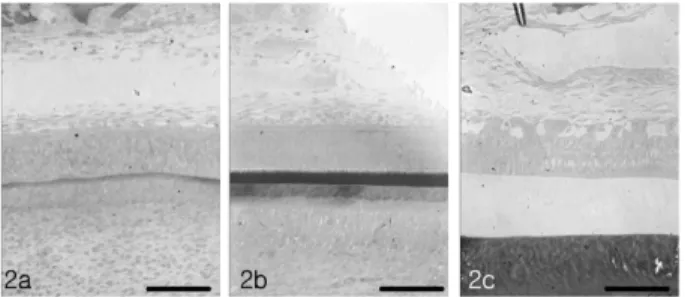
대조군의전분비대에서중간층은편평형의세포로구성 되어 있었으며
,
법랑모세포층에 위치한 세포들은 입방형 또는 키가 낮은 원주세포의 형태를 취하고 있었다.
세포질의대부분은핵으로가득차있었고
,
세포의핵은mod-
ified multiple stain
에 진하게 염색되는 인이 관찰되었다.
이 세포들은여러 층으로 구성된 중층을 형성하였고
,
입방형의 세포들은주로 기저부위에 그리고원주형의 세포 들은 세포층의 기저 위쪽에서 관찰되었다
(Fig. 2a).
분비대를 구성하는 법랑모세포층의 세포들은 전반적으로 길 게 신장된 원주형의세포들로 이루어져 있었다
.
이 세포들의핵은 난형으로기저부위쪽에분포하고 있었으며
,
핵 주변의 근위부에는 세포질과 비교하여 이질염색성을 띄 는 과립들이규칙적인양상으로 분포하였다.
법랑질과경 계를 이루는 법랑모세포들의 세포질은 톱니바퀴의 형태 를 이루고 있었다.
법랑모세포들의 원위부의 세포질에는불규칙한 형태의 공포들이다수 법랑질 방향으로 집적되
Fig. 1. Diagram of the sagittal sectioned mandibular incisor.
RT: Root tip, B: Band, D: Distance, IT: Incisal tip, RT-1st B: Distance from root tip to 1st band, Final B-IT: Distance from
final band to incisal tip
어 있었다
(Fig. 2b).
성숙대에서는 법랑질성숙에따른 평 탄끝 법랑모세포와 주름끝 법랑모세포가 관찰되었다.
성 숙대를구성하고 있는세포들은 분비대를구성하고있는 법랑모세포들과 비교하여 단위면적당 세포의 수가 감소 되어 관찰되었다.
또한 법랑모세포들은modified multi-
ple stain
염색에서 대부분은 약한염색성을 보였으나,
일부세포들의세포질은다른세포질과비교하여높은염색 성을보였다
.
분비대의 세포들과동일하게 신장된원주형의세포로관찰되었으나세포의길이는현저하게짧게관 찰되었다
.
또한부분적으로기저막과근접한세포질에커 다란공포를지니고있는세포들이관찰되었다(Fig. 2c).
불소농도
100 ppm
투여군의 경우,
전분비대의 법랑모세포층을 구성하고 있는세포들은 대조군과 비교하여 세 포의수적인감소가관찰되었다
.
또한법랑모세포층을구성하는 세포들은
modified multiple stain
에 약하게 염색된 세포들과 강하게 염색된 세포들로 이루어져 있었다
.
주로약하게염색된세포들은기저막을따라배열되어있 었고
,
이들 세포들 사이에 강하게 염색된 세포들이 위치하고 있었다
(Fig. 3a).
분비대에서는 법랑모세포는 대조군과 비교하여세포의수적인감소가관찰되었다
.
법랑모세포들은세포질전반에공포를다수지니고있었으며 세포 의 핵들은 정상조직과 비교하여 좀 더 신장된 형태로 확인되었다
.
정상조직에서 핵의 근위부에서 관찰되었던 과립들의 염색성은 다소 저하되어 관찰되었다.
또한 법랑질과 접하는 법랑모세포의 세포질은 뚜렷한 톱니바퀴 모양으로 관찰되었다
(Fig. 3b).
성숙대에서는 법랑모세포들의 세포간격이 떨어진상태로 관찰되었다
.
법랑모세포들의 세포질에는 대부분 공포가 산재되어 있었으며 특 히
,
기저막 근위부위에 커다란 공포들이 확인되는 등,
대조군에 비하여 전반적으로 세포 변형이 관찰되었다
(Fig. 3c).
한편
,
불소농도200 ppm
투여군의 경우는 전분비대의법랑모세포층은전반적으로
modified multiple stain
에고른 염색양상을나타내었다
.
이층을 구성하고있는 세포들은
100 ppm
과 비교하여수적으로 다소 증가되어 관찰되었다
(Fig. 4a).
또한 분비대의 법랑모세포들은100 ppm
과 비교하여 더욱 신장된 형태를 하고 있었고
modified
multiple stain
에 염색성 저하를 나타내었다.
세포질 내에서 관찰되는공포들은주로법랑질과 경계하고있는세포 질쪽에분포되어관찰되었으나
, 100 ppm
실험군에비해수적으로감소되었다
(Fig. 4b).
그리고성숙대의법랑모세포들 은대조군과비교하여세포의조직학적구성은동일한것으 로 확인되었고, 100 ppm
투여군에서 전반적으로산재되어 있던공포들은수적으로감소되어관찰되었다(Fig. 4c).
2. 전자현미경 소견
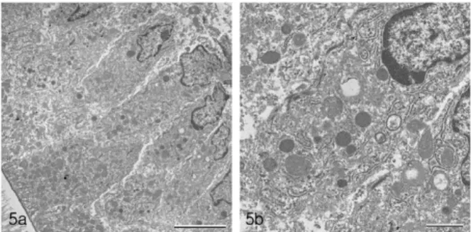
대조군의 경우
,
성숙대의법랑모세포들은 기저부위에서원위부위로갈수록세포의형태를구분할수없을정도로 혼재되어 있었다
.
세포의 핵은 기저부위 쪽으로 치우쳐있었으며
,
핵의주변을따라다수의간상형미토콘드리아 가 위치하고있었다.
세포의원위부에는간상형의미토콘드리아가 집적되어있었다
.
법랑질과인접한부위의세포 질은 세포막이 함입되어 불규칙하게 주름잡힌 미로구조Fig. 2. a: Photomicrograph of control presecretory zone stained by modified multiple stain. This layer consists of cuboidal type cells and shows the beginning of ameloblast differentiation. b:
Photomicrograph of control secretory zone. The ameloblasts are more differentiated than presecreting cells. The interdigitating portions of the process have not yet developed. c: Photomicrograph of control maturation zone. The ameloblasts are reduced in height, but two staggered levels of nuclei are persisting. The apical surface of ameloblasts is relatively smooth.
Fig. 3. a: After injection of 100ppm fluoride, region of presecretory zone. Longitudinal section in the region of ameloblasts facing pulp posterior portion. There are at least three or four levels of nuclei. b:
After injection of 100ppm fluoride, region of secretory zone. A thin layer of lightly stained initial enamel defines the dentin enamel junction. c: After injection of 100 ppm fluoride, region of maturation zone. Large intracellular space separates adjacent ameloblasts.
Fig. 4. a: After injection of 200ppm fluoride, region of presecretory
zone. The ameloblasts remain poorly differentiated. b: After injection
of 200ppm fluoride, region of secretory zone. Ameloblasts are long
columnar cell. The cells are taller, the nuclei are still in two staggered
levels and Tome’s processes are characteristically arranged in picket
fence configuration. c: After injection of 200 ppm fluoride, region of
maturation zone.
가관찰되었으며
,
법랑질경계부위는전자밀도가법랑질 과비교하여높게관찰되었다.
또한 이부위의세포질내에는다수의분비과립들이분포되어있었다
(Figs. 5a, b).
실험군의 경우
,
불소농도100 ppm
투여군 법랑모세포의 세포질은 대조군에 비해 전자밀도가 높게 나타났고
,
세포핵의형태는원주형에가깝게관찰되었다
.
또한조면 소포체의경우는잘정돈된형태로핵의위쪽에서관찰되었으나 대조군에 비해 팽창되어 관찰되었다
.
이 시기에미토콘드리아는 핵 위로부터 원위부위까지 전반적으로 분포되어 있었으나
,
이들은 대부분 막성구조가 유실되어붕괴되었거나 세포질에 미토콘드리아 기질 내에서 관찰 된 라멜라형태의동심원상 구조가팽창됨을 동반하였다
.
또한 이 시기의 법랑모세포내에는 전반적으로 전자밀도 가 다소 낮은 형태와거의전자밀도가 나타나지 않은커 다란공포가형성되었다
.
전자의경우,
붕괴된미토콘드리아들이 융합된 형태로서 관찰되었고
,
후자는 조면소포체 의 팽창에의한 공포임이 확인되었다(Fig. 6a, b).
그리고불소농도
200 ppm
투여군의 법랑모세포질의 전자밀도는대조군에 비해 높게 관찰되었으나
, 100 ppm
투여군과는거의 동일한 양상으로 확인되었다
.
특히 조면소포체는100 ppm
투여군과 비교하였을 때,
극도로팽창되어 세포내에불규칙한형태의공포를형성하였다
.
원위부의 세포질은 대조군과
100 ppm
불소투여군에 비해 전자밀도가낮게관찰되었으며
,
또한이 부위에서미토콘드리아의변형양상은
100 ppm
불소투여군과유사하였으나조면세포체는 팽창되거나 붕괴되어심한 막성구조의 변화가 관찰 되었다
(Fig. 7a, b).
3. 성숙기 법랑모세포의 전환주기 변화
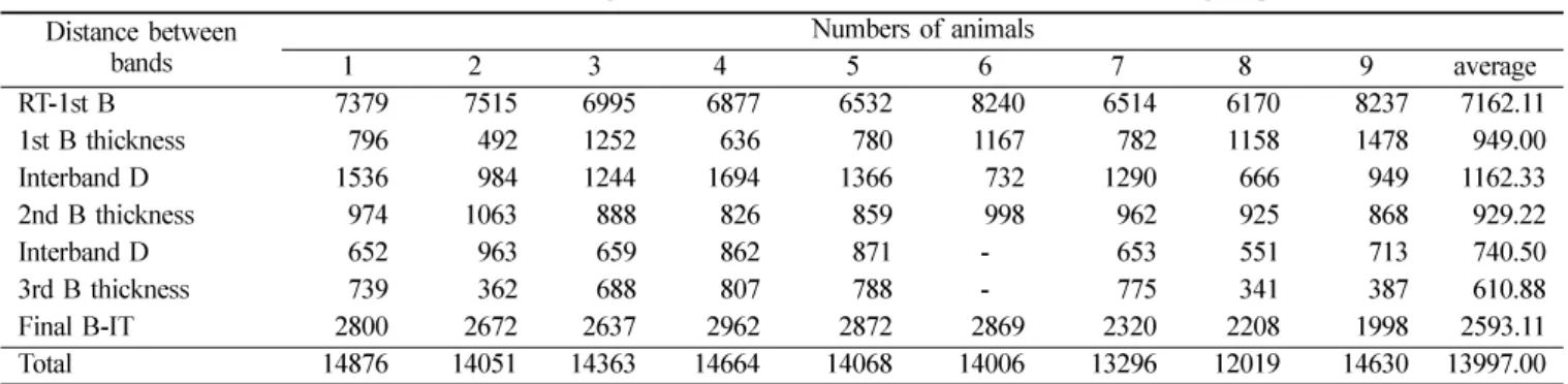
대조군에서나타나는첫번째평탄끝법랑모세포의평균
두께는
949.00
µm,
두 번째는929.22
µm,
세 번째는610.88
µm
였다(Table 1). 100 ppm
불소투여군에서는 각각890.77
µm, 594.66
µm, 392.50
µm
이었고(Table 2), 200 ppm
불소투여군은
770.56
µm, 475.00
µm, 345.00
µm
로 측정되었다
(Table 3).
또한대조군과실험군에서평탄끝법랑모세포의평균두께는절단연으로갈수록좁아지는경향을나타 내었고특히
,
불소를 투여한경우고농도일수록 평탄끝법 랑모세포의두께가좁아지는양상을보였다(Table 4).
주름끝 법랑모세포의 평균두께는 대조군에서는 각각
1162.33
µm, 740.50
µm(Table 1)
이었고, 100 ppm
불소투여군은
1454.44
µm, 799.00
µm(Table 2)
로나타났고, 200 ppm
불소투여군에서는
1299.89
µm, 756.00
µm
이었다(Table 3).
대조군과 실험군에서주름끝 법랑모세포의 길이도 절단연 으로갈수록좁아지는경향을나타내었으나
,
불소투여군에서농도가높아짐에따른유의한차이는없었다
(Table 4).
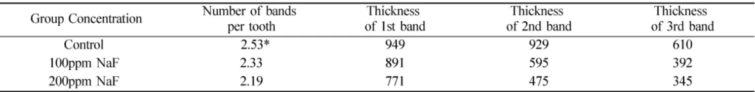
한편
,
평탄끝 법랑모세포의 전환주기의 수는 대조군에 서는2.53
회, 100 ppm
불소투여군은2.33
회, 200 ppm
불 소투여군은2.19
회로 나타났다.
또한 대조군에서는 평균3
회의전환주기를 보이는반면,
실험군에서는평균2
회의법랑모세포의 전환주기를 보였다
.
그러므로 불소의 영향 으로대조군에비해실험군에서평균1
회의법랑모세포의전환주기가감소되었다
(Table 5, Fig. 1).
4. 치아의 성장 변화
치아의 평균 총길이는 대조군에서는
13997.00
µm(Table
Fig. 5. Transmission electron micrographs of ameloblasts in control maturation zone. a: Low magnification of ameloblasts.
Mitochondria concentrated in distal cytoplasm of ameloblast. b:
High magnification of distal region of ameloblast. Cell surface was ruffled and contains numerous mitochondria.
Fig. 6. Cytoplasm of ameloblast in maturation zone after injection of 100 ppm fluoride. a: Nucleus of ameloblasts are elongated shape and ameloblast contain large vacuoles. b: Distal region of ameloblast. Electron dense materials are scattered in the cytoplasm.
Fig. 7. Transmission electron micrographs of ameloblast
cytoplasm after injection of 200 ppm fluoride in maturation
zone. a: Most ameloblasts show severe changes in their distal
cytoplasm. b: Distal region of ameloblast cytoplasm. The
arrowheads indicate distension of the rough surfaced endoplasmic
reticulum.
1), 100ppm
불소투여군에서13665.33
µm(Table 2), 200ppm
불소투여군은
11670.88
µm(Table 3)
이였다.
따라서 대조군 에서치아의평균총길이가가장길었고,
불소투여농도가높아질수록작아지는경향을나타내었다
(Table 4).
성숙기 법랑질 영역이 치아 총길이에서 차지하는비율 은 대조군에서
48.8%, 100 ppm
불소투여군은41.8%, 200 ppm
불소투여군은39.4%
이었다(Table 4).
이결과는 치아의총길이에서 성숙기 법랑질영역이 차지하는비율 은대조군에서가장많이차지하고불소투여농도가높아 질수록작아지는경향을나타내었다.
고 찰
흰쥐 절치의 법랑모세포는 크게 전분비대
,
분비대 및성숙대의
3
단계를 거쳐 성장,
소멸한다.
전분비대에서는 아직 분비활동에 관여하지않는 미분화세포들이 주를이 루며,
다시이 세포들은 치수에접한 부위와상아질에접 하는 부위로 나뉜다18).
본 실험에서도 정상 치아기의 조 직학적 구성은 크게 나누어 전분비대,
분비대 및 성숙대로 관찰되었다
.
전분비대에서 중간층은 편평형의 세포로 구성되어 있었으며,
법랑모세포층에 위치한 세포들은 입Table 1. Measurement of the rat incisor which was given calcein 1 hour before sacrifice in the control group ( µ m) Distance between
bands Numbers of animals
1 2 3 4 5 6 7 8 9 average
RT-1st B 7379 7515 6995 6877 6532 8240 6514 6170 8237 7162.11
1st B thickness 796 492 1252 636 780 1167 782 1158 1478 949.00
Interband D 1536 984 1244 1694 1366 732 1290 666 949 1162.33
2nd B thickness 974 1063 888 826 859 998 962 925 868 929.22
Interband D 652 963 659 862 871 - 653 551 713 740.50
3rd B thickness 739 362 688 807 788 - 775 341 387 610.88
Final B-IT 2800 2672 2637 2962 2872 2869 2320 2208 1998 2593.11
Total 14876 14051 14363 14664 14068 14006 13296 12019 14630 13997.00
RT: Root tip, B: Band, D: Distance, IT: Incisal tip, RT-1st B: Distance from root tip to 1st band Final B-IT: Distance from final band to incisal tip, 1 - 9: Different animals
Table 2. Measurement of the rat incisor which was given calcein 1 hour before sacrifice in the experimental group of 100ppm sodium fluoride injection
( µ m) Distance between
bands Numbers of animals
1 2 3 4 5 6 7 8 9 average
RT-1st B 9573 8120 9005 7970 6509 6530 9351 7527 7034 7957.66
1st B thickness 959 652 627 1424 861 677 793 1096 928 890.77
Interband D 1146 1121 1221 1641 1086 2384 899 1938 1654 1454.44
2nd B thickness 537 487 604 582 565 692 539 526 820 594.66
Interband D - - - - - - - 774 824 799.00
3rd B thickness - - - - - - - 387 398 392.50
Final B-IT 2378 2497 2616 2739 2479 2763 2841 1887 2327 2503.00
Total 14593 12877 14073 14356 11500 13046 14423 14135 13985 13665.33
RT: Root tip, B: Band, D: Distance, IT: Incisal tip, RT-1st B: Distance from root tip to 1st band Final B-IT: Distance from final band to incisal tip, 1 - 9: Different animals
Table 3. Measurement of the rat incisor which was given calcein 1 hour before sacrifice in the experimental group of 200ppm sodium fluoride injection
( µ m) Distance between
bands Numbers of animals
1 2 3 4 5 6 7 8 9 average
RT-1st B 5737 7421 6641 7556 6169 6430 8136 8815 6743 7072.00
1st B thickness 397 798 660 740 701 1110 883 657 989 770.56
Interband D 1859 1124 1234 1135 1283 1529 1179 710 1646 1299.89
2nd B thickness 636 681 443 461 443 518 282 336 475 475.00
Interband D - - - - - - - - 756 756.00
3rd B thickness - - - - - - - - 345 345.00
Final B-IT 2694 1468 1759 1811 1788 2199 2381 1839 1441 1931.11
Total 11323 11492 10737 11703 10384 11786 12861 12357 12395 11670.88
RT: Root tip, B: Band, D: Distance, IT: Incisal tip, RT-1st B: Distance from root tip to 1st band
Final B-IT: Distance from final band to incisal tip, 1 - 9: Different animals
방형또는키가낮은원주세포의형태를취하고있었다
.
분비대에서는 분화된 법랑모세포들이 미성숙된 법랑기질을 분비하며법랑모세포에서
Tome ’ s
돌기가관찰된다.
분비대는 다시 초기 법랑질 분비부위
(region of initial enamel
secretion),
내법랑질분비부위(region of inner enamel secre- tion),
외법랑질분비부위(region of outer enamel secretion),
말기 법랑질 분비부위
(region of final enamel secretion)
로나뉜다고하였다19,20)
.
본연구결과에서도분비대를구성하는 법랑모세포층의세포들은 전반적으로 길게신장된 원 주형의세포들로이루어져있었다
.
법랑질과인접한법랑모세포들은 부착반에 의해 결합되어 있었고 불규칙한
Tome ’ s
돌기가 확인되었다. Tome ’ s
돌기 내에는 세포질막을 따라 작은 분비과립들이 다수 관찰되었다
.
또한Tome ’ s
돌기와인접한부위의법랑질내에는무정형의섬유성 물질들과 규칙적으로 배열되어있는 광화되는법랑 기질이관찰되었다
.
Leblond
와Warshawsky
19)의연구결과,
성숙대에서는초기에 법랑모세포들의 높이가 줄어 들고
1/4
에서1/2
정도 의법랑모세포들이소실되며,
그이후법랑모세포들은평활한주름진표면이
2-3
회반복되는 전환양상을보이다가 착색된다.
성숙대의 법랑모세포는 법랑모세포 전환부위(region of ameloblastic modulation),
법랑모세포 착색부위
(region of ameloblastic pigmentation)
로나뉘거나19),
분 비 후 이행부위(region of postsecretory transition),
고유성숙부위
(region of maturation proper),
착색부위(region of pigmentation),
퇴축법랑모세포부위(region of reduced
ameloblast)
로 구분된다고하였다18).
본 연구에서도 성숙대에서는 법랑질 성숙에 따른 평탄끝 법랑모세포와 주름끝 법랑모세포가관찰되었다
.
성숙대를구성하고있는세포들은분비대를구성하고있는법랑모세포들과비교하여단위 면적당세포의수가감소되어관찰되었다
.
한편
,
치아의법랑질형성기간동안섭취한 불소는우식에 높은저항성을갖게한다
.
그러나불소를장기간에걸 쳐과다섭취를 할경우법랑질에구조및성분변화를초 래한다21).
즉 동물의 경우 절치 맹출부위 법랑질 색소의 부분적또는 전체적소실22),
성숙기법랑질에서 단백질제거의감소23,24)
,
법랑질형성기간중성숙기에나타나는평탄끝 법랑모세포 전환주기 감소25,26)
,
성숙기법랑질의 광화27,28) 등이 초래된다
.
하지만 현재까지 이러한 변화율의원인과결과에관한상호관계는확실히알려져있지않다
.
법랑모세포의 전환주기는 법랑질 발생에서 성숙기의 두
드러진특징이다29-32)
.
이러한세포성주기는 45Ca
과형광물질 등의 주입에의하여 관찰할수 있다33,34)
.
또한 고농도의 불소를 투여할경우 법랑질 성숙기에서 법랑모세포의
주기수가감소함을관찰할수있다
. Fejerskov
등25)은고농도의 불소가함유된 음용수를흰쥐에 투여하여 법랑모 세포의 전환주기가 감소하였다고 보고하였다
.
이러한 세 포성주기의 변화는 불소의 농도에 따라 증가한다. Smith
등16)은
100 ppm
농도의 불소가 함유된 음용수를 흰쥐에게
6
주 동안 투여한 결과 법랑질형성에 있어 전분비대,
분비대 및 성숙대에서 각각의 평균시상길이는 대조군과 유사하였다
.
하지만 평탄끝 법랑모세포의 전환주기가 대Table 4. Comparison of average measurement of the rat incisor which was given calcein 1 hour before sacrifice among control and experimental group
( µ m)
Distance between bands Group
control 100 ppm NaF 200 ppm NaF
RT-1st B 7162 7958 7072
1st B thickness 949 891 771
Interband D 1162 1454 1300
2nd B thickness 929 595 475
Interband D 741 799 756
3rd B thickness 611 392 345
Final B-IT 2593 2503 1931
Total 13997 ± 877 13665 ± 1005 11670 ± 799
RT: Root tip, B: Band, D: Distance, IT: Incisal tip
RT-1st B: Distance from root tip to 1st band, Final B-IT: Distance from final band to incisal tip
Table 5. Comparison of the number and the thickness of calcein bands among control and experimental groups in the case of calcein injection 1 hour before sacrifice
*( µ m) Group Concentration Number of bands
per tooth Thickness
of 1st band Thickness
of 2nd band Thickness
of 3rd band
Control 2.53* 949 929 610
100ppm NaF 2.33 891 595 392
200ppm NaF 2.19 771 475 345
조군보다 약
2
회가 더감소되었다고 하였다.
즉,
시상길이에서다소 넓어진 평탄끝법랑모세포의 전환주기로 인 하여 나타나는 효과로 실험군은 대조군에 비해 약
25%
적은전환주기를보인다고하였다
.
이러한결과는법랑질 형성을마치고성숙기법랑질표면을덮고있는법랑모세 포와법랑질형성과정중분비대에서성숙대로이행된법 랑모세포의 전환주기중 주름끝 법랑모세포 시기보다 평 탄끝법랑모세포의시기에서더많은시간을소모하기때 문이라고하였다.
또한치아의시상총길이는불소음용수 를 먹인실험군에서 다소줄어드는 경향이었으나유의한 차이는없었다고하였다.
그리고평탄끝법랑모세포의두 께와 주름끝법랑모세포의 두께가절단연으로 갈수록 좁 아지는경향을나타내며,
성숙대가 절단연으로갈수록짧 아진다고보고하였다16).
본실험은전환주기가대조군에비하여불소투여군에서 평균
1
회가 감소되었는데불소농도가 증가할수록 평탄끝 법랑모세포의 두께는 감소하는 양상을 나타내었다.
또한대조군에 비해 불소투여군에서 시상총길이에 대한 주름 끝 법랑모세포의 두께비율보다 평탄끝법랑모세포의 두
께 비율이 증가함을 볼 수 있었다
.
이러한 결과는Smith
등16)의결과와일치하였다
.
또한치아의총길이에 있어서100 ppm
불소투여군은 대조군과 유사하였으나200 ppm
불소투여군에서는 다소 짧아지는 경향을 나타내었다
.
그 리고 불소투여군의 평탄끝 법랑모세포의 두께와 주름끝 법랑모세포의 두께가 절단연으로 갈수록 좁아지는 경향 을 띄었고성숙대의 길이도절단연으로 갈수록 짧아지는 양상을나타내었다.
따라서 본 실험결과 흰쥐태아치아기의 조직학적구성 은 전분비대
,
분비대 및 성숙대로관찰되었으며,
특히 성 숙대에서는 법랑질에서 물과 유기물질을 선택적으로 제 거하는 평탄끝 법랑모세포(smooth-ended ameloblast)
와무기이온을 추가로 공급하는 주름끝 법랑모세포
(ruffle-
ended ameloblast)
가관찰되었다.
그리고형광물질검사결과 대조군에비해 실험군에서 불소투여 농도가 높아짐에 따라 전환주기가 감소되고이것은치아의 총길이도감소 하게되어
,
결국치아성장을저해함을확인하였다.
감사의 글
이 논문은
2008
년도 조선대학교 학술연구비의 지원을받아연구되었음
.
요 약
태아형성 시기에 투여된 불소가 법랑모세포의 법랑질 형성과정에미치는효과를알아보고자생후
11
일된흰쥐의하악절치를대상으로대조군과두그룹의실험군으로 나누어 실험하였다
.
전자현미경을 이용한 형태학적 분석결과
,
흰쥐태아치아기의조직학적 구성은전분비대,
분비대 및 성숙대로 관찰되었으며특히 성숙대에서는 법랑질 에서 물과 유기물질을 선택적으로 제거하는 평탄끝 법랑
모세포
(smooth-ended ameloblast)
와 무기이온을 추가로공급하는 주름끝 법랑모세포
(ruffle-ended ameloblast)
가 관찰되었다.
이러한 조직학적 구성은 흰쥐태아에서도 성체에서관찰되는 구조들과동일한것으로확인되었다
.
한 편,
법랑모세포의 전환주기를 알아보기 위한 형광물질(calcein)
을이용한검사결과,
전환주기가대조군에비하여실험군에서 평균
1
회가 감소되었는데 불소농도가 증가할수록 평탄끝법랑모세포의 두께는 감소하는 양상을 나타 내었다
.
또한대조군에 비해실험군에서시상총길이에대한 주름끝 법랑모세포의두께 비율보다 평탄끝법랑모세 포의두께 비율이증가함을볼 수있었다
.
그리고치아의총길이에 있어서
100 ppm
불소투여군은대조군과유사하였으나
200 ppm
불소투여군에서는다소짧아지는경향을나타내었다
.
그리고 실험군의 평탄끝 법랑모세포의 두께와 주름끝 법랑모세포의두께가 절단연으로 갈수록 좁아 지는 경향을띄었고 성숙대의 길이도 절단연으로 갈수록 짧아지는 양상을나타내었다
.
따라서대조군에비해 실험 군에서 불소투여농도가높아짐에 따라 전환주기가 감소 되고 이것은 치아의 총길이도 감소하게 되어,
결국 치아성장을저해함을확인하였다