• 검색 결과가 없습니다.

All-trans Retinoic Acid Induces Expression and Secretion of Carboxypeptidase D in THP-1 Cells

N/A
N/A
Protected

Academic year: 2021

Share "All-trans Retinoic Acid Induces Expression and Secretion of Carboxypeptidase D in THP-1 Cells"

Copied!
11
0
0

로드 중.... (전체 텍스트 보기)

전체 글

(1)

Biomedical Science Letters 2020, 26(4): 256~266 https://doi.org/10.15616/BSL.2020.26.4.256 eISSN : 2288-7415

All-trans Retinoic Acid Induces Expression and Secretion of Carboxypeptidase D in THP-1 Cells

Hang Thi Thu Nguyen

*

and Jae Young Kim

,**

Department of Life Science, Gachon University, Seongnam, Kyeonggi-Do 13120, Korea

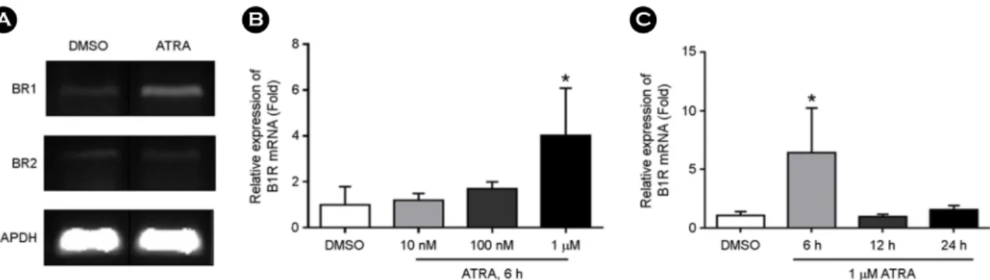
Carboxypeptidase D (CPD) is a zinc-dependent protease, which is highly expressed in macrophages, and is thought to participate in inflammatory processes. In the present study, we investigated the possible regulatory effect of all-trans retinoic acid (ATRA), which is an active form of vitamin A and plays a critical regulatory role in both the innate and adaptive immunity, on CPD expression and secretion in human monocytic THP-1 cells. CPD mRNA expression first increased, from a concentration as low as 10 nM ATRA to a maximum level of expression, at 1 μM. ATRA enhanced intracellular CPD expression in a time- and concentration-dependent manner but did not affect cell surface CPD expression.

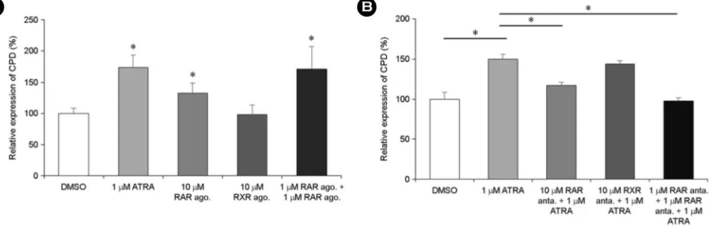
Interestingly, 9-cis-RA did not affect CPD expression. Additionally, an experiment with RAR/RXR selective agonist or antagonists demonstrated that ATRA-induced enhancement of CPD expression was RAR/RXR dependent. ATRA also enhanced CPD secretion from THP-1 cells; however, this enhancement was RAR/RXR-independent. The anti-inflammatory agent dexamethasone reversed ATRA-induced enhancement of CPD expression and secretion. Our results suggest ATRA exerts regulatory effects on expression and secretion of CPD in human monocytes, and ATRA-induced CPD secretion may be associated with inflammatory response.

Key Words: All-trans retinoic acid, Carboxypeptidase D, THP-1, Monocytes, Vitamin A, Secretion, Inflammation

INTRODUCTION

All-trans retinoic acid (ATRA), a physiologically active form of vitamin A, plays a crucial role in embryonic devel- opment and cell fate determination (Niederreither and Dolle, 2008). In addition, ATRA plays an important regulatory role in both the innate and adaptive immunity (Kim, 2018).

ATRA shapes early intestinal immune responses by pro- moting interleukin (IL)-22 synthesis by γδ T cells and innate lymphoid cells (Mielke et al., 2013) and regulates dendritic cell differentiation (Klebanoff et al., 2013). ATRA controls gut homing of effector T cells, survival and effector func-

tions of CD8

+

T cells, and immune responses of Th17 cells (Iwata et al., 2004; Hall et al., 2011; DePaolo et al., 2011).

Moreover, ATRA suppresses IgE synthesis and secretion (Scheffel et al., 2005), and induces the differentiation of regulatory B cells (Eriksen et al., 2015). ATRA is able to induce and promote the development and function of in- ducible regulatory T cells (Elias et al., 2008). ATRA mediates conversion of monocyte-derived macrophages into tissue- resident macrophages (Gundra et al., 2017). ATRA inhibits the production of pro-inflammatory mediators, such as tumor necrosis factor (TNF)-α, nitric oxide, and IL-12 in macro- phages (Mehta et al., 1994; Kang et al., 2000; Wang et al., 2007). In contrast, ATRA enhances production of IL-10 in

Original Article

Received: October 21, 2020 / Revised: December 12, 2020 / Accepted: December 14, 2020

*Graduate student, **Professor.

Corresponding author: Jae Young Kim. Department of Life Science, Gachon University, Seongnam, Kyeonggi-Do 13120, Korea.

Tel: +82-31-750-4762, Fax: +82-31-750-5389, e-mail: jykim85@gachon.ac.kr

CThe Korean Society for Biomedical Laboratory Sciences. All rights reserved.

CCThis is an Open Access article distributed under the terms of the Creative Commons Attribution Non-Commercial License (http://creativecommons.org/licenses/by-nc/3.0/) which permits unrestricted non-commercial use, distribution, and reproduction in any medium, provided the original work is properly cited.

(2)

THP-1 (Wang et al., 2007) and contributes to murine M2 macrophage polarization synergistically with transforming growth factor (TGF)-β, prostaglandin E2, and IL-4 (Ho et al., 2016), suggesting an immune modulatory effect of ATRA on macrophages.

Proteases functions in various biological processes, such as cell growth, death, migration, and morphogenesis and protein catabolism and activation of coagulation. Proteases also play a critical regulatory role in immune system; they are involved in complement activation (Sim and Laich, 2000), antigenic peptide processing (Manoury, 2013), the activation or inactivation of immune mediators (Marshall et al., 2017). For instance, innate immune cells, such as monocytes/macrophages and neutrophils, contribute to in- flammation during the initial phase of infection by secreting metalloproteinases, which cleave cytokines or chemokines, thereby activating or inactivating them (Parks et al., 2004).

Carboxypeptidase D (CPD) is a zinc-dependent protease, which specifically cleaves C-terminal arginine or lysine. CPD is broadly expressed in mammalian tissues and especially high in macrophages (Skidgel and Erdos, 1998). CPD is pri- marily found in the trans-Golgi network, where it participates in protein processing in the constitutive secretory pathway (Varlamov and Fricker, 1998). However, CPD is also found to some extent on the plasma membrane, where it can cleave extracellular substrates (Varlamov et al., 1999; Hadkar and Skidgel, 2001; Kalinina et al., 2002). By re- moving the C- terminal arginine or lysine residues of proteins and poly- peptide, CPD is suggested to participate in inflammatory processes; CPD mRNA and protein expression was markedly increased in murine macrophages following treatment with LPS and interferon (IFN)-γ (Hadkar and Skidgel, 2001).

Despite its high expression in macrophages, no studies have investigated the possible regulatory effects of ATRA on CPD expression of human macrophages. The present study investigated the effect of ATRA on CPD expression and secretion in human monocyte THP-1 cells.

MATERIALS AND METHODS

Reagents and chemicals

ATRA, RARα agonist BMS753, RXRα agonist LG-

100268, RARα antagonist BMS195614, 9-cis-retinoic acid (9cis-RA), and dexamethasone were obtained from Sigma- Aldrich (St. Louis, MO, USA). RXRα antagonist UVI3003, anti-β-actin antibody, and secondary antibodies for western blot were purchased from Santa Cruz Biotechnology (Dallas, TX, USA). RPMI-1640 medium for cell culture was obtained from Welgene Inc. (Gyongsan, Korea). Fetal bovine serum (FBS) was purchased from Invitrogen (Gibco BRL, MD, USA).

ATRA was prepared as a 20 mM stock in dimethyl sul- foxide (DMSO) and stored at -20℃ before being diluted to the desired concentration in medium in each experiment.

Purified anti-CPD antibody (ab184960) used in flow cyto- metric analysis was purchased from Abcam (Cambridge, MA, USA).

Cell culture and treatment

The human monocytic cell line THP-1 was obtained from Korean cell line bank. THP-1 cells were cultured in RPMI-1640 supplemented with 10% heat-inactivated FBS, 1% antibiotic-antimycotic, 10 mM HEPES, and 50 μM β- mercaptoethanol (Invitrogen). Cells were maintained at 37℃

in a 5% CO

2

humidified incubator. Around 6 to 12 h before treatment, cells were seeded into T25 flasks and cultured in RPMI media without FBS.

To determine agonist effects, cells were treated with agonist for 6 h. Additionally, to assess the effects of ATRA antagonists, cells were pre-incubated with 1 μM RARα or RXRα antagonist for 2 h and then with 1 μM ATRA for 6 h.

Quantitative real-time PCR (qRT-PCR)

To analyze the expression of CPD in mRNA level, 10

6

cells were treated with various concentration of ATRA or with 1 μM ATRA and incubate at different times. Then, total mRNA was isolated and analyzed by qRT-PCR. The extrac- tion of total RNA was performed using Qiagen RNeasy mini kit (Qiagen, Seoul, Korea). RNA concentrations were deter- mined with a SD2000 micro spectrophotometer (Bioprince, Atlanta, GA, USA). cDNA was synthesized from 2 μg of total RNA using MMLV reverse transcriptase (Bioprince) and an oligo dT primer (Bioprince) at 65℃ for 1 h.

qRT-PCR was performed on a iQ5 Multicolor Real-Time

(3)

PCR Detection System (Bio-Rad, Hercules, CA, USA) using the iQ SYBR Green Supermix (Bio-Rad). The sequences of the specific primers were as follows: CPD 5'-ccacca- ccatttccctgata-3', 5'-ccaaggaataaagccgggta-3'; and β-actin 5'-caccattggcaatgagcggttc-3', 5'-aggtctttgcggatgtccacgt-3'.

Normalization was performed using the human β-actin gene as an endogenous control. For each sample, the relative abundance of target mRNA was calculated from the cycle threshold (Ct) values for the target and endogenous reference gene β-actin.

Flow cytometric analysis

Flow cytometric analysis was performed to check the expression of intracellular and cell surface CPD expression in different treatment conditions. For determining surface expression, 10

6

cells were incubated with 1 μM ATRA for 48 h. Then, cells were collected and incubated with purified anti-CPD antibody (ab184960) for 30 min at 4℃. After washing twice with phosphate buffered saline (PBS), the cells were incubated with the phycoerythrin-conjugated secondary antibody for 30 min at 4℃. After washing, cells were re- suspended in PBS, and analyzed on a Cytomics FC500 MLP (Beckman Coulter Inc., Fullerton, CA, USA).

To determine intracellular CPD expression, cells were incubated with 4% formaldehyde fixation solution for 30 min at room temperature and then permeabilized with 0.1%

Triton X-100 in PBS for 30 min. Following procedures are the same as those for detection of surface CPD expression by flow cytometry.

Western blot analysis

Cells were lysed with Triton X-100 lysis buffer (Santa Cruz Biotechnology). The supernatants from whole cell lysates were collected by centrifugation at 14,000 × g for 15 min. Cell lysate protein concentrations were measured using a Qubit Fluorometer (Invitrogen). Ten microgram of total protein was subjected to 10% sodium dodecyl sulfate- polyacrylamide gel electrophoresis and blotted onto a poly- vinylidene difluoride membrane (Millipore Corp., Billerica, MA, USA). Membranes were then blocked with 5% bovine serum albumin in 1 × tris-buffered saline (2.68 mM KCl, 137 mM NaCl, 25 mM Tris-HCl, and 0.05% Tween 20).

Subsequently, the membrane was probed with an anti-CPD (ab153874; Abcam) or anti-β-actin antibody (sc-1616). Pro- teins of interest were detected with HRP-conjugated goat anti-rabbit IgG antibody (sc-2055) or donkey anti-goat IgG antibody (sc-2020) and visualized with ECL Solution from GenDEPOT (Barker, TX, USA) using the ChemiDoc MP system (Bio-Rad) according to the provided protocol. β- actin was used as internal control.

To measure the secreted CPD proteins, the culture su- pernatants were collected by centrifugation at 180 × g for 5 min and then concentrated by using Amicon Ultra-15 centrifugal filter (Millipore Corp.) at 4,000 × g for 30 min at 4℃. Thus, concentrated protein samples (32 times) were recovered and were subjected to western blot analysis.

Statistical analysis

Data were analyzed by one-way analysis of variance (ANOVA) followed by post hoc comparison with either the Tukey HSD (honestly significant difference test) for groups of data with equal variances or the Games-Howell test in one-way ANOVA with unequal variances using SPSS 12.0 for Windows. Values are expressed as mean ± standard deviation (SD). Statistical significance was defined as P <

0.05.

RESULTS

ATRA, but not 9-cis-RA, enhances CPD expression in THP-1 cells

We examined effect of ATRA on CPD expression in THP-1 cells by qRT-PCR analysis. As shown in Fig. 1A, CPD mRNA expression first increased, from a concentration as low as 10 nM ATRA to a maximum level of expression (approximately seven folds higher than that in the DMSO- treated control), at 1 μM ATRA, and decreased thereafter.

CPD mRNA expression sharply increased 6 h after 1 μM

ATRA treatment and gradually decreased thereafter; however,

the expression of CPD mRNA at 48 h was still statistically

higher than that in the DMSO-treated control (Fig. 1B). CPD

protein expression levels, as determined by western blotting,

were also enhanced (by approximately two folds) upon treat-

ment with 1 μM ATRA (Fig. 1C). Because CPD is known

(4)

to be expressed on both the intracellular compartments and cell surfaces (Garcia-Pardo et al., 2017), CPD expression was also analyzed by flow cytometry. Intracellular CPD expression gradually increased over time (Fig. 1E) and was enhanced in an ATRA dose-dependent manner (Fig. 1F);

however, cell surface CPD expression was not significantly affected by ATRA treatment (Fig. 1D).

9-cis-RA is the second most abundant retinoid in the

human body and can bind and activate both RAR and RXR, while ATRA selectively binds RAR only (Bastien and Rochette-Egly, 2004). To determine whether ATRA and 9- cis-RA exert a similar regulatory effect on CPD expression, we examined the effect of 9-cis-RA on CPD expression in THP-1 cells. As shown in Fig. 2A, 9-cis-RA did not affect CPD mRNA expression in THP-1 cells. Protein expression, as determined by western blotting, was also not affected by Fig. 1. ATRA upregulates CPD mRNA and intracellular protein expression but does not affect cell surface CPD protein expression in THP-1 cells. (A) THP-1 cells were treated with various concentrations of ATRA for 6 h. (B) THP-1 cells were treated with 1 µM ATRA for various time periods, as indicated above. (C) CPD mRNA expression was examined by qRT-PCR. Cells were treated with 1 µM ATRA for 48 h. CPD protein levels were analyzed by western blotting, and densitometric analysis of CPD bands was performed using a ChemiDoc MP System.

(D) Cells were treated with 1 µM ATRA for 48 h. Cell surface CPD expression was analyzed by flow cytometry. (E) THP-1 cells were treated

with 1 µM ATRA for various time periods or (F) treated with various concentrations of ATRA, as indicated above, for 48 h. (E and F)

Intracellular CPD expression was measured by flow cytometry. Bar graphs present data as mean of relative expression ± SD. *P < 0.05

vs. DMSO.

(5)

9-cis-RA treatment (Fig. 2B).

ATRA-induced CPD expression depends on RAR/RXR pathway

Since multiple biological functions of ATRA are mediated by the RAR/RXR pathway (Tsai and O'Malley, 1994), we wanted to confirm whether ATRA exerts its effect on CPD expression via this classical pathway. To this end, we used selective RAR-α/RXR-α agonists and antagonists and found that 10 μM of the RAR agonist alone increases CPD protein expression. Moreover, combined treatment with 1 μM RAR agonist and 1 μM RXR agonist induced CPD expression

comparable to that induced by 1 μM ATRA (Fig. 3A).

Additionally, treatment with 10 μM RAR antagonist alone suppressed ATRA-induced CPD expression, but treatment with 10 μM RXR antagonist alone did not (Fig. 3B). Com- bined treatment with 1 μM RAR antagonist and 1 μM RXR antagonist completely reversed the induction of CPD ex- pression by ATRA (Fig. 3B)

ATRA-induced CPD secretion depends on incubation time and ATRA concentration, but does not depend on RAR/RXR

Since CPD is located in the secretory pathway (Varlamov

Fig. 2. 9-cis-RA does not affect CPD expression in THP-1 cells. (A) THP-1 cells were treated with 1 µM 9-cis-RA for various time periods.

Expression of CPD mRNA was examined by qRT-PCR and was normalized to that of β-actin mRNA. Data are presented as mean of relative mRNA expression ± SD. (B) Cells were treated with 1 µM 9-cis-RA for 48 h, and CPD protein levels were examined by western blotting.

Fig. 3. ATRA enhances CPD expression through the RAR/RXR pathway. (A) THP-1 cells were incubated with 10 µM RAR agonist or

RXR agonist alone or a combination of 1 µM RAR agonist and 1 µM RXR agonist. (B) Cells were pretreated with 10 µM RAR or RXR

antagonist alone, or a combination of 1 µM RAR/RXR antagonists for 2 h, followed by 1 µM ATRA treatment. To measure intracellular

CPD protein expression, after 48 h of incubation, cells were fixed, permeabilized, and analyzed by flow cytometry. Data are presented as

mean of relative expression ± SD. *P < 0.05 vs. DMSO.

(6)

and Fricker, 1998), we wanted to investigate the possibility of CPD secretion by THP-1 cells. To this end, THP-1 cells were treated with 1 μM ATRA for 48 h and the culture supernatants were collected, concentrated by filtration, and subjected to western blotting (Fig. 4A). Secreted CPD levels were markedly increased by more than six folds upon ATRA treatment (Fig. 4B). After treatment with 1 μM ATRA,

secreted CPD levels sharply increased at 24 h, reaching approximately 800 % of the control level at 48 h and slightly decreasing thereafter (Fig. 4C and D). Secreted CPD levels were markedly increased by 100 nM ATRA treatment, with a maximum increase of 2200 % at 1 μM ATRA (Fig. 4E and F).

To determine whether ATRA-induced CPD secretion is

Fig. 4. ATRA induces the secretion of CPD in a concentration- and time-dependent manner. (A and B) THP-1 cells were treated with 1 μM ATRA for 48 h. (C and D) Cells were treated with 1 µM ATRA for various time periods, as indicated above. (E and F) Cells were treated with various concentrations of ATRA, as indicated above, for 48 h. (A, C, and E) The culture supernatants were collected, concentrated by filtration, and subjected to western blotting. (B, D, and F) Densitometry analysis of CPD bands shown in (A, C, and E) was performed using a ChemiDoc MP System. Data are presented as mean of relative secretion ± SD. *P < 0.05 vs. DMSO.

Fig. 5. ATRA induces the secretion of CPD through an RAR/RXR-independent pathway. THP-1 cells were treated with a combinations of

1 µM RAR/RXR agonists or with a combination of 1 µM RAR/RXR antagonists in the presence of 1 µM ATRA for 48 h. (A) The culture

supernatants were collected, concentrated by filtration, and subjected to western blotting. (B) Densitometry analysis of CPD bands shown

in (A) was performed using a ChemiDoc MP System. Bar graphs present data as mean of relative secretion ± SD. *P < 0.05 vs. DMSO.

(7)

mediated by the action of the RAR/RXR heterodimer, we used selective RARα/RXRα agonists and RARα/RXRα antagonists. As shown in Fig. 5, ATRA-induced CPD secre- tion was not inhibited by pretreatment of THP-1 cells with both the 1 μM RARα antagonist as well as the RXRα antagonist. Similarly, combined treatment with 1 μM RARα agonist and 1 μM RXRα agonist did not induce the secretion

of CPD, suggesting that ATRA-induced CPD secretion does not depend on RAR/RXR.

Dexamethasone inhibits ATRA-induced CPD expression and secretion

Since it has been suggested that CPD is upregulated in macrophages under inflammatory conditions (Hadkar and

Fig. 6. Dexamethasone inhibits ATRA-induced CPD expression and secretion. (A) THP-1 cells were pretreated with 1 µM dexamethasone for 2 h, followed by treatment with 1 µM ATRA for different time periods, as indicated above. CPD mRNA expression was measured by qRT- PCR. (B) THP-1 cells were pretreated with 1 µM dexamethasone for 2 h and then treated with 1 µM ATRA for 48 h. CPD protein expression was analyzed by western blotting. (C) Cells were subjected to the treatment described in (B) and, the culture supernatants were then collected, concentrated by filtration, and subjected to western blotting. Bar graphs present data as mean of relative expression ± SD. *P < 0.05.

Fig. 7. ATRA induces bradykinin B1 receptor mRNA expression but does not affect bradykinin B2 receptor mRNA expression in THP-1

cells. (A) THP-1 cells were treated with 1 µM ATRA for 6 h. The mRNA expression of bradykinin B1 and B2 receptors was examined by

semi-quantitative PCR. (B) Cells were treated with different concentrations of ATRA, as indicated, for 6 h. (C) Cells were treated with 1 µM

ATRA for different time periods, as indicated. Bradykinin B1 receptor mRNA expression was examined by qRT-PCR. Bar graphs present

data as mean ± SD. *P < 0.05 vs. DMSO.

(8)

Skidgel, 2001) and our previous study demonstrated that dexamethasone, an anti-inflammatory drug, inhibits ATRA- induced MMP-2 secretion in THP-1 cells (Vu et al., 2018), we tested whether this drug can also suppress ATRA-induced CPD expression and secretion in THP-1 cells. ATRA- induced enhancement of CPD mRNA expression (Fig. 6A), protein expression (Fig. 6B), and secretion (Fig. 6C) were markedly inhibited by a 2-h pretreatment of THP-1 cells with 1 μM dexamethasone.

ATRA induces bradykinin B1 receptor mRNA expres- sion but not B2 receptor

Next, we examined effect of ATRA on bradykinin receptor expression in THP-1 cells by qRT-PCR analysis. As shown in Fig. 7A, bradykinin B1 receptor mRNA expression was enhanced by ATRA treatment but B2 receptor expression was not affected. More specifically, 1 μM ATRA induced B1 receptor expression, but 10 and 100 nM ATRA did not (Fig. 7B). Moreover, B1 receptor expression was induced at 6 h and decreased to a basal level by 12 h after 1 μM ATRA treatment (Fig. 7C).

DISCUSSION

The present study demonstrated that ATRA, but not 9- cis-RA, enhanced CPD expression in THP-1 cells. Several previous studies have reported the enhancement of cellular CPD expression by exogenous stimuli. Lipopolysaccharide and IFN-γ upregulated CPD expression in murine macro- phages and vascular endothelial cells (Hadkar and Skidgel, 2001; Hadkar et al., 2004). Moreover, prolactin, 17β-estradiol, and the synthetic androgen R1881 increased CPD expression in breast cancer cells (Abdelmagid and Too, 2008; Koirala et al., 2014), while prolactin and testosterone enhanced CPD expression in prostate cancer cells (Thomas et al., 2012). In- terestingly, CPD was downregulated in CD14

+

cells isolated from patients with lupus erythematosus, suggesting a pos- sible role for CPD in the pathogenesis of this autoimmune disease (Hoff et al., 2007). All these studies indicate a role of the proteolytic regulation of CPD in immune responses.

In our study, ATRA-induced CPD expression was depen- dent on the RAR/RXR pathway. Previous studies have shown

that the synergistic activation of both partners of the RAR- RXR complex is necessary to induce ATRA-responsible target gene expression (Brooks et al., 1996; Botling et al., 1997). Similarly, our results revealed that the use of an RARα agonist moderately induces CPD protein expression, even at higher concentrations, whereas combined treatment with an RARα agonist and an RXRα agonist significantly induced CPD expression, suggesting that the RAR-RXR complex is required to induce the transcriptional activation of CPD expression. Similar to current observations, our pre- vious study showed that ATRA enhances MMP-2 expression and secretion in THP-1 cells in an RAR/RXR-dependent manner (Vu et al., 2018).

Based on its cellular localization, CPD is thought to play a role in the processing of proteins and peptides within different cellular compartments, such as the secretory com- partments, endocytic pathway compartments, and the cell surface (Garcia-Pardo et al., 2017). However, the secretion of CPD from cells has not been reported. In the present study, our results revealed that CPD secretion does not depend on the RAR/RXR pathway, suggesting a distinct mechanism for CPD secretion. Ca

2+

is typically present at millimolar concentrations in the secretory pathway and is necessary to induce rapid membrane fusion to allow for secretion (Jeremic et al., 2004). In our previous study, ATRA increased intracel- lular calcium levels, and a calcium channel blocker inhibited ATRA-induced MMP-2 secretion in THP-1 cells, suggesting that calcium influx is necessary for ATRA-induced MMP-2 secretion. Another study also showed that ATRA increased intracellular calcium levels (Gao et al., 1998). Based on these reports, we suggest that ATRA-induced calcium in- flux may be involved in the induction of CPD secretion in THP-1 cells. However, it is necessary to uncover the precise mechanisms of ATRA-induced CPD secretion in THP-1 cells in future studies.

In the present study, pretreatment of THP-1 cells with

dexamethasone significantly inhibited the ATRA-induced

expression and secretion of CPD. Since it is known that

ATRA can induce NF-κB activation in THP-1 cells (Hoang

et al., 2019), resulting in CPD expression (Koirala et al.,

2014), and that dexamethasone suppresses NF-κB activity in

THP-1 cells (Steer et al., 2000), we suggest that the inhibitory

(9)

action of dexamethasone on ATRA-induced CPD expression and secretion may be due to a suppressive effect on NF-κB activation.

Due to its localization and substrate specificity, it has been proposed that intracellular CPD primarily functions in the maturation of receptors and growth factors (Reznik and Fricker, 2001). However, the function of secreted CPD has not been studied. Several studies have suggested potential substrates of CPD as being soluble immune mediators, such as bradykinin (Timblin et al., 2002) and complement frag- ments C3a, C4a, and C5a (Skidgel, 1988; Skidgel, 1996).

With respect to the former, CPD has been suggested to either inactivate or alter the specificity of the bradikynin by cleaving its C-terminal arginine (Skidgel, 1988). There are two types of receptors for bradykinin on the cell surface:

bradykinin B1 and B2 receptors. While the bradykinin B2 receptor is constitutively expressed, the bradykinin B1 re- ceptor is only expressed when cells are stimulated by inflam- matory mediators (deBlois et al., 1991; Bhoola et al., 1992).

Under normal conditions, bradykinin binds to the bradykinin B2 receptor, whereas cleaved des-arg9-bradykinin, which can be produced from bradykinin by proteolytic removal of the C-terminal arginine, binds to the bradykinin B1 receptor, and acts as an inflammatory mediator, inducing inflammatory responses, such as the enhanced production and release of NO or cytokines (Naffah-Mazzacoratti et al., 2014). Our study revealed that ATRA induces the mRNA expression of the bradykinin B1 receptor but does not affect B2 receptor expression in THP-1 cells. Based on our results and those of a previous study (Naffah-Mazzacoratti et al., 2014), we cautiously speculate that CPD activity may play a role in determining the fate of bradykinin-bradykinin receptor- mediated signaling.

With respect to the complement system, C3a, C4a, and C5a, all have C-terminal arginine residues (Pasupuleti et al., 2007). Monocytes/macrophages express both the C3a re- ceptor (C3aR) and C5aR to bind their respective ligands C3a and C5a (Stephan et al., 2012). Previous structural studies have proposed that the C-termini of C3a (Lu et al., 1984) and C5a (Huber-Lang et al., 2003) contribute to their binding to their corresponding receptors. Thus, proteolytic cleavage of the C-terminal arginines of both C3a and C5a

by cell surface or secreted CPD may play a critical role in the regulation of C3a(5a)-C3a(5a) receptor interactions.

Based on the essential role of ATRA in immune regulation as well as previous studies showing inflammatory stimuli- induced CPD expression (Hadkar and Skidgel, 2001; Hadkar et al., 2004), we suggest that ATRA-induced CPD secretion may be involved in the regulation of inflammatory responses by cleavage of inflammatory mediators, such as C3a, C5a, and/or bradykinin. To test this hypothesis, it is necessary to examine whether the ATRA-induced secretion of CPD cleaves the substrates mentioned above in a future study.

In summary, this study revealed, for the first time, that ATRA enhances CPD expression in THP-1 cells in an RAR /RXR-dependent manner and CPD secretion in a RAR/

RXR-independent manner. This enhanced expression and secretion of CPD were inhibited by dexamethasone treat- ment. We suggest that increased expression and secretion of CPD induced by ATRA treatment may be involved in regulating inflammatory responses.

ACKNOWLEDGEMENT

This research was supported by Basic Science Research Program through the National Research Foundation of Korea (NRF) funded by the Ministry of Education (NRF- 2018R1D1A1A09083908).

CONFLICT OF INTEREST

The authors declare that they have no competing interests.

REFERENCES

Abdelmagid SA, Too CK. Prolactin and estrogen up-regulate carboxypeptidase-d to promote nitric oxide production and survival of mcf-7 breast cancer cells. Endocrinology. 2008.

149: 4821-4828.

Bastien J, Rochette-Egly C. Nuclear retinoid receptors and the transcription of retinoid-target genes. Gene. 2004. 328: 1-16.

Bhoola KD, Figueroa CD, Worthy K. Bioregulation of kinins:

kallikreins, kininogens, and kininases. Pharmacol Rev. 1992.

44: 1-80.

Botling J, Castro DS, Oberg F, et al. Retinoic acid receptor/

retinoid X receptor heterodimers can be activated through both

subunits providing a basis for synergistic transactivation and

(10)

cellular differentiation. J Biol Chem. 1997. 272: 9443-9449.

Brooks SC 3

rd

, Kazmer S, Levin AA, et al. Myeloid differentiation and retinoblastoma phosphorylation changes in HL-60 cells induced by retinoic acid receptor- and retinoid X receptor- selective retinoic acid analogs. Blood. 1996. 87: 227-237.

deBlois D, Bouthillier J, Marceau F. Pulse exposure to protein synthesis inhibitors enhances vascular responses to des-Arg9- bradykinin: possible role of interleukin-1. Br J Pharmacol.

1991. 103: 1057-1066.

DePaolo RW, Abadie V, Tang F, et al. Co-adjuvant effects of retinoic acid and IL-15 induce inflammatory immunity to dietary antigens. Nature. 2011. 471: 220-224.

Elias KM, Laurence A, Davidson TS. Retinoic acid inhibits Th17 polarization and enhances FoxP3 expression through a Stat-3/

Stat-5 independent signaling pathway. Blood. 2008. 111: 1013 -1020.

Eriksen AB, Berge T, Gustavsen MW, et al. Retinoic acid enhances the levels of IL-10 in TLR-stimulated B cells from patients with relapsing-remitting multiple sclerosis. J Neuroimmunol.

2015. 278: 11-18.

Gao ZY, Xu G, Stwora-Wojczyk MM, et al. Retinoic acid induction of calcium channel expression in human NT2N neurons.

Biochem Biophys Res Commun. 1998. 247: 407-413.

Garcia-Pardo J, Tanco S, Diaz L, et al. Substrate specificity of human metallocarboxypeptidase D: Comparison of the two active carboxypeptidase domains. PLoS One. 2017. 12:

e0187778.

Gundra UM, Girgis NM, Gonzalez MA, et al. Vitamin A mediates conversion of monocyte-derived macrophages into tissue- resident macrophages during alternative activation. Nature Immunology. 2017. 18: 642-653.

Hadkar V, Sangsree S, Vogel SM, et al. Carboxypeptidase-mediated enhancement of nitric oxide production in rat lungs and microvascular endothelial cells. Am J Physiol Lung Cell Mol Physiol. 2004. 287: L35-L45.

Hadkar V, Skidgel RA. Carboxypeptidase D is up-regulated in raw 264.7 macrophages and stimulates nitric oxide synthesis by cells in arginine-free medium. Mol Pharmacol. 2001. 59: 1324 -1332.

Hall, JA, Cannons JL, Grainger JR, et al. Essential role for retinoic acid in the promotion of CD4(+) T cell effector responses via retinoic acid receptor alpha. Immunity. 2011. 34: 435-447.

Ho VW, Hofs E, Elisia I, et al. All trans retinoic acid, transforming growth factor β and prostaglandin E2 in mouse plasma syn- ergize with basophil-secreted interleukin-4 to M2 polarize

murine macrophages. PLoS One. 2016. 11: article e0168072.

Hoang TX, Jung JH, Kim JY. All-trans retinoic acid enhances bacterial flagellin-stimulated proinflammatory responses in human monocyte THP-1 cells by upregulating CD14. BioMed Res Int. 2019. 2019: article ID 8059312.

Hoff NP, Degrandi D, Hengge U, et al. Carboxypeptidase D: a novel TGF-beta target gene dysregulated in patients with lupus erythematosus. J Clin Immunol. 2007. 27: 568-579.

Huber-Lang MS, Sarma JV, McGuire SR, et al. Structure-function relationships of human C5a and C5aR. J Immunol. 2003. 170:

6115-6124.

Iwata M, Hirakiyama A, Eshima Y, et al. Retinoic acid imprints gut- homing specificity on T cells. Immunity. 2004. 21: 527-538.

Jeremic A, Kelly M, Cho JA, et al. Calcium drives fusion of SNARE-apposed bilayers. Cell Biol Int. 2004. 28: 19-31.

Kalinina E, Varlamov O, Fricker LD. Analysis of the carboxy- peptidase D cytoplasmic domain: Implications in intracellular trafficking. J Cell Biochem. 2002. 85: 101-111.

Kang BY, Chung SW, Kim SH, et al. Retinoid-mediated in- hibition of interleukin-12 production in mouse macrophages suppresses Th1 cytokine profile in CD4(+) T cells. Brit J Pharmacol. 2000. 130: 581-586.

Kim CH. Control of innate and adaptive lymphocytes by the RAR-retinoic acid axis. Immune Netw. 2018. 18: e1.

Klebanoff CA, Spencer SP, Torabi-Parizi P, et al. Retinoic acid controls the homeostasis of pre-cDC-derived splenic and intestinal dendritic cells. J Exp Med. 2013. 210: 1961-1976.

Koirala S, Thomas LN, Too CK. Prolactin/Stat5 and androgen R1881 coactivate carboxypeptidase-D gene in breast cancer cells. Mol Endocrinol. 2014. 28: 331-343.

Lu ZX, Fok KF, Erickson BW, et al. Conformational analysis of COOH-terminal segments of human C3a. Evidence of ordered conformation in an active 21-residue peptide. J Biol Chem.

1984. 259: 7367-7370.

Manoury B. Proteases: essential actors in processing antigens and intracellular toll-like receptors. Front Immunol. 2013. 4: 299.

Marshall NC, Finlay BB, Overall CM. Sharpening host defenses during infection: proteases cut to the chase. Mol Cell Pro- teomics. 2017. 16: S161-S171.

Mehta K, McQueen T, Tucker S, et al. Inhibition by all-trans- retinoic acid of tumor necrosis factor and nitric oxide pro- duction by peritoneal macrophages. J Leuk Biol. 1994. 55:

336-342.

Mielke LA, Jones SA, Raverdeau M, et al. Retinoic acid expression

associates with enhanced IL-22 production by gammadelta T

(11)

cells and innate lymphoid cells and attenuation of intestinal inflammation. J Exp Med. 2013. 210: 1117-1124.

Naffah-Mazzacoratti Mda G, Gouveia TL, Simoes PS, et al.

What have we learned about the kallikrein-kinin and renin- angiotensin systems in neurological disorders? World J Biol Chem. 2014. 5: 130-140.

Niederreither K, Dolle P. Retinoic acid in development: towards an integrated view. Nat Rev Genet. 2008. 9: 541-553.

Parks WC, Wilson CL, López-Boado YS. Matrix metallopro- teinases as modulators of inflammation and innate immunity.

Nat Rev Immunol. 2004. 4: 617-629.

Pasupuleti M, Walse B, Nordahl EA, et al. Preservation of anti- microbial properties of complement peptide C3a, from in- vertebrates to humans. J Biol Chem. 2007. 282: 2520-2528.

Reznik SE, Fricker LD. Carboxypeptidases from A to Z: impli- cations in embryonic development and Wnt binding. Cell Mol Life Sci. 2001. 58: 1790-1804.

Scheffel F, Heine G, Henz BM, et al. Retinoic acid inhibits CD40 plus IL-4 mediated IgE production through alterations of sCD23, sCD54 and IL-6 production. Inflamm Res. 2005. 54:

113-118.

Sim RB, Laich A. Serine proteases of the complement system.

Biochem Soc Trans. 2000. 28: 545-550.

Skidgel RA, Erdos EG. Cellular carboxypeptidases. Immunol Rev.

1998. 161: 129-141.

Skidgel RA. Basic carboxypeptidases: regulators of peptide hor- mone activity. Trends Pharmacol Sci. 1988. 9: 299-304.

Skidgel RA. Structure and function of mammalian zinc carboxy- peptidases. In zinc metalloproteases in health and disease.

CRC Press. 1996. 261-304.

Steer JH, Kroeger KM, Abraham LJ, et al. Glucocorticoids suppress tumor necrosis factor-alpha expression by human monocytic THP-1 cells by suppressing transactivation through adjacent NF-kappa B and c-Jun-activating transcription factor-2 binding sites in the promoter. J Biol Chem. 2000. 275: 18432-18440.

Stephan AH, Barres BA, Stevens B. The complement system: an unexpected role in synaptic pruning during development and disease. Annu Rev Neurosci. 2012. 35: 369-389.

Thomas LN, Morehouse TJ, Too CK. Testosterone and prolactin increase carboxypeptidase-D and nitric oxide levels to promote survival of prostate cancer cells. Prostate. 2012. 72: 450-460.

Timblin B, Rehli M, Skidgel RA. Structural characterization of the human carboxypeptidase D gene and its promoter. Int Immunopharmacol. 2002. 2: 1907-1917.

Tsai MJ, O'Malley BW. Molecular mechanisms of action of steroid /thyroid receptor superfamily members. Annu Rev Biochem.

1994. 63: 451-486.

Varlamov O, Eng FJ, Novikova EG, et al. Localization of metal- locarboxypeptidase D in AtT-20 cells. Potential role in pro- hormone processing. J Biol Chem. 1999. 274: 14759-14767.

Varlamov O, Fricker LD. Intracellular trafficking of metallocar- boxypeptidase D in AtT-20 cells: localization to the trans- Golgi network and recycling from the cell surface. J Cell Sci.

1998. 111: 877-885.

Vu HT, Hoang TX, Kim JY. All-trans retinoic acid enhances matrix metalloproteinase 2 expression and secretion in human mye- loid leukemia THP-1 cells. Biomed Res Int. 2018. 2018: article ID 5971080.

Wang X, Allen C, Ballow M. Retinoic acid enhances the pro- duction of IL-10 while reducing the synthesis of IL-12 and TNF-α from LPS-stimulated monocytes/macrophages. J Clin Immunol. 2007. 27: 193-200.

https://doi.org/10.15616/BSL.2020.26.4.256

Cite this article as: Nguyen HTT, Kim JY. All-trans

Retinoic Acid Induces Expression and Secretion of

Carboxypeptidase D in THP-1 Cells. Biomedical Science

Letters. 2020. 26: 256-266.

수치

Fig. 3. ATRA enhances CPD expression through the RAR/RXR pathway. (A) THP-1 cells were incubated with 10 µM RAR agonist or  RXR agonist alone or a combination of  1 µM RAR agonist and 1 µM RXR agonist
Fig. 5. ATRA induces the secretion of  CPD through an RAR/RXR-independent pathway. THP-1 cells were treated with a combinations of  1 µM RAR/RXR agonists or with a combination of  1 µM RAR/RXR antagonists in the presence of  1 µM ATRA for 48 h
Fig. 7. ATRA induces bradykinin B1 receptor mRNA expression but does not affect bradykinin B2 receptor mRNA expression in THP-1  cells

참조

관련 문서

포에서 We s t e r nbl ot 을 통해 HJ ME가 i NOS의 단백질 발현을 농도의존적으로 저해 하는 것을 확인하였다( Fi g.6) .그러므로 환삼덩굴은 i NOS의 발현을

qRT-PCR for VSMC synthetic phenotype protein, cellular retinol binding protein 1(CRBP-1) and connexin 43, from thoratic aorta showed that CRBP-1 expression was increased by 70%

As for the research method, RBL-2H3 cells, a mast cell line, were treated with supercritical carbon dioxide extracted Chamaecyparis obtusa oil (C oil), German chamomile oil (G

Quantitation of procollagen synthesis by Hs68 cells treated with fish collagen hydrolysates (FCHs), their &lt;1 kDa fraction, and their &gt;1 kDa fraction for 24 hrs.

Control- and ASTRIN- depleted HeLa cells were untreated or treated with 5 Gy. IR and were then fixed at

Dombret H, Sutton L, Duarte M, Daniel MT, Leblond V, Castaigne S, Degos L: Combined therapy with all-trans retinoic acid and high-dose chemotherapy in

Effect of caloric restriction on the expression of PGC-1 and PPARs mRNA in liver of Otsuka Long-Evans Tokushima Fatty Rats.. Long-lived growth hormone

4 &gt; Effects of Taro on COX-2 expression and iNOS expression(hot water) in human thyroid cancer cells. The cells were pretreated for 48hours with either