INTRODUCTION
Hypertensive encephalopathy is an acute disorder character- ized by severe hypertension, headache, vomiting, altered mental status, visual changes, and seizures. Syndromes of hypertensive encephalopathy are usually reversible if quickly diagnosed and treated. The most common finding of hypertensive encephalop- athy is bilateral edema in the supratentorial white matter, espe- cially in the parieto-occipital area. However, atypical feature of hypertensive encephalopathy involving the frontal lobes, brain- stem, cerebellum, cortical watershed zones, and basal ganglia has been sporadically reported. Brainstem and/or cerebellar le- sions may be presented as the only abnormalities in unusual cases (1). Here we report three cases of hypertensive encepha- lopathy involving brainstem showing characteristic alternating linear bright and low signals in the pons on T2-weighted mag- netic resonance imaging (MRI), although many brainstem le-
sions showed no peripheral sparing compared to those of os- motic demyelination (2). This retrospective study has been approved by our Institutional Review Board. The requirement for obtaining informed consent from patients was waived.
CASE REPORT
Case 1
A 36-year-old woman suffered from headache and dizziness one week before her admission. She had a previous history of poorly controlled hypertension without antihypertensive medi- cation. On admission, she showed alert mental status. Blood pressure was of 270/170 mm Hg with body temperature of 36°C.
Neurological examination revealed no abnormal finding. He- matologic examination showed increased blood levels of urea nitrogen (29.3 mg/dL) and creatinine (2.53 mg/dL). Urine ex- amination revealed proteinuria. Her blood electrolyte level and
J Korean Soc Radiol 2014;71(6):314-319 http://dx.doi.org/10.3348/jksr.2014.71.6.314
Received June 21, 2014; Accepted September 11, 2014 Corresponding author: Sung-Tae Park, MD Department of Radiology, Soonchunhyang University Hospital, Soonchunhyang University School of Medicine, 59 Daesagwan-ro, Yongsan-gu, Seoul 140-743, Korea.
Tel. 82-2-709-9396 Fax. 82-2-709-9066 E-mail: [email protected]
This is an Open Access article distributed under the terms of the Creative Commons Attribution Non-Commercial License (http://creativecommons.org/licenses/by-nc/3.0) which permits unrestricted non-commercial use, distri- bution, and reproduction in any medium, provided the original work is properly cited.
Hypertensive encephalopathy typically presents with bilateral parietooccipital vaso- genic edema. Brainstem and cerebellar edema are uncommon in association with typ- ical supratentorial changes. We experienced three cases of atypical hypertensive en- cephalopathy involving brainstem and cerebellum as well as cerebral white matter, which showed characteristic alternating linear bright and low signals in the pons, the so-called “stripe sign”. We report these cases here with a brief literature review.
Index terms
Malignant Hypertension Hypertensive Encephalopathy
Posterior Leukoencephalopathy Syndrome Brainstem
Brain Edema
Predominant Hypertensive Brainstem Encephalopathy with Supratentorial Involvement: Case Report and Literature Review
1 비전형적 대뇌 침범을 동반한 고혈압성 뇌간뇌병증: 증례 보고 3예 및 문헌고찰1Ji Hee Kim, MD
1, Sung-Tae Park, MD
1, Hyun Kyung Lim, MD
1, Sung Tae Kim, MD
2, Jihoon Cha, MD
21Department of Radiology, Soonchunhyang University Hospital, Soonchunhyang University School of Medicine, Seoul, Korea
2Department of Radiology, Samsung Medical Center, Sungkyunkwan University School of Medicine, Seoul, Korea
Case 2
A 42-year-old man admitted to our hospital with a history of headache for three months. One week before the admission, he developed bilateral blurred vision and gait disturbance. He was diagnosed with hypertension about two years ago. However, he was not treated with any medication.
Neurologic examination revealed that he was normal with alert metal status. Physical examination revealed his blood pres- sure at 236/170 mm Hg with body temperature of 36.3°C. Fun- duscopic examination revealed suspicious retinal microhemor- rhage. His blood creatinine and electrolyte levels were normal.
Cerebrospinal fluid study showed normal finding except mildly elevated white blood cell count of 1/mm3 and protein concen- tration of 79.7 mg/dL (normal: 12–60 mg/dL).
MRI revealed extensive hyperintensity in the brainstem, thala- mus and cerebellum on T2-weighted and FLAIR images. We also observed the alternating bright and low signals in the pons as seen in case 1. The pons and cerebellum appeared swollen with com- pression of the fourth ventricle and mild obstructive hydrocepha- lus. These lesions showed increased ADC value with no enhance- ment. There was no peripheral sparing of the pons. After 6 months of antihypertensive treatment, his headache and blurred vision improved. There was no available follow-up image.
Case 3
A 55-year-old woman was presented to our hospital with head- cerebrospinal fluid study were within the normal limits.
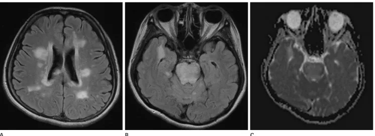
Brain computed tomography (CT) and MRI were performed immediately after her arrival. CT showed diffuse low density ar- eas in the midbrain, pons, cerebellum, and periventricular white matter. MRI after the CT on the same day revealed extensive hy- perintensity in the whole midbrain, pons and upper medulla on T2-weighted and fluid-attenuated inversion recovery (FLAIR) images. We also observed scattered and irregular shaped lesions in the cerebellum, bilateral thalami and bilateral periventricular cerebral white matter. Alternating linear bright and low signals were found on T2-weighted and FLAIR images without periph- eral sparing in the pons (Fig. 1A, B). There was no mass effect.
These lesions showed increased apparent diffusion coefficient (ADC) value (Fig. 1C) as slight hypointense without enhance- ment on T1-weighted images. Magnetic resonance (MR) angi- ography was normal. She immediately received intravenous in- fusion of a calcium channel blocker (nicardipine hydrochloride, 40 mg) which controlled her systemic pressure to 140–170/70–
90 mm Hg. Antihypertensive medications were followed by an oral calcium channel blocker (nifedipine, 66 mg) and an angio- tensin II receptor blocker (candesartan cilexetil, 16 mg). Two days after the admission, her elevated blood pressure was con- sistently reduced to the normal range with disappearance of headache and dizziness. She was discharged on the 11th day post-admission with prescribed antihypertensive medication.
Her lesions were completely resolved within 3 weeks.
Fig. 1. A 36-year-old female presented with headache. MR images showed hyperintensity involving the periventricular cerebral white matter (A), pons and cerebellum bilaterally, with the stripe sign in the pons on fluid-attenuated inversion recovery (FLAIR) images (B). Corresponding apparent diffusion coefficient map revealed increased diffusion in the brainstem (C). Follow-up MR images 3 weeks after initial MRI showed sig- nificant diminution of hyperintensity on FLAIR images.
B
A C
tle or considerable supratentorial deep white matter involve- ment. Patient 3 showed peripheral sparing of pontine lesion known as typical pattern in osmotic demyelination.
Characteristic MRI findings of pontine lesions revealed alter- nating linear bright and low signals in all our three cases, proba- bly signifying fluid between the transverse pontine bundles (3).
We defined this specific pattern of the pons as the “stripe sign”.
A similar finding was described in osmotic demyelination syn- drome that showed symmetric triangular-shaped T2-hyperin- tensity with stripes in the central pons (4). The transverse pon- tine fibers were seen as lines of preserved brain passing from one side to the other. In literatures, osmotic demyelination syn- dromes show the sparing of peripheral pons and pontine por- tion of corticospinal tracts. However, hypertensive encephalopa- thy does not. Acute lesions might reveal restricted diffusion in osmotic demyelination (4).
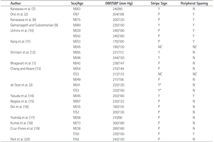
We reviewed 17 articles on hypertensive encephalopathy with brainstem involvement for the evaluation of either presence or absence of the stripe sign and other indicative MR findings. A to- tal of 26 patients were reviewed in 17 articles. Two patients were excluded due to no images given. Of the remaining 24 cases, the stripe sign was shown in 5 patients on MRI images and 2 cases with descriptions. However, the stripe sign was not shown or mentioned in other cases. It has been widely accepted that hyper- tensive brainstem encephalopathy does not spare the peripheral pons (4). However, the periphery of pons was spared in 8 of 24 cases. Only one case by Bhagavati et al. (1) showed mild obstruc- tive hydrocephalus by brainstem swelling. Diffusion weighted imaging (DWI) was evaluated in 15 cases by either imaging or literature. All 15 cases had normal DWI, suggesting vasogenic edema (Table 1). In our cases, case 3 revealed sparing of the pe- ripheral pons whereas case 2 showed mass effect by brainstem swelling.
Hypertensive encephalopathy involving brainstem should be differentiated from acute infarction, central pontine myelinoly- sis, brainstem glioma, acute disseminated encephalomyelopathy, and infectious encephalitis. Acute infarction could be ruled out by the absence of acute ischemic findings on initial DWI and resolution of MR finding with a recovery of clinical symptom.
Acute disseminated encephalomyelopathy, infectious encephali- tis, and central pontine myelinolysis could be differentiated read- ily based on laboratory examination and rapid recovery. Glioma ache and vomiting. She visited a local hospital previously with
high arterial blood pressure. Although she received oral medica- tion for arterial hypertension, she suffered progressive headache after that. Her metal state was alert. There was no abnormal find- ing on neurologic or funduscopic examinations. Her blood pres- sure was at 215/109 mm Hg with body temperature of 36.8°C on admission. Her blood creatinine and blood electrolyte levels were normal. Cerebrospinal fluid evaluation was not performed.
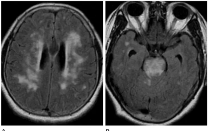
MRI showed extensive hyperintensity in the brainstem, cere- bellum and periventricular deep cerebral white matter on T2- weighted and FLAIR images (Fig. 2). We also observed the sym- metric T2-hyperintensity in the pons with stripes as seen in case 1 and 2. Tendency of the peripheral sparing was observed in the pons. ADC maps revealed increased diffusion in the brainstem le- sion. Multiple microbleedings were presented in the cerebellum, suggesting hypertensive microangiopathy. There was no evidence of abnormal enhancement in the brain parenchyma. MR angiog- raphy revealed no abnormality. After 2 weeks of antihypertensive treatment, her headache subsided. Follow-up MRI showed com- plete disappearance of abnormal hyperintense lesions.
DISCUSSION
T2-hyperintensity lesions are uncommonly seen in the brain- stem, cerebellum, or basal ganglia in hypertensive encephalopa- thy. In our three cases, the main MR finding was hyperintensity on T2-weighted image in the brainstem and cerebellum with lit-
Fig. 2. A 55-year-old female suffered from headache and vomiting.
MR images showed symmetric hyperintensity in the periventricular cerebral white matter (A), brainstem and cerebellum on fluid-attenu- ated inversion recovery images with the stripe sign in the pons. The periphery of the pons was relatively spared (B).
A B
showed particular MR pattern of the hypertensive encephalopa- thy, thus allowing a better understanding of the changes associ- ated with arterial hypertension.
We concluded that the presence of diffuse T2-hyperintensity with stripe sign in the pons in patients with severe paroxysmal hypertension could be helpful for the diagnosis of hypertensive encephalopathy. Tumor or inflammatory conditions could be easily excluded by their non-enhancing nature. However, the peripheral sparing of pontine lesions on T2-weighted image can occur in both osmotic demyelination syndrome and hyperten- sive encephalopathy.
REFERENCES
1. Bhagavati S, Chum F, Choi J. Hypertensive encephalopathy presenting with isolated brain stem and cerebellar edema.
J Neuroimaging 2008;18:454-456 involves expansion and mass effect. The presence of stripe sign
in the pons without obvious mass effect or contrast enhance- ment could be used to exclude the possibility of glioma, enceph- alitis, and demyelinating disease.
Pathophysiologic changes of hypertensive encephalopathy is associated with vasogenic edema without evidence of cytotoxic edema or infarction. Vessels in posterior circulation are sparsely innervated by sympathetic nerves, therefore poorly initiating vasoconstriction in response to suddenly increased arterial pres- sure (5). Deep regions such as thalamus, basal ganglia, and brainstem are supplied from the arterioles branches of the mid- dle cerebral artery or basilar artery. Cortex and subcortex are supplied from terminal branch arteries (6). Deep gray matter and brain stem are affected by more intense arterial blood pres- sure than the cerebral cortex and subcortex. Therefore, severe and lasting acceleration of elevated arterial pressure may con- tribute greatly to the involvement of deep structures. Our cases
Table 1. Clinical and MR Features of Patients with Hypertensive Encephalopathy
Author Sex/Age DBP/SBP (mm Hg) Stripe Sign Peripheral Sparing
Karasawa et al. (7) M/63 240/80 Y N
Ono et al. (2) F/67 204/106 P Y
Kanazawa et al. (8) M/75 200/120 P Y
Gamanagatti and Subramanian (9) M/60 220/150 Y Y
Uchino et al. (10) M/29 240/160 P Y
M/42 240/160 P Y
Kang et al. (11) M/53 170/100 P Y
M/45 190/130 NC NC
Shintani et al. (12) M/85 221/112 Y N
M/46 244/150 Y N
Bhagavati et al. (1) M/42 238/147 P N
Chang and Keane (13) M/54 210/144 P N
F/22 213/133 NC NC
M/49 211/156 P N
de Seze et al. (3) M/41 220/120 Y* N
F/52 220/150 Y* N
Yasuda et al. (14) M/45 250/160 Y Y
Nagata et al. (15) M/67 230/122 P N
Doi et al. (16) M/35 180/118 P N
F/52 200/130 P N
Yoshida et al. (17) M/58 210/90 P N
Kumai et al. (18) M/73 300/160 P N
Cruz-Flores et al. (19) M/38 280/160 P N
F/30 230/150 P Y
Park et al. (20) F/42 245/150 P N
Note.-DBP = diastolic blood pressure, N = the finding was negative, NC = non-contributory, P = poor image quality, SBP = systolic blood pressure, Y = the finding was positive, Y* = the finding was described as positive in the literature
cephalopathy involving the brainstem. J Clin Neurol 2007;
3:50-52
12. Shintani S, Hino T, Ishihara S, Mizutani S, Shiigai T. Revers- ible brainstem hypertensive encephalopathy (RBHE): clini- coradiologic dissociation. Clin Neurol Neurosurg 2008;110:
1047-1053
13. Chang GY, Keane JR. Hypertensive brainstem encephalop- athy: three cases presenting with severe brainstem edema.
Neurology 1999;53:652-654
14. Yasuda Y, Akiguchi I, Imai T, Sonobe M, Kage M. Hyperten- sive brainstem encephalopathy. Intern Med 2003;42:1131- 1134
15. Nagata M, Maeda M, Tsukahara H, Maier SE, Takeda K.
Brain stem hypertensive encephalopathy evaluated by line scan diffusion-weighted imaging. AJNR Am J Neuroradiol 2004;25:803-806
16. Doi Y, Kimura F, Fujiyama T, Fujimura C, Nishina T, Sato T, et al. Hypertensive brainstem encephalopathy without pa- rieto-occipital lesion--two case reports. Neurol Med Chir (Tokyo) 2006;46:75-79
17. Yoshida K, Yamamoto T, Mori K, Maeda M. Reversible pos- terior leukoencephalopathy syndrome in a patient with hypertensive encephalopathy--case report. Neurol Med Chir (Tokyo) 2001;41:364-369
18. Kumai Y, Toyoda K, Fujii K, Ibayashi S. Hypertensive en- cephalopathy extending into the whole brainstem and deep structures. Hypertens Res 2002;25:797-800
19. Cruz-Flores S, de Assis Aquino Gondim F, Leira EC. Brain- stem involvement in hypertensive encephalopathy: clinical and radiological findings. Neurology 2004;62:1417-1419 20. Park JH, Kim SM, Shin HW, An SJ. Hypertensive brainstem
encephalopathy involving deep supratentorial regions:
does only blood pressure matter? Neurol Int 2010;2:e9 2. Ono Y, Manabe Y, Hamakawa Y, Murakami T, Omori N,
Hayashi Y, et al. Localized lesions on MRI in a case of hy- pertensive brainstem encephalopathy. Intern Med 2005;
44:1002-1005
3. de Seze J, Mastain B, Stojkovic T, Ferriby D, Pruvo JP, Des- tée A, et al. Unusual MR findings of the brain stem in ar- terial hypertension. AJNR Am J Neuroradiol 2000;21:391- 394
4. Osborn AG. Acquired Metabolic and Systemic Disorders. In Osborn AG. Osborn’s Brain: Imaging, Pathology, and Anat- omy. Salt Lake City, UT: Amirsys, 2012:907-957
5. Beausang-Linder M, Bill A. Cerebral circulation in acute arterial hypertension--protective effects of sympathetic nervous activity. Acta Physiol Scand 1981;111:193-199 6. Sadoshima S, Fujii K, Yao H, Kusuda K, Ibayashi S, Fujishi-
ma M. Regional cerebral blood flow autoregulation in normotensive and spontaneously hypertensive rats--ef- fects of sympathetic denervation. Stroke 1986;17:981- 984
7. Karasawa S, Kawanami T, Kimura H, Kurita K, Kato T. An unusual case of hypertensive encephalopathy involving the brain stem. Intern Med 2004;43:448-449
8. Kanazawa M, Sanpei K, Kasuga K. Recurrent hypertensive brainstem encephalopathy. J Neurol Neurosurg Psychiatry 2005;76:888-890
9. Gamanagatti S, Subramanian S. Hypertensive encephalop- athy: isolated pons involvement mimicking central pon- tine myelinolysis. Korean J Radiol 2006;7:218-219 10. Uchino M, Haga D, Nomoto J, Mito T, Kuramitsu T. Brain-
stem involvement in hypertensive encephalopathy: a re- port of two cases and literature review. Eur Neurol 2007;
57:223-226
11. Kang SY, Choi JC, Kang JH. Two cases of hypertensive en-
비전형적 대뇌 침범을 동반한 고혈압성 뇌간뇌병증: 증례 보고 3예 및 문헌고찰1
김지희
1· 박성태
1· 임현경
1· 김성태
2· 차지훈
2고혈압성 뇌병증은 전형적으로 양측 두정엽 및 후두엽 부위의 부종으로 나타난다. 뇌간과 소뇌의 부종은 천막상부 병변과 연관되어 드물게 나타날 수 있다. 본 저자들은 대뇌 피질뿐만 아니라 뇌간과 소뇌를 침범하는 비전형적 고혈압성 뇌병증을 보여주는 3예를 경험하였으며, 특히 뇌간 병변은 고신호강도와 저신호강도의 띠가 교차로 나타나는 특징적인 “줄무늬 신 호(stripe sign)”를 보였다. 이에 저자들은 이러한 증례들을 문헌고찰과 함께 보고하는 바이다.
1순천향대학교 의과대학 서울병원 영상의학과, 2성균관대학교 의과대학 삼성의료원 영상의학과