Radiation Dose from Whole-Body F-18 Fluorodeoxyglucose Positron Emission Tomography/Computed Tomography:
Nationwide Survey in Korea
The purpose of this study was to estimate average radiation exposure from
18F-fluorodeoxyglucose (FDG) positron emission tomography/computed tomography (PET/
CT) examinations and to analyze possible factors affecting the radiation dose. A nation- wide questionnaire survey was conducted involving all institutions that operate PET/CT scanners in Korea. From the response, radiation doses from injected FDG and CT examination were calculated. A total of 105 PET/CT scanners in 73 institutions were included in the analysis (response rate of 62.4%). The average FDG injected activity was 310 ± 77 MBq and 5.11 ± 1.19 MBq/kg. The average effective dose from FDG was estimated to be 5.89 ± 1.46 mSv. The average CT dose index and dose-length product were 4.60 ± 2.47 mGy and 429.2 ± 227.6 mGy∙cm, which corresponded to 6.26 ± 3.06 mSv. The radiation doses from FDG and CT were significantly lower in case of newer scanners than older ones (P < 0.001). Advanced PET technologies such as time-of-flight acquisition and point-spread function recovery were also related to low radiation dose (P < 0.001). In conclusion, the average radiation dose from FDG PET/CT is estimated to be 12.2 mSv. The radiation dose from FDG PET/CT is reduced with more recent scanners equipped with image-enhancing algorithms.
Keywords: FDG PET/CT; Radiation Exposure; National Survey; Dose Reference Level Hyun Woo Kwon,1,2* Jong Phil Kim,1*
Hong Jae Lee,1 Jin Chul Paeng,1,3 Jae Sung Lee,1 Gi Jeong Cheon,1,3 Dong Soo Lee,1,2,3 June-Key Chung,1,3 and Keon Wook Kang1,3
1Department of Nuclear Medicine, Seoul National University Hospital, Seoul, Korea; 2Department of Molecular Medicine and Biopharmaceutical Science, Graduate school of Convergence Science and Technology, Seoul National University, Seoul, Korea;
3Institute of Radiation Medicine, Medical Research Center, Seoul National University College of Medicine, Seoul, Korea
* Hyun Woo Kwon and Jong Phil Kim equally contributed to this work.
Received: 3 November 2015 Accepted: 3 February 2016 Address for Correspondence:
Jin Chul Paeng, MD
Department of Nuclear Medicine, Seoul National University Hospital, 103 Daehak-ro, Jongno-gu, Seoul 03080, Korea E-mail: [email protected]
Funding: This research was partly supported by a grant of the Korea Health Technology R&D Project through the Korea Health Industry Development Institute (KHIDI), funded by the Ministry of Health & Welfare, Republic of Korea (grant number:
HI14C1072).
http://dx.doi.org/10.3346/jkms.2016.31.S1.S69 • J Korean Med Sci 2016; 31: S69-74
INTRODUCTION
In the current medical practice, radiological imaging studies are of critical importance in every aspect of patient management, and thus, they have been dramatically expanded in recent years.
Most commonly used radiological imaging methods are planar X-ray and computed tomography (CT), which cause radiation exposure of patients (1). Although the benefit from medical im- aging far surpasses the potential risk of radiation, medical doc- tors need to properly understand the risk and benefit of radia- tion exposure in decision making of imaging studies.
18F-fluorodeoxyglucose (FDG) positron emission tomogra- phy (PET) is a molecular imaging method that visualizes glu- cose metabolism in vivo. Currently, a hybrid imaging of FDG PET/CT is widely used in clinical practice for diverse diseases such as cancers, inflammatory diseases, neurological disorders, and myocardial metabolic disorders. Due to its usefulness, FDG
PET/CT has been rapidly expanded; in Korea, more than 500,000 examinations were performed in 2014 (2). With the increase of FDG PET/CT examinations, a concern has been raised with re- gard to the radiation exposure by PET/CT, because it causes both internal and external radiation from radiopharmaceutical administration and CT acquisition.
The radiation dose of FDG PET/CT depends on both injected activity of FDG and CT protocol. Notably, radiation dose may be reduced with recent PET/CT scanners, which enable sensi- tive gamma ray detection for PET and dose-reduction algorithms for CT. Thus, radiation dose from FDG PET/CT should be esti- mated separately in each society that has different conditions regarding scanner equipment and cultural background of med- ical imaging. However, there have been scarce data on radiation dose of FDG PET/CT based on a real world survey in Korea.
In this study, a nation-wide survey was conducted in Korea to estimate the average radiation dose of FDG PET/CT exami-
nations. Additionally, correlations were investigated between the radiation dose and possible affecting factors.
MATERIALS AND METHODS Questionnaire survey
The study design was exempted from the ethical review by the decision of the Institutional Review Board of Seoul National Uni- versity Hospital (E-1511-003-713). This survey aimed to include all working PET/CT scanners in Korea, which were known to be 154 scanners in 117 institutions according to a survey in 2013 (3). In July 2015, a survey questionnaire was e-mailed to the per- sons in charge of PET/CT examinations in all institutions where PET/CT was in operation. The questionnaire was designed for dosimetry-related information in usual FDG PET/CT examina- tions covering torso (from the skull base to the upper thigh) area.
The questionnaire was composed of 3 parts (Table 1); the first part was related to the equipment information such as manu- facturer, model name, and installation date; the second part was related to the examination protocol in terms of FDG injec- tion and PET/CT acquisition, including image-enhancing meth- ods such as time-of-flight (TOF) acquisition and point spread function (PSF)-recovery algorithms. In the third part, patient dosimetry data of PET/CT in real practice were requested, for the most recent 10 patient results per each scanner, including age, sex, body weight, scan-covered area, scan length, injected activity of FDG, CT parameters of volume CT dose index (CTDI-
vol) and dose-length product (DLP).
Estimation of radiation dose
Effective dose from FDG PET was calculated from the injected FDG activity using the conversion factor presented by the Inter- national Commission on Radiological Protection (4). Effective dose from CT was calculated from CT parameters using the CT- Expo method (version 2.4, Institut fűr Diagnostische und Inter- ventionelle Radiologie, Hannover, Germany) with tissue weight- ing factors defined in the publication 60 of the ICRP (5,6). When
additional contrast-enhanced CT scans were obtained after con- ventional FDG PET/CT scan, only the CT scan for attenuation correction and lesion localization was included in the analysis.
Statistical analysis
Radiation dose of FDG PET/CT was calculated from the inject- ed FDG activity and DLP in real practice. Additionally, the in- fluence of equipment characteristics on radiation dose was as- sessed in terms of equipment age (installation year) and use of dose-reduction software, TOF acquisition, and PSF-recovery algorithm. All values were expressed as mean ± standard devia- tion. In comparison of continuous variables, χ2 test and one-way ANOVA test with Bonferroni’s post-hoc correction were used and P values less than 0.05 were regarded significant. All statis- tical analyses were conducted using a commercial statistical software package (SPSS version 22, IBM SPSS statistics, Chica- go, IL, USA).
Ethics statement
The study design was exempted from the ethical review by the institutional review board of Seoul National University Hospital (E-1511-003-713). Informed consent was also waived.
RESULTS
Collection of questionnaires
The questionnaires were returned from 73 institutions and in- formation of 105 PET/CT scanners was collected. The response rate was 62.4% on institution-basis and 68.2% on scanner-basis.
Regional distribution of the institutions that responded in this
Table 1. Surveyed items and questions in the questionnaire
Part Surveyed item or question
Equipment Model name, manufacturer, installation date Image protocol of
each scanner
Is TOF acquisition used in PET acquisition?
Is PSF-recovery algorithm used in PET acquisition?
Is FDG activity adjusted by body weight or fixed?
Is there any other consideration point in activity determination?
What is injected FDG activity?
How many times of CT scan are acquired?
Is contrast agent is used in CT acquisition?
Is CT dose is reported routinely?
Is dose-reduction software is used in CT imaging?
What are CT acquisition parameters (kVp, mA, rotation time, slice thickness, pitch factor)?
Dosimetry of real
patients Age, sex, body weight, scan coverage, injected FDG activity, CT factors (total mAs, CTDIvol, DLP)
3
15 3 26
1 2
3
3 2
1 1
1
1 6
5
Fig. 1. Geometric distribution of the 73 institutions included in this survey.
Table 2. Characteristics of the dedicated PET/CT scanners included in the survey
Characteristics No.
Manufacturer and model (No.)
GE 45
Discovery 600 (5), 690 (8) 710 (7) ST (4), STE (12), STE8 (1), STE16 (6), VCT (2)
Philips 18
GXL6 (1), 16POWER (1), TF (4), TF16 (4), TF64 (8)
Siemens 41
DUO (2), TruePoint (1), TruePoint2 (2), TruePoint6 (4), TruePoint16 (2), TruePoint40 (12), TruePoint64 (1), mCT20 (2), mCT40 (1), mCT 64 (5), mCT128 (6), mCT X4R (1), mCT FLOW (2)
No information 1
Scintillator crystal of PET scanners
BGO 30
GSO 3
LBS 15
LSO 42
LYSO 14
No information 1
Installation year
2000-2005 13
2006-2010 50
2011-2015 41
No information 1
Total 105
BGO, bismuth germanium oxide; CT, computed tomography; GSO, cerium-doped gad- olinium oxyorthosilicate; LBS, lutetium based scintillators (not otherwise specified);
LSO, cerium-doped lutetium oxyorthosilicate; LYSO, cerium-doped lutetium-yttrium oxyorthosilicate; PET, positron emission tomography.
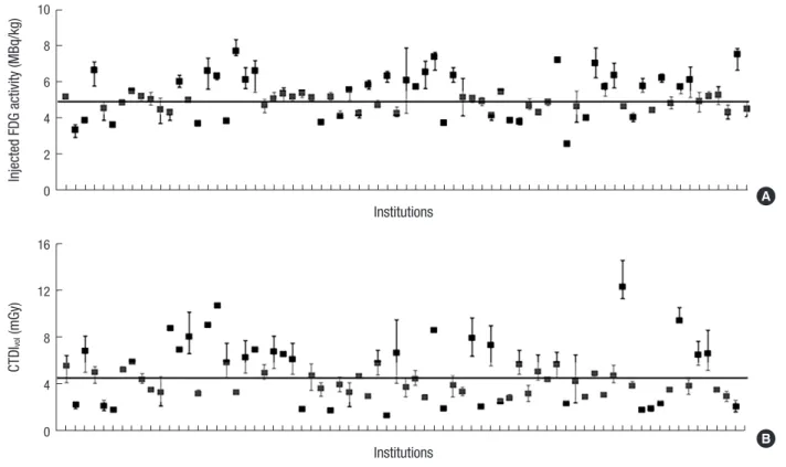
Fig. 2. Distribution of injected FDG activity and CTDIvol values of CT examinations in each institution. (A) Median value of injected FDG activity is 5.00 MBq/kg (25th-75th per- centile, 4.24-5.62 MBq/kg; gray box). (B) Median value of CTDIvol is 4.10 mGy (25th-75th percentile, 2.80-5.96 mGy; gray box).
Injected FDG activity (MBq/kg)
Institutions 10
8 6 4 2
0 A
CTDIvol (mGy)
Institutions 16
12
8
4
0 B
nation-wide survey is shown in Fig. 1. The response included PET/CT results of 1,041 adults (M:F = 633:408, age 60 ± 13 years, body weight 61.4 ± 11.4 kg) and 3 children. One responder re- turned only FDG PET results without CT information, and only the PET results were included in the analysis. Characteristics of the enrolled PET/CT scanners are summarized in Table 2.
Injected FDG activity and radiation dose
The distributions of FDG activity and CTDIvol are shown in Fig.
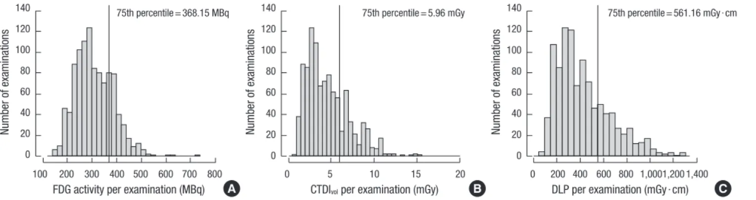
2. In adults, mean injected activity was 310 ± 77 MBq (range 126-729 MBq). In 90.2% of responding institutions, the injected activity was determined primarily based on body weight of a patient; mean value of injected activity per body weight was 5.11 ± 1.19 MBq/kg (range, 2.56-11.40 MBq/kg) (Fig. 2A). When radiation dose was calculated from the injected activity of all real practice data, 75th percentile of injected activity was 368 MBq (Fig. 3A) and mean effective dose from FDG was estimat- ed to be 5.89 ± 1.46 mSv (range 2.39-13.85 mSv).
In children, the injected activity was determined primarily based on body weight of a patient in 75.5% of the surveyed in- stitutions. Mean value of injected activity per body weight was 4.47 ± 1.20 MBq/kg, which was slightly lower than that of adults (P = 0.01). Among the collected data of real examinations, 3 were results of children, in which the injected dose was 4.37, 4.81, and 4.93 MBq/kg.
With regard to CT scan, mean CTDIvol was 4.60 ± 2.47 mGy (range 0.97-15.19 mGy) in adults. Mean CTDIvol of the surveyed 73 institutions are shown in Fig. 2B and 75th percentile was 5.96 mGy (Fig. 3B). Mean DLP was 429.2 ± 227.6 mGy∙cm (range, 99.0-1,274.0 mGy∙cm) and 75th percentile was 561 mGy∙cm (Fig.
3C). Based on the results, radiation dose from CT component in adult patients was estimated to be 6.26 ± 3.06 mSv. In 9 insti- tutions (12.3%), additional CT scan was routinely performed with contrast-enhancement or breath-holding, for which radia- tion dose was not evaluated.
Factors affecting radiation dose of FDG PET/CT
Among the surveyed PET/CT scanners, 73 were equipped with software for CT dose reduction. Mean DLP was not significantly different between scanners equipped with the software and those without the software (436.1 ± 217.1 mGy∙cm vs. 412.9 ± 250.4 mGy∙cm, P = 0.14).
When PET/CT scanners were classified into 3 groups accord- ing to installation year, 42 were less than 5 years old, 50 were 5-10 years old, and 13 were more than 10 years old. Injected FDG activity was significantly reduced in more recently installed scanner groups (P < 0.001, Table 3). In addition, radiation dose from CT was also significantly lower in more recently installed scanner groups (P < 0.001 for both CTDIvol and DLP).
In PET acquisition, TOF technology was available in 45 PET scanners. Mean injected FDG activity for the TOF-available scan- ners was lower than that for TOF-unavailable scanners (4.76 ± 0.96 MBq/kg vs. 5.37 ± 1.28 MBq/kg, P < 0.001). PSF-recovery algorithm was equipped in 36 PET scanners. Mean injected FDG
activity for these scanners was also lower than that for PSF re- covery-unavailable scanners (4.64 ± 0.85 MBq/kg vs. 5.36 ± 1.27 MBq/kg, P < 0.001).
DISCUSSION
In this nation-wide survey, which covered approximately 55%
of the total PET/CT scanners in operation in Korea, the average radiation dose from FDG PET/CT was estimated to be 12.2 mSv;
5.89 mSv from FDG PET and 6.26 mSv from CT. It was also dem- onstrated that more recent PET/CT scanners equipped with certain image-enhancing methods are related to lower radia- tion dose.
With the recent expansion of radiological imaging procedures, medical radiation exposure has been rapidly increased during the last 3 decades; in the United States, annual per capita medi- cal radiation exposure has been increased from 0.53 mSv in 1980 to 3.0 mSv in 2006, the largest source of which was CT (7). The proportion of CT examination has become more considerable in medical radiation exposure because the amount of CT ex- amination is related to economic development (8). However, a concern recently has been raised that FDG PET/CT would be another large source of medical radiation exposure because it has been increased rapidly over the last 10 years. In Korea, a to- tal of 308,663 PET/CT examinations were performed in 2009 (3), and approximately 513,000 FDG PET/CT examinations, in 2014 (9). Additionally, FDG PET/CT is a source of both internal and external radiations; internal radiation from intravenously injected FDG, and external radiation from CT imaging. On the other hand, a single examination of FDG PET/CT may substi- tute several CT scans and nuclear imaging studies because it covers whole body in a single scan. Thus, medical doctors need to understand the radiation dose from FDG PET/CT and to make a decision for diagnostic imaging based on appropriate risk-ben- efit assessment.
There have been nation-wide surveys of radiation dose from FDG PET/CT and its quality control in some European coun- tries (10-12). However, radiation dose can vary widely accord- Table 3. Radiation dose of FDG PET/CT according to installation year
Installation year No. FDG activity,
MBq/kg CTDIvol, mGy DLP, mGy ∙ cm 2000-2005 13 6.10 ± 1.19 6.04 ± 2.58 514.5 ± 246.5 2006-2010 50 5.30 ± 1.22 4.87 ± 2.85 460.7 ± 241.2 2011-2015 42 4.60 ± 0.85 3.95 ± 1.97 369.6 ± 185.4
P value < 0.001 < 0.001 < 0.001
CTDIvol, volume computed tomography dose index; DLP, dose-length product; FDG, F-18 fluorodeoxyglucose.
Fig. 3. Distribution of injected FDG activity (A), CTDIvol (B), and DLP (C) of each PET/CT examination.
Number of examinations
FDG activity per examination (MBq) 100 200 300 400 500 600 700 800 140
120 100 80 60 40 20 0
75th percentile = 368.15 MBq
A
Number of examinations
CTDIvol per examination (mGy)
0 5 10 15 20
140 120 100 80 60 40 20 0
75th percentile = 5.96 mGy
B
Number of examinations
DLP per examination (mGy ∙ cm) 0 200 400 600 800 1,000 1,200 1,400 140
120 100 80 60 40 20 0
75th percentile = 561.16 mGy ∙ cm
C
ing to imaging protocols and scanner models. Additionally, be- cause recent PET/CT scanners are equipped with highly sensi- tive detectors and dose reduction algorithms, FDG PET/CT can be performed with lower radiation dose than before. In Korea, many PET/CT scanners have been installed in recent years with expansion of its use. Thus, a real world survey is required to es- timate overall radiation dose of FDG PET/CT.
The average radiation dose demonstrated in this study is gross- ly similar to the previously reported results. In a nation-wide survey in France, mean radiation dose from FDG PET/CT was estimated to be 14.3 mSv; 5.6 mSv from FDG PET and 8.7 mSv from CT (10). In our study, the dose from FDG PET was slightly higher whereas the dose from CT was lower than that in the French survey. The injected FDG activity recommended by the European Association of Nuclear Medicine (EANM) is 2.5-5.0 MBq/kg (13), which is slightly lower than the mean injected FDG activity surveyed in our study (mean 5.11 MBq/kg). It is speculated to be caused by different imaging protocol. In the EANM guideline, the injected activity is based on a protocol us- ing a fixed scan time of 5 minutes/bed; however, in Korea, scan time is usually less than 2-3 minutes/bed, chiefly for patients’
convenience and high throughput. In a recent guideline, inject- ed activity is determined considering scan time; 7-14 MBq∙min∙
bed-1∙kg-1 (14). Although the current FDG activity is within a rea- sonable range, more efforts should be made in the future based on the balance of risk and benefit.
An intriguing point of this study is the relationship between equipment characteristics and radiation dose. Both the radia- tion doses from FDG and CT were significantly reduced by us- ing more recently installed scanners equipped with image-en- hancing methods. TOF acquisition algorithms can reduce back- ground signal noise and cause an increase in sensitivity (15). As PSF-recovery algorithms can enhance image resolution and overall image quality (16), TOF technology combined with PSF- recovery algorithm would be a main cause of the reduced in- jected activity. The optimal injected FDG activity is determined in each institution by considering image quality and patients’
radiation safety. The present study demonstrated in a real world study that injected FDG activity is reduced by using more re- cent scanners equipped with these algorithms based on the improved image quality. Additionally, the radiation dose from CT was also lower in more recent scanners, although mean DLP was not significantly different between scanners with and with- out dose reduction software. It is speculated that hardware fac- tors such as multi-detectors are more important than software factors. Additionally, the influence of specific CT protocol in each institution should be investigated in further studies. Con- sidering the results of the current study, use of obsolete scan- ners should be discouraged by health insurance reimbursement system or healthcare policy, for patients’ radiation safety.
Quality control programs of imaging equipment and proto-
col are also important for maintaining the performance of diag- nostic tests and reducing unnecessary radiation exposure. In a quality control program, various steps of image acquisition and reconstructions are checked up and authoritative recommen- dations for standardized quality control protocols have been reported regarding daily procedures, calibration of PET/CT scan- ners and image quality evaluation (13,14). Quality control and standardization of imaging procedures are necessary not only for radiation safety but also for comparing image results between different institutions in case of multicenter clinical trials (17,18).
The International Commission on Radiological Protection recommended constitution of the national diagnostic reference levels (DRL) to achieve evidence-based medical radiation pro- tection (19). DRLs are defined as dose levels in medical radio- logical diagnostic practices or typical levels of radiopharmaceu- tical activity for groups of standard-sized patients or standard phantom (20). In terms of radiation protection and standard procedure, DRLs are recommended to be implemented for med- ical radiation diagnostic procedures. The present study is ex- pected to be a basis for establishing the national DRLs for FDG PET/CT scan; the DRL for CT component of whole body FDG PET/CT may be suggested as 560 mGy∙cm (75th percentile of DLP in Fig. 3C), which is lower than the value of 750 mGy∙cm proposed in a French survey (10). DRL for FDG activity may be suggested as 370 MBq (75th percentile of injected activity in Fig.
3A), which is similar to proposed values of 350-385 MBq in oth- er countries (10,21,22).
The present study has a limitation that it is based on a ques- tionnaire survey without actual evaluation of radiation dose in each scanner. However, this is the first study that conducted a nation-wide survey on radiation dose of FDG PET/CT in Korea, and more than 50% of scanners were included in this study. Fur- ther studies are required regarding actual measurement of ra- diation dose.
In conclusion, the average radiation dose from FDG PET/CT is estimated to be 12.2 mSv from this nation-wide survey in Ko- rea. The radiation dose is reduced with more recent scanners equipped with image-enhancing algorithms. The results are ex- pected to be a basis for establishing the national DRLs for FDG PET/CT scan.
DISCLOSURE
The authors declare that they have no conflict of interest.
AUTHOR CONTRIBUTION
Conception and design: Paeng JC, Cheon GJ, Lee JS. Acquisi- tion of data: Kwon HW, Kim JP, Lee HJ. Analysis and interpreta- tion of data: Kwon HW, Kim JP. First draft of manuscript: Kwon HW, Paeng JC. Revision and critical review of the manuscript:
Lee JS, Cheon GJ, Lee DS, Chung JK, Kang KW. Manuscript ap- proval: all authors.
ORCID
Hyun Woo Kwon http://orcid.org/0000-0002-0597-837X Jong Phil Kim http://orcid.org/0000-0002-3359-2933 Hong Jae Lee http://orcid.org/0000-0002-7400-5934 Jin Chul Paeng http://orcid.org/0000-0002-7464-9342 Jae Sung Lee http://orcid.org/0000-0001-7623-053X Gi Jeong Cheon http://orcid.org/0000-0002-1360-5186 Dong Soo Lee http://orcid.org/0000-0003-1627-6557 June-Key Chung http://orcid.org/0000-0002-6866-8571 Keon Wook Kang http://orcid.org/0000-0003-2622-9017 REFERENCES
1. Bly R, Jahnen A, Järvinen H, Olerud H, Vassileva J, Vogiatzi S. Collective effective dose in Europe from X-ray and nuclear medicine procedures.
Radiat Prot Dosimetry 2015; 165: 129-32.
2. Lee MC, Oh SW, Chung JK, Lee DS. The current status and future per- spectives of nuclear medicine in Korea. Nucl Med Mol Imaging 2010; 44:
95-101.
3. Korean Society of Nuclear Medicine. Annual statistics of nuclear medi- cine procedures in Korea. Available at http://www.ksnm.or.kr/education/
sub2_5.php [accessed on 1 September 2015].
4. ICRP. Radiation dose to patients from radiopharmaceuticals. Addendum 3 to ICRP Publication 53. ICRP Publication 106. Approved by the Com- mission in October 2007. Ann ICRP 2008; 38: 1-197.
5. Stamm G, Nagel HD. CT-expo--a novel program for dose evaluation in CT. Rofo 2002; 174: 1570-6.
6. ICRP. Recommendations of the International Commision on Radiologi- cal Protection. ICRP Publication 60. Ann ICRP 1991; 21: 1-201.
7. Linet MS, Slovis TL, Miller DL, Kleinerman R, Lee C, Rajaraman P, Ber- rington de Gonzalez A. Cancer risks associated with external radiation from diagnostic imaging procedures. CA Cancer J Clin 2012; 62: 75-100.
8. Jahnen A, Järvinen H, Olerud H, Vassilieva J, Vogiatzi S, Shannoun F, Bly R.
Analysis of factors correlating with medical radiological examination fre- quencies. Radiat Prot Dosimetry 2015; 165: 133-6.
9. Health Insurance Review and Assessment Service of Korea. Statistics of medical procedures. Available at https://www.hira.or.kr/rd/dissdic/in- foMdfeeList.do?pgmid=HIRAA020044030000 [accessed on 1 September 2015].
10. Etard C, Celier D, Roch P, Aubert B. National survey of patient doses from whole-body FDG PET-CT examinations in France in 2011. Radiat Prot Dosimetry 2012; 152: 334-8.
11. Avramova-Cholakova S, Ivanova S, Petrova E, Garcheva M, Vassileva J.
Patient doses from PET-CT procedures. Radiat Prot Dosimetry 2015; 165:
430-3.
12. Rausch I, Bergmann H, Geist B, Schaffarich M, Hirtl A, Hacker M, Beyer T.
Variation of system performance, quality control standards and adher- ence to international FDG-PET/CT imaging guidelines. A national sur- vey of PET/CT operations in Austria. Nuklearmedizin 2014; 53: 242-8.
13. Boellaard R, O’Doherty MJ, Weber WA, Mottaghy FM, Lonsdale MN, Stro- obants SG, Oyen WJ, Kotzerke J, Hoekstra OS, Pruim J, et al. FDG PET and PET/CT: EANM procedure guidelines for tumour PET imaging: version 1.0. Eur J Nucl Med Mol Imaging 2010; 37: 181-200.
14. Boellaard R, Delgado-Bolton R, Oyen WJ, Giammarile F, Tatsch K, Eschner W, Verzijlbergen FJ, Barrington SF, Pike LC, Weber WA, et al. FDG PET/
CT: EANM procedure guidelines for tumour imaging: version 2.0. Eur J Nucl Med Mol Imaging 2015; 42: 328-54.
15. Karp JS, Surti S, Daube-Witherspoon ME, Muehllehner G. Benefit of time- of-flight in PET: experimental and clinical results. J Nucl Med 2008; 49:
462-70.
16. Akamatsu G, Ishikawa K, Mitsumoto K, Taniguchi T, Ohya N, Baba S, Abe K, Sasaki M. Improvement in PET/CT image quality with a combination of point-spread function and time-of-flight in relation to reconstruction parameters. J Nucl Med 2012; 53: 1716-22.
17. Boellaard R, Oyen WJ, Hoekstra CJ, Hoekstra OS, Visser EP, Willemsen AT, Arends B, Verzijlbergen FJ, Zijlstra J, Paans AM, et al. The Netherlands protocol for standardisation and quantification of FDG whole body PET studies in multi-centre trials. Eur J Nucl Med Mol Imaging 2008; 35: 2320- 33.
18. Graham MM, Wahl RL, Hoffman JM, Yap JT, Sunderland JJ, Boellaard R, Perlman ES, Kinahan PE, Christian PE, Hoekstra OS, et al. Summary of the UPICT protocol for 18F-FDG PET/CT imaging in oncology clinical trials. J Nucl Med 2015; 56: 955-61.
19. ICRP. International Commission on Radiological Protection, Committee I. The evaluation of risks from radiation. Health Phys 1966; 12: 239-302.
20. European Commission. Group of Advisers on the Ethical Implications of Biotechnology. Report to the European Commission: ethical aspects of cloning techniques. Politics Life Sci 1997; 16: 309-12.
21. Botros GM, Smart RC, Towson JE. Diagnostic reference activities for nu- clear medicine procedures in Australia and New Zealand derived from the 2008 survey. ANZ Nucl Med 2009; 40: 2-11.
22. Korpela H, Bly R, Vassileva J, Ingilizova K, Stoyanova T, Kostadinova I, Slavchev A. Recently revised diagnostic reference levels in nuclear medi- cine in Bulgaria and in Finland. Radiat Prot Dosimetry 2010; 139: 317-20.