Changes in Anti-Group A Rotavirus Antibody Seroprevalence and Levels in the Western Gyeongnam Province of Korea Over 16 Years
To observe how anti-group A rotavirus antibody seropositivity rates and levels have changed in the western region of Gyeongnam Province, 2,030 serum samples collected at four collection periods (1989-1990, 1994-1995, 1999-2000, and 2004-2005) were tested by Enzyme-Linked Immunosorbent Assay for IgG, and IgA antibodies reacting to
recombinant VP6 protein. The seroprevalences exhibit no regular patterns over a 16-yr period. For all four collection periods, the anti-rVP6 IgG levels rose steadily during the first 5 months of life, after which they remained high. However, the 2-9 yr and 10-39 yr groups had significantly higher IgG levels in 1999-2000 and 2004-2005, respectively, than in the other collection periods. The 1-5 mo, 40- ≥ 60 yr, and 4-29 yr groups had
significantly higher IgA levels in 1989-1990, 1999-2000, and 2004-2005, respectively. The 4 yr (25.0%), 5-9 yr (18.8%), 10-14 yr (41.1%), 20-29 yr (35.0%), and 30-39 yr (20.0%) groups in 2004-2005 had significant higher IgA seropositivity rate compared to the other three collection periods. These observations suggest that in the western region of Gyeongnam Province since the late 1990s, rotavirus reinfection has occurred more frequently than previously, with all ages being at risk.
Key Words: Rotavirus; VP6; ELISA; Seroepidemiology Ji-Hyun Seo,1 Jung Je Park,2
Jae-Young Lim,1 Jin-Su Jun,1 Chan-Hoo Park,1 Hyang-Ok Woo,1 Hee-Shang Youn,1 Young-Cheol Kwon,3 Hyung-Lyun Kang,3 Seung-Chul Baik,3 Woo-Kon Lee,3 Myung-Je Cho,3 Kwang-Ho Rhee,3 and Wonyong Kim4 Departments of 1Pediatrics, 2Otorhinolaryngology, and 3Microbiology, Gyeongsang National University School of Medicine, Gyeongsang Institute of Health Science, Jinju; 4Department of Microbiology, Chung-Ang University College of Medicine, Seoul, Korea
Received: 29 June 2012 Accepted: 24 October 2012 Address for Correspondence:
Hee-Shang Youn, MD
Department of Pediatrics, Gyeongsang National University School of Medicine, 79 Gangnam-ro, Jinju 660-702, Korea Tel: +82.55-750-8158, Fax: +82.55-752-9339 E-mail: [email protected]
This work was supported by a Korea Research Foundation Grant (KRF-2007-313-E00267) that is funded by the Korean Government (MOEHRD).
http://dx.doi.org/10.3346/jkms.2013.28.1.55 • J Korean Med Sci 2013; 28: 55-61
INTRODUCTION
Symptomatic rotavirus infections are common among infants and young children all over the world, both in developed and developing countries having similar overall incidences of rota- virus infection (1). In Korea, it was found that since 1980, rotavi- rus is the main cause of diarrhea in hospitalized children (2, 3).
Since 2001, outbreaks of rotavirus in newborns at the postpar- tum care centers that care for postpartum mothers and their healthy newborn babies have been reported (4). In addition, there has been an increase in the incidence of symptomatic in- fections in Korean children older than five years of age (5, 6).
While most individuals are exposed to rotavirus at least five times during their lifetime, only the first infection causes severe acute gastroenteritis. Subsequent exposures, even to different rotavirus serotypes, only induce minor symptoms at worst. Thus, acquired immunity seems to play an important role in prevent- ing the ill effects of rotavirus infections (7-9).
Most of the seroepidemiological studies on rotavirus infec- tion are cross-sectional or cohort studies of natural infections in childhood. A cross sectional study examining the natural rota- virus infection of children over 10 yr of age and adults in the same geographical area has not been performed. To address natural rotavirus infection in children and adults, we examined the anti-group A rotavirus antibodies of Koreans living in the same city in serum samples collected from 1989 to 2005, cover- ing the period before introduction of the rotavirus vaccine. For this purpose, purified recombinant group A rotavirus VP6 pro- tein (rVP6) was generated and purified and used in enzyme- linked immunosorbent assays (ELISA).
MATERIALS AND METHODS Serum samples
Gyeongsang National University Hospital, as a member of the National Biobank of Korea, collects randomly serum samples
from patients and stores them at -20ºC. Two thousands and thirty serum samples collected between 1989 to 2005 from pa- tients without acute gastroenteritis were recruited. Of the sam- ples collected for 16 yr, the serum samples in the Gyeongsang National University Hospital of 1989-1990, 1994-1995, 1999- 2000, and 2004-2005 were examined. The sera were stratified into 16 age groups, namely, < 7 days, 1-5 months, 6-11 months, 12-17 months, 18-23 months, 2 yr, 3 yr, 4 yr, 5-9 yr, 10-14 yr, 15-19 yr, 20-29 yr, 30-39 yr, 40-49 yr, 50-59 yr, and ≥ 60 yr. The serum sam- ples were tested for antibodies against rVP6 by ELISA (Table 1).
Expression and purification of rVP6
The rVP6 protein was expressed and the quality of the prepara- tion was validated as described elsewhere (10). Briefly, the full- length rVP6 DNA clone (1,194 base pairs) that was donated by one of us was amplified by polymerase chain reaction (PCR), after which the PCR products were recloned into the pGEM-T vector (Promega, Madison, WI, USA). The pGEM-T/VP6 clone was purified and digested by the deI and BamHI restriction en- zymes and ligated into a deI- and BamHI-digested pET-15b ex- pression vector. Escherichia coli Rosetta II strain was transformed with the recombinant plasmid and grown overnight at 37ºC in LB medium containing 100 μg/mL of ampicillin. The saturated culture was then diluted 1:1,000 and, when the absorbance at 600 nm reached 0.6 to 0.8, isopropyl thiogalactopyranoside was added to a final concentration of 1 mM and the culture was main- tained for 4 hr. The cell inclusion bodies containing the recom- binant VP6 proteins were isolated from the sonicated cell lysate by centrifugation at 13,000 rpm for 30 min. The inclusion bod- ies were extracted by being stirred overnight in 8 M urea in 50 mM Tris-HCl (pH 8.0). The extracted proteins were refolded by being stirred overnight in 0.8 M L-arginine. Affinity chromatog- raphy was performed by using Probond Nickel Agarose Resin (Invitrogen, Carlsbad, CA, USA) and the purified recombinant VP6 proteins were eluted with 50 mM Tris-HCl (pH 8.0), 200 mM NaCl, and 320 mM imidazole, and then dialyzed. The quality of the rVP6 preparation was confirmed by 12% sodium dodecyl sulfate polyacrylamide gel electrophoresis, after which the prep- aration was preserved at -70ºC.
Enzyme-Linked Immunosorbent Assay (ELISA)
To detect rVP6-specific serum IgG and IgA antibodies, the wells of 96-well flat-bottomed EIA plates (Costar, Bloomington, MN, USA) were coated overnight at 4ºC with 50 μL of a 10 μg/mL solution of rVP6 in 0.05 M carbonate, pH 8.0. The plates were washed once with 0.05% Tween 20-PBS (PBST), after which 150 μg of 3% bovine serum albumin-PBST were added to each well and incubated at 37ºC for 3 hr. After three washes with PBST, 50 μL of diluted serum samples (for IgG, and IgA, the dilutions were 1:500, and 1:100, respectively) and positive and negative control serum samples were added to the wells and incubated
for 1 hr at 37ºC. After three washes with PBST, 50 μL of perox- ide-conjugated goat anti-human IgG, or IgA (Bethyl Lab., Mont- gomery, TX, USA; diluted 1:10,000) were added to the wells and incubated at 37ºC for 1 hr. The plates were then washed five times with PBST and 50 μL of o-phenylene diamine was added to each well. The reaction was stopped after 30 min at room tempera- ture by adding 50 μL of 2 N H2SO4. Optical density (OD) was measured at 492 nm. The IgG, and IgA levels in each specimen were tested in duplicate and the mean ODs were obtained. The normal cutoff levels for anti-rVP6 IgG, and IgA were determined by obtaining the mean OD of all negative controls and adding three standard deviations. As a result, the normal cutoff levels for IgG, and IgA were 0.130, and 0.062, respectively. The inter- plate coefficients of variance for the IgG and IgA ELISAs were 10.7%, and 8.5%, respectively (10).
Statistical analysis
The data were analyzed by using SAS statistical software, version 9.1 (SAS Institute, Cary, NC, USA). How the anti-rVP6 IgG, and IgA levels and seropositivity rates in the population varied de- pending on age, gender, and the collection period was examined.
Statistically significant differences in seroprevalence between age groups were determined by using the chi-squared test. Non- parametric tests (Kruskal-Wallis Test and Wilcoxon Scores) were used to analyze the mean anti-rVP6 IgG, and IgA antibody levels at different collection periods. P values of < 0.05 were consid- ered to be statistically significant. Post hoc analysis using Schef- fe method was performed when the difference among 4 peri- ods was significant.
Ethics statement
This study was performed after the institutional review board reviewed and approved the research protocols of the present study (GNUHI- RB-5413). The agreement exemption was ap- plied due to all serum samples had no genetic information and derived from the National Biobank of Korea were obtained with informed consent under institutional review board-approved protocols.
RESULTS
Gender differences and longitudinal changing pattern Males and females did not differ in their seropositivity rates when they were divided according to age or collection period.
However, compared to males, females had higher median anti- rVP6 IgG antibody levels in the 2004-2005 (P = 0.012, data not shown). By contrast, males had higher median anti-VP6 IgA levels in the 1999-2000 (P < 0.001, data not shown). No specific patterns according to age were observed during the 16-yr with regard to the anti-rVP6 IgG and IgA antibody levels and sero- positivity rates.
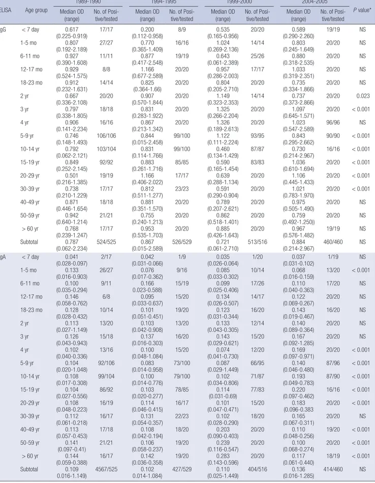
Table 1. Anti-rVP6 IgG and IgA antibody levels and seropositivity rates over 16 yr from 1989 to 2005.
ELISA Age group
1989-1990 1994-1995 1999-2000 2004-2005
P value*
Median OD
(range) No. of Posi-
tive/tested Median OD
(range) No. of Posi-
tive/tested Median OD
(range) No. of Posi-
tive/tested Median OD
(range) No. of Posi- tive/tested IgG < 7 day
1-5 mo 6-11 mo 12-17 mo 18-23 mo 2 yr 3 yr 4 yr 5-9 yr 10-14 yr 15-19 yr 20-29 yr 30-39 yr 40-49 yr 50-59 yr
> 60 yr Subtotal
0.617 (0.225-0.919)
0.807 (0.192-2.189)
0.927 (0.390-1.608)
0.929 (0.524-1.575)
0.912 (0.232-1.631)
0.667 (0.336-2.108)
0.797 (0.338-1.805)
0.906 (0.141-2.234)
0.746 (0.148-1.493)
0.792 (0.062-2.121)
0.849 (0.252-2.145)
0.501 (0.216-1.385)
0.738 (0.210-1.229)
0.871 (0.446-1.654)
0.942 (0.640-1.214)
0.768 (0.239-1.247)
0.787 (0.062-2.234)
17/17 27/27 11/11 8/8 14/14 20/20 18/18 16/16 106/106 103/104 92/92 19/19 17/17 18/18 21/21 17/17 524/525
0.200 (0.112-0.958)
0.770 (0.365-1.409)
0.877 (0.417-2.548)
1.166 (0.677-2.589)
0.825 (0.364-1.66)
0.907 (0.570-1.844)
0.831 (0.283-1.922)
0.867 (0.213-1.342)
0.844 (0.015-2.458)
0.831 (0.114-1.766)
0.883 (0.261-1.716)
1.166 (0.406-2.022)
0.812 (0.511-1.277)
0.881 (0.351-1.570)
0.755 (0.240-1.213)
0.953 (0.535-1.703)
0.867 (0.015-2.589)
8/9 16/16 19/19 20/20 20/20 20/20 20/20 20/20 99/100 99/100 85/85 17/17 23/23 20/20 20/20 20/20 526/529
0.535 (0.165-0.956)
1.024 (0.269-2.136)
0.643 (0.061-2.389)
0.957 (0.286-2.003)
0.804 (0.205-2.710)
1.149 (0.323-2.353)
1.325 (0.266-2.204)
1.326 (0.189-2.613)
1.122 (0.111-2.224)
0.460 (0.134-1.429)
0.590 (0.165-1.454)
0.639 (0.288-1.134)
0.591 (0.290-0.904)
0.789 (0.207-2.621)
0.862 (0.518-1.401)
0.885 (0.426-1.643)
0.721 (0.061-2.710)
20/20 14/14 25/26 17/17 20/20 14/14 20/20 20/20 93/95 87/87 83/83 20/20 20/20 20/20 20/20 20/20 513/516
0.589 (0.290-2.260)
0.803 (0.245-1.649)
0.880 (0.318-2.535)
1.033 (0.319-2.351)
0.735 (0.334-1.866)
0.737 (0.373-2.866)
1.097 (0.645-1.571)
1.023 (0.547-2.589)
0.843 (0.295-2.662)
0.730 (0.214-2.967)
1.036 (0.610-1.694)
1.106 (0.445-1.433)
1.021 (0.783-1.970)
0.975 (0.505-1.490)
0.759 (0.492-1.250))
0.967 (0.576-1.482)
0.884 (0.214-2.967)
19/19 20/20 20/20 20/20 20/20 20/20 20/20 96/96 90/90 16/16 20/20 20/20 20/20 20/20 20/20 19/19 460/460
NS NS NS NS NS 0.023
< 0.001 NS
< 0.001
< 0.001
< 0.001
< 0.001
< 0.001 NS NS NS NS
IgA < 7 day 1-5 mo 6-11 mo 12-17 mo 18-23 mo 2 yr 3 yr 4 yr 5-9 yr 10-14 yr 15-19 yr 20-29 yr 30-39 yr 40-49 yr 50-59 yr
> 60 yr Subtotal
0.041 (0.028-0.097)
0.133 (0.016-0.903)
0.100 (0.035-0.294)
0.146 (0.058-0.762)
0.128 (0.028-0.432)
0.113 (0.027-1.149)
0.126 (0.043-0.943)
0.102 (0.040-0.336)
0.104 (0.020-1.048)
0.108 (0.017-0.308)
0.104 (0.027-0.556)
0.108 (0.048-0.223)
0.112 (0.061-0.218)
0.113 (0.057-0.453)
0.141 (0.097-0.41)
0.144 (0.059-0.388)
0.109 0.016-1.149)
2/17 26/27 9/11 6/8 10/14 13/20 15/18 13/16 92/106 99/104 86/92 16/19 16/17 17/18 21/21 16/17 4567/525
0.042 (0.031-0.066)
0.076 (0.017-0.362)
0.166 0.023-0.588)
0.095 (0.033-0.637)
0.101 (0.051-0.451)
0.103 (0.042-0.908)
0.137 (0.016-0.303)
0.100 (0.048-1.084)
0.083 (0.014-0.958)
0.100 (0.014-0.776)
0.103 (0.020-0.277)
0.114 (0.046-0.415)
0.131 (0.054-0.357)
0.108 (0.042-0.194)
0.106 (0.058-0.237)
0.142 (0.036-0.358)
0.102 0.014-1.084)
1/9 9/16 15/19 15/20 19/20 13/20 16/20 15/20 73/100 79/100 78/85 16/17 22/23 18/20 19/20 19/20 427/529
0.035 (0.026-0.064)
0.085 (0.033-0.302)
0.099 (0.025-0.406)
0.134 (0.026-0.507)
0.123 (0.031-0.344)
0.133 (0.043-0.305)
0.143 (0.029-0.621)
0.074 (0.041-0.730)
0.087 (0.029-1.449)
0.102 (0.034-0.806)
0.114 (0.031-0.69)
0.101 (0.047-0.471)
0.102 (0.028-0.290)
0.203 (0.090-0.403)
0.239 (0.116-0.547)
0.283 (0.143-0.596)
0.110 (0.025-1.449)
1/20 10/14 17/26 14/17 16/20 12/14 15/20 12/20 66/95 71/87 77/83 15/20 18/20 20/20 20/20 20/20 404/516
0.037 (0.031-0.102)
0.068 (0.016-0.159)
0.110 (0.040-0.363)
0.122 (0.069-0.267)
0.143 (0.019-0.467)
0.140 (0.089-0.364)
0.167 (0.092-1.285)
0.169 (0.097-0.971)
0.140 (0.046-0.480)
0.193 (0.049-0.783)
0.220 (0.097-0.462)
0.183 (0.096-0.383
0.165 (0.067-0.311)
0.110 (0.048-0.256)
0.100 (0.068-0.274)
0.117 (0.061-0.440)
0.136 (0.016-1.285)
1/19 13/20 17/20 20/20 16/20 20/20 20/20 20/20 87/96 87/90 16/16 20/20 20/20 19/20 20/20 18/19 414/460
NS
< 0.001 NS NS NS NS NS
< 0.001
< 0.001
< 0.001
< 0.001
< 0.001 NS
< 0.001
< 0.001
< 0.001 NS
*Statistically significant differences between the four time periods in terms of optical density. OD, optical density.
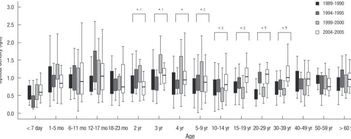
Anti-rVP6 IgG antibody levels and seropositivity rates As shown in Table 1 and Fig. 1, the < 2 yr age groups had simi- lar anti-rVP6 IgG levels to the ≥ 40 yr age groups. For all collec- tion periods, the anti-rVP6 IgG levels started rising in the 0-5 months of age, after which they remained high in all age groups.
However, the 1999-2000 collection period was associated with differences between the 2-39 yr age groups with regard to anti- rVP6 IgG levels. In particular, the 2-9 yr age groups had signifi- cantly higher median ODs than the 10-39 yr groups (P < 0.001).
In addition, in the 2004-2005 collection period, the median ODs of the 10-39 yr groups were significantly higher than the median ODs of the same groups in the other three collection periods (P = 0.05) (Fig. 1). Moreover, the 1994-1995 period was associated with lower neonatal anti-rVP6 IgG levels than the
other three periods, although this trend did not show statistical significance. The anti-rVP6 IgG seropositivity rates for all age groups in all collection periods were almost 100% (Fig. 2).
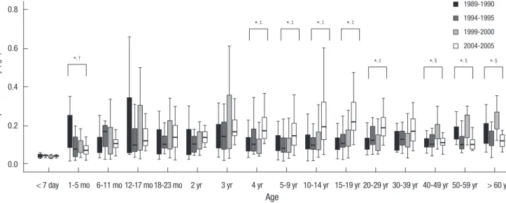
Anti-rVP6 IgA antibody levels and seropositivity rates For all collection periods, all age groups had lower median anti- rVP4 IgA ODs than anti-rVP6 IgG ODs (Table 1 and Fig. 3). The median IgA OD of the 1-5 months age group was significantly higher in the 1989-1990 collection period than in the other three periods (P < 0.001), while the 4-29 yr age groups had significant- ly higher ODs in 2004-2005 than in other periods (P < 0.001).
Moreover, the 40- ≥ 60 yr age groups had significantly higher ODs in 1999-2000 than in the other periods (P < 0.001).
In terms of anti-rVP6 IgA seropositivity rates, the 1989-1990,
Optical density (IgG)
Age
< 7 day 1-5 mo 6-11 mo 12-17 mo 18-23 mo 2 yr 3 yr 4 yr 5-9 yr 10-14 yr 15-19 yr 20-29 yr 30-39 yr 40-49 yr 50-59 yr > 60 yr 3.0
2.5 2.0 1.5 1.0 0.5 0.0
1989-1990 1994-1995 1999-2000 2004-2005
Fig. 1. Anti-recombinant VP6 protein IgG antibodies at four serum collection periods between 1989 and 2005. The IgG levels of each age group at each time point are ex- pressed as median optical densities. For all collection periods, the optical density began to increase in the 0-5 mo group, after which it remained continuously high. *Statistically significant differences between the four time periods in terms of optical density (P < 0.05); †The median optical density (OD) in 1999-2000 was higher than the median ODs in 1989-1990, 1994-1995, and 2004-2005; ‡The median OD in 1999-2000 was higher than the median ODs in 1989-1990, 1994-1995, and 2004-2005; §The median OD in 1999-2000 was lower than the median ODs in 1989-1990, 1994-1995, and 2004-2005; ¶The median ODs in 1994-1995 and 2004-2005 were higher than the median ODs in 1989-1990 and 1999-2000.
*, †
*, §
*, †
*, §
*
*, ¶
*, ‡
*, ¶
Seropositivitry rate (IgG)
100.0 80.0 60.0 40.0 20.0 0.0
Age
< 7 day
18-23 mo
5-9 yr
30-39 yr
6-11 mo 3 yr
15-19 yr
50-59 yr
1-5 mo 2 yr
10-14 yr
40-49 yr
12-17 mo 4 yr
20-29 yr
> 60 yr
Fig. 2. Anti-recombinant VP6 protein IgG seroposivity rates at four serum collection periods between 1989 and 2005. At all collection periods, all age groups exhibited IgG se- ropositivity rates of almost 100%.
1989-1990 Group
1989-1990 1994-1995
1994-1995 1999-2000
1999-2000 2004-2005
2004-2005
1994-1995, and 1999-2000 periods exhibited bimodal patterns, where seroprevalence first peaks in early childhood (between 1 month and 2 yr); this is followed by a reduction in seropreva- lence until a trough appears at 2-4 yr. Thereafter, seroprevalence climbs before reaching a high value at 10-19 yr that is similar to the values of older age groups. The remaining period, 2004- 2005, showed consistently high seropositivity rates after the age of 1 yr (Fig. 4). The 1-5 months group had a higher seropositivity rate in the 1989-1990 period than in the other periods (P = 0.067).
Moreover, the 2-14 yr age groups had significantly higher sero- positivity rates in 2004-2005 than in the other periods (P < 0.001).
The high seropositivity rates of the 1-yr-olds in 2004-2005 were thus also observed in older age groups of that collection period.
Optical density (IgA)
Age
< 7 day 1-5 mo 6-11 mo 12-17 mo 18-23 mo 2 yr 3 yr 4 yr 5-9 yr 10-14 yr 15-19 yr 20-29 yr 30-39 yr 40-49 yr 50-59 yr > 60 yr 0.8
0.6
0.4
0.2
0.0
1989-1990 1994-1995 1999-2000 2004-2005
Fig. 3. Anti-recombinant VP6 protein IgA levels at four serum collection periods between 1989 and 2005. The IgA levels of each age group at each time point are expressed as median optical densities. Compared to the other periods, the 1-5 mo age group had higher IgA levels in 1989-1990, the 4-29 yr age groups had higher IgA levels in 2004- 2005, and the 40- ≥ 60 yr age groups had higher IgA levels in 1999-2000. *Statistically significant differences between the four time periods in terms of optical density (P <
0.05); †The median optical density (OD) in 1989-1990 was higher than the median ODs in 1994-1995, 1999-2000, and 2004-2005; ‡The median OD in 2004-2005 was higher than the median ODs in 1989-1990, 1994-1995, and 1999-2000; §The median OD in 1999-2000 was higher than the median ODs in 1989-1990, 1994-1995, and 2004-2005.
*, ‡
*, †
*, ‡ *, ‡ *, ‡
*, ‡ *, § *, § *, §
Seropositivitry rate (IgA)
100.0 80.0 60.0 40.0 20.0 0.0
Age
< 7 day
18-23 mo
5-9 yr
30-39 yr
6-11 mo 3 yr
15-19 yr
50-59 yr
1-5 mo 2 yr
10-14 yr
40-49 yr
12-17 mo 4 yr
20-29 yr
> 60 yr
Fig. 4. Anti-recombinant VP6 protein IgA seropositivity rates at four collection periods between 1989 and 2005. The IgA seropositivity rates showed a similar bimodal pattern in 1989-1990, 1994-1995, and 1999-2000, namely high seropositivity rates in young children and 10-40 yr-olds and low seropositivity rates in 2-4 yr-olds.
1989-1990 Group
1989-1990 1994-1995
1994-1995 1999-2000
1999-2000 2004-2005
2004-2005
DISCUSSION
In terms of group A rotavirus seroprevalence, no regular chang- ing patterns were seen over the 16-yr study period (1989-2005).
This suggests that rotavirus infections are random events that depend on environmental or social factors at each specific time period. In addition, no specific changes in group A rotavirus se- roprevalence were observed throughout the study period, de- spite the fact that symptomatic neonatal rotavirus infections have been increasing steadily in Korea since 2001 (4-6).
All age groups in the all periods had anti-VP6 IgG seroposi- tivity rates of almost 100%, which indicates that all age groups had been exposed repeatedly to rotavirus during the 16-yr study period. Since the serum samples were from patients without
acute gastroenteritis, this repeated rotavirus exposure appears to boost anti-rotavirus immunity in children and adults rather than causing disease. Notably, a study analyzing cord blood from Indian infants revealed that their anti-rotavirus IgG levels declined at 6 months of age (11). A serological analysis in Brazil also showed clearly that the anti-rotavirus IgG levels decrease during the first 6-9 months of age (12). By contrast, our study showed no significant decline in the anti-rVP6 IgG levels dur- ing the first year of life; indeed, the high IgG levels that were ob- served in the 6-11 months age group were sustained in all sub- sequent age groups. This discrepancy may relate to the fact that the VP6-specific IgG levels produced by the Koreans participat- ing in this study are higher than those generated by the Indians and Brazilians (11, 12). The surveillance of acute gastroenteritis in Seoul, Korea showed rotavirus is the most common patho- gen in infants with viral gastroenteritis (42.7%, 496/1,161) (13).
In a study of rotavirus surveillance testing on all the newborns who were admitted to the nursery in Korea, 47 of 61 neonates had no symptoms of gastroenteritis such as fever, vomiting and diarrhea (14). Both results supported that the higher level of IgG in 6-11 months of infants in the present study. It may also be due to the fact that Koreans are earlier exposed and more fre- quently re-exposed to rotavirus. Infants who did not have rota- virus infections had significantly higher IgG level in cord blood and serum samples at 6 months than infants who had symp- tomatic/asymptomatic rotavirus infections. This result suggest- ed that fewer rotavirus infections occur when cord blood re- tains higher level of anti-rotavirus IgG antibodies (11). In the present study, the newborns (< 7 day) in all four collection pe- riods had lower anti-rVP6 IgG levels than most of the other age groups, yet concomitant decreases in anti-rVP6 IgG levels were not observed in the women of reproductive age (20-39 yr of age). Thus, our data do not support the notion that the recent in- creases in symptomatic neonatal infection are due to low ma- ternal serum IgG levels. Indeed, we observed that compared to men, women had significantly higher anti-rVP6 IgG levels in 2004-2005. This may reflect greater exposure of mothers to their children than fathers and thus their greater chance of re-expo- sure to rotavirus, which boosts their anti-rotavirus IgG levels.
However, this theory does not explain why women did not dif- fer from men in their anti-rVP6 IgA levels; indeed, in several collection periods, men had higher anti-rVP6 IgA levels than women (data not shown). Further studies are needed to resolve this issue.
The prevalence of rotavirus infection is generally underesti- mated because asymptomatic infections occur frequently and adults who present with diarrhea are not routinely tested for ro- tavirus infection. The anti-rVP6 IgA antibody pattern in the pres- ent study suggest that, compared to other collection periods, 1) rotavirus re-exposure of the adult population occurred less fre- quently in the late 1980s, and 2) the entire population was less
frequently re-exposed until the late 1990s. In the 2004-2005 pe- riod, the adults had very high IgA levels and seropositivity rates, which is suggestive of frequent re-exposure to rotavirus infec- tion after a long period of limited exposure. Notably, long-term hospitalization is a risk factor for rotavirus-induced illnesses because such patients tend to live in closed communities which may impair immune response (15). Gyeongsang National Uni- versity Hospital is located in Jinju, which is a city of 350,000 peo- ple in the western part of Gyeongnam Province. Jinju is a rela- tively small city by Korean standards and is surrounded by rural communities. The annual growth of the population is 0.4% since 1995. Jinju is known as the city of education and there are 6 uni- versities, 26 high schools, 20 middle schools and 45 elementary schools. In Korea, most of the middle and high school students stay in the same class from early in the morning until late in the evening; this can also be true for college students. Thus, many students live in closed-type communities, which may make them more vulnerable to rotavirus, which in turn could result in epidemics of rotavirus infections.
Mucosal and serum IgA antibodies protect against rotavirus infection (16, 17). A cohort study of 200 infants also found that serum anti-rotavirus IgA antibodies are a stronger marker of protection than serum anti-rotavirus IgG antibodies (17). Thus, the IgA seropositivity rates may be reflective of recent rotavirus infections and epidemics. In the present study, the first three collection periods were associated with bimodal patterns of an- ti-rVP6 IgA seropositivity. This suggests that during these peri- ods, the first infection occurred in very young children and re- infection occurred in over the age of 10-19 yr. By contrast, in the 2004-2005, most of the age groups showed high anti-rVP6 IgA seropositivity rates. This suggests that, by this time, most people had been exposed repeatedly to rotavirus.
The present study suffers from two limitations. First, the rota- virus infection history of the patients was not known. The sec- ond is that laboratory-based surveillance of stool rotaviruses was not performed when the sera were obtained. As a consequence, it is not clear whether other rotavirus groups were prevalent, which could potentially affect the immune responses to the rVP6 antigen of group A rotaviruses. Alternatively, the antibod- ies raised by the other rotavirus groups may be crossreactive and could have led to false positives in the ELISA used in the present study. However, it has also been shown that the anti- bodies elicited by different rotavirus strains are not crossreac- tive in VP6-based ELISAs (18).
In late 2008, the rotavirus vaccine started to be used in Kore- an infants on a selective basis. A recent review of the efficacy of this vaccine has shown that it was more efficacious in developed and middle-income countries than in lower income countries (19). It was suggested that the impact of national rotavirus vacci- nation should be assessed by postmarketing surveillance. More- over, the circulating rotavirus strains should be monitored con-
tinually.
In summary, the anti-rVP6 IgG and IgA antibody responses examined in this study show that people in the southern central part of Korea have been frequently and repeatedly exposed to rotaviruses since the late 1990s despite socioeconomic, hous- ing, and environmental-sanitation conditions improve.
ACKNOWLEDGMENTS
The biospecimens used in this study were provided by the Gyeongsang National University Hospital, which is a member of the National Biobank of Korea, which is supported by the Ministry of Health, and Welfare. All samples derived from the National Biobank of Korea were obtained with informed con- sent under institutional review board approved protocols. The authors have no conflicts of interest to disclose.
REFERENCES
1. Centers for Disease Control and Prevention. Rotavirus. In: Atkinson WHJ, McIntyre L, Wolfe S, eds. Epidemiology and Prevention of Vaccine- preventable Diseases, 10th ed. Washington, DC, Public Health Founda- tion, 2007: 295-306.
2. Seo JK, Sim JG. Overview of rotavirus infections in Korea. Pediatr Int 2000; 42: 406-10.
3. Chae JH, Kim MJ, Kim DH, Lee KY, Kang JH, Lee JS. Epidemiologic study of rotavirus gastroenteritis, in Daejeon, Korea, 2001-2005. Korean J Pe- diatr Infect Dis 2007; 14: 155-61.
4. Kim JS, Lee HS, Choi JH, Shin YJ, Koo ML, Kim SS, Kim HS, Kim EA, Yoon SW, Kwon JH, et al. A study of acute gastroenteritis in neonates transferred from postpartum care centers. Korean J Pediatr Infect Dis 2003; 10: 186-92.
5. Park SI, Kwon HO, Lee JH, Jung SJ. Clinical features of rotaviral gastro- enteritis in neonates. Korean J Pediatr 2005; 48: 1121-5.
6. Seo HJ, Jung YJ, Park SK, Choi SH, Lee JH, Kim MJ, Chang YS, Park WS.
Rotavirus-associated neonatal necrotizing enterocolitis. Korean J Pedi- atr 2009; 52: 56-60.
7. Veláquez FR, Matson DO, Guerrero ML, Shults J, Calva JJ, Morrow AL, Glass RI, Pickering LK, Ruiz-Palacios GM. Serum antibody as a marker of protection against natural rotavirus infection and disease. J Infect Dis
2000; 182: 1602-9.
8. Griffin DD, Fletcher M, Levy ME, Ching-Lee M, Nogami R, Edwards L, Peters H, Montague L, Gentsch JR, Glass RI. Outbreaks of adult gastro- enteritis traced to a single genotype of rotavirus. J Infect Dis 2002; 185:
1502-5.
9. Ward RL, Bernstein DI, Shukla R, Young EC, Sherwood JR, McNeal MM, Walker MC, Schiff GM. Effects of antibody to rotavirus on protection of adults challenged with a human rotavirus. J Infect Dis 1989; 159: 79-88.
10. Seo JH, Kim SY, Park JS, Lim JY, Park CH, Woo HO, Youn HS, Kim W, Kang HL, Baik SC, et al. Usefulness of Escherichia coli-expressed recom- binant VP6 proteins of group A rotavirus in serodiagosis of rotavirus in- fection. Korean J Pediatr Gastroenterol Nutr 2010; 13: 134-45.
11. Ray PG, Kelkar SD, Walimbe AM, Biniwale V, Mehendale S. Rotavirus immunoglobulin levels among Indian mothers of two socio-economic groups and occurrence of rotavirus infection among their infants up to six months. J Med Virol 2007; 79: 341-9.
12. Cox MJ, Azevedo RS, Nokes DJ, Beards GM, McCrae MA, Massad E, Medley GF. Seroepidemiolgy of group A rotavirus in suburban São Pau- lo, Brazil. Epidemiol Infect 1998; 120: 327-34.
13. Lee JI, Park SH, Kim MS, Oh YH, Yu IS, Choi BH, Lee GC, Kim MS, Jang SY, Lee CH. Surveillance of acute gastroenteritis in Seoul, Korea, during May 2004 and June 2007. J Bacteriol Virol 2009; 39: 363-71.
14. Kim CR, Oh JW, Yun MK, Lee JH, Kang JO. Rotavirus infection in neo- nates at a university hospital in Korea. Infect Control Hosp Epidemiol 2009; 30: 893-5.
15. Iijima Y, Iwamoto T, Nukuzuma S, Ohishi H, Hayashi K, Kobayashi N.
An outbreak of rotavirus infection among adults in an institution for re- habilitation: long-term residence in a closed community as a risk factor for rotavirus illness. Scand J Infect Dis 2006; 38: 490-6.
16. Burns JW, Siadat-Pajouh M, Krishnaney AA, Greenberg HB. Protective effect of rotavirus VP6-specific IgA monoclonal antibodies that lack neu- tralizing activity. Science 1996; 272: 104-7.
17. Grimwood K, Lund JCS, Coulson BS, Hudson IL, Bishop RF, Barnes GL.
Comparison of serum and mucosal antibody responses following severe acute rotavirus gastroenteritis in young children. J Clin Microbiol 1988;
26: 732-8.
18. Tsunemitsu H, Jiang B, Saif LJ. Detection of group C rotavirus antigens and antibodies in animals and humans by ELISA. J Clin Microbiol 1992;
30: 2129-34.
19. O’Ryan M, Linhares AC. Update on Rotarix: an oral human rotavirus vaccine. Expert Rev Vaccines 2009; 8: 1627-41.