Infection &
Chemotherapy
Received: March 9, 2016 Accepted: April 1, 2016 Published online: February 7, 2017 Corresponding Author : Youn Jeong Kim, MD
Department of Internal Medicine, Seoul St. Mary’s Hospital, The Catholic University of Korea, 222 Banpo-daero, Seocho-gu, Seoul 06592, Korea
Tel: +82-2-2258-6073, Fax: +82-2-2258-1254 E-mail : [email protected]
This is an Open Access article distributed under the terms of the Creative Commons Attribution Non-Commercial License (http://creativecommons.org/licenses/by-nc/3.0) which permits unrestricted non-commercial use, distribution, and repro- duction in any medium, provided the original work is properly cited.
Copyrights © 2017 by The Korean Society of Infectious Diseases | Korean Society for Chemotherapy
www.icjournal.org
Emphysematous Osteomyelitis due to Escherichia coli
Jinhee Lee, Chai Ho Jeong, Myun Hee Lee, Eun-Gyo Jeong, Youn Jeong Kim, Sang Il Kim, and Yang Ree Kim
Department of Internal Medicine, College of Medicine, The Catholic University of Korea, Seoul, Korea
Emphysematous osteomyelitis, especially that involving the extra-axial skeleton, is an extremely rare presentation but associat- ed with significant morbidity and mortality. Here, we report a case in which a 58-year-old female patient with diabetes mellitus presented with emphysematous osteomyelitis that involved the sternum, clavicle, and pelvic bone and was caused by Esche- richia coli via hematogenous spread of urinary tract infection. We successfully treated her with urgent and aggressive surgical drainage with prolonged antibiotics therapy. Early diagnosis and immediate surgical intervention are required for better out- comes in cases of emphysematous osteomyelitis.
Key Words: Emphysematous osteomyelitis; Escherichia coli; Urinary tract infection https://doi.org/10.3947/ic.2017.49.2.151
Infect Chemother 2017;49(2):151-154
ISSN 2093-2340 (Print) · ISSN 2092-6448 (Online)
Case Report
Introduction
Although intraosseous gas in the vertebral bodies or disc space is mostly associated with a non-infectious cause, in- traosseous gas in the extra-axial skeleton in the absence of di- rect communication is virtually pathognomonic for emphy- sematous osteomyelitis [1]. The first case of emphysematous osteomyelitis was reported in 1981 [2], and only 29 cases have been reported globally thus far. To the best of our knowledge, no case of emphysematous osteomyelitis has been reported from Korea.
Here, we report the case of a 58-year-old female patient with poorly controlled diabetes mellitus, who presented with em- physematous osteomyelitis involving the sternum, clavicle,
and pelvic bone due to Escherichia coli that had spread hema- togenously from a urinary tract infection.
Case Report
A 58-year-old woman was admitted to the emergency de- partment owing to generalized weakness, chilling sensation and myalgia since the previous 10 days. She had a medical history of uncontrolled diabetes mellitus and hypertension.
She took a prescription for diabetes mellitus, but did not take well. She had no history of trauma or surgery. Her vital signs were as follows: blood pressure, 90/40 mmHg; pulse rate, 100/
min; respiratory rate, 20/min; and body temperature, 36.5℃.
Lee J, et al. • Emphysematous osteomyelitis by E. coli www.icjournal.org
152
Laboratory data showed that a white blood cell count 24,290/
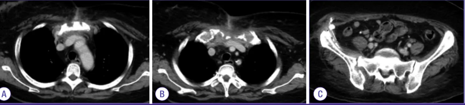
mm3 with 93% neutrophils, hemoglobin 11.7 g/dL, platelet count 57,000/mm3 and C-reactive protein 33.2 mg/dL. Liver function enzymes showed aspartate aminotransferase 22 U/L, alanine aminotransferase 17 U/L, alkaline phosphatase 1,303 U/L, gamma glutamyl transferase 135 U/L, and total bilirubin 1.11 mg/dL. The HbA1c level was 11.9%, and serum glucose level was 665 mg/dL. Urinalysis showed many bacteria. Blood and urine cultures were obtained prior to initiation of empiri- cal antibiotics. The chest radiograph showed a prominent heart size but no apparent lung parenchymal lesion. The chest and abdominal computed tomography (CT) scan revealed in- traosseous gas in the sternum and left clavicle and intra-mus- cular gas in the left pectoralis major muscle as well as soft tis- sue emphysema involving the left shoulder and both chest walls (Fig. 1A and 1B). Additionaly, intraosseous gases were noted in the right iliac bone and sacroiliac joint (Fig. 1C).
Right pyelonephritis with abscess at the right lower kidney was observed as well (Fig. 1D).
Empirical antibiotic therapy with intravenous meropenem (1 g q8h, iv) and teicoplanin (800 mg q24h for 3 days and then 400 mg q24h, iv) was initiated and urgent surgical drainage was performed. At the time of surgical decompression, the pus from sternum, left clavicle and pelvic bone had a foul odor. The pus revealed extended-spectrum ß-lactamase non-producing E. coli, and both blood and urine culture grew E. coli with same antimicrobial susceptibility. Antibiotics were changed to piperacillin/tazobactam (4.0/0.5 g q8h, iv).
A follow-up CT conducted 4 weeks after admission showed marked regression of intraosseous gas in the sternum, left
clavicle and adjacent extensive soft tissue emphysema in the left side of neck, left axilla, and both anterior chest walls. How- ever, slightly increased size of abscess pocket involving the right iliacus muscle and osteomyelitis involving the right iliac bone were still noted. Therefore, catheter drainage of right ili- ac area was maintained for 8 weeks, and pus drainage at the right iliacus muscle was performed again and antibiotic thera- py was continued. Follow-up CT scan after 8 weeks revealed emphysematous osteomyelitis of the sternum, left clavicle and right iliac bone with adjacent cellulitis almost resolved (Fig. 2).
After 13 weeks of intravenous antibiotic therapy, the patient showed clinical improvement and was discharged.
Discussion
Intraosseous gas in the extra-axial skeleton is rare and pathognomonic for emphysematous osteomyelitis. Although intravertebral gas is considered to derive from degenerative disease, or less commonly, osteonecrosis or neoplasm [3-5], serious infection should be considered in cases of extensive intravertebral gas, bone edema or adjacent fluid collections.
In 1981, for the first time, Ram and colleagues described three patients with emphysematous osteomyelitis involving the tibia, pelvis, and fibula, respectively [2]. Until now, only 29 cases have been reported in the English literature globally [1, 6-8]. The involving sites of osteomyelitis of reported cases were vertebrae, pelvis, sacrum, femur, tibia, fibula, and mid- foot [1, 6-8]. To the best of our knowledge, emphysematous osteomyelitis has not previously been described in Korea.
Figure 1. Chest and abdominal computed tomography (CT) scan at admission showed intraosseous gases in- volving the sternum (arrow) (A), left clavicle with swelling and intra-muscular gases in the adjacent muscle and soft tissues (arrow) (B). Intraosseous gases are noted in the right iliac bone and sacroiliac joint (arrow) (C). The CT scan showed a hypodense wedge-shaped lesion in the right lower kidney,(arrow) which indicated acute pyelone- phritis with abscesses (D).
A
C
B
D
https://doi.org/10.3947/ic.2017.49.2.151 • Infect Chemother 2017;49(2):151-154
www.icjournal.org 153
In most cases, the infection occurs by hematogenous spread, however may also be related to contagious spread from an in- traabdominal source of infection, or from a skin or soft tissue source of infection; or following intra-abdominal or spinal sur- gery [7]. In this case, a urinary tract infection was the possible source of hematogenous spread that resulted in emphysema- tous osteomyelitis. Underlying comorbidities such as diabetes mellitus and malignancy are known to compromise immune function [1]. In this case, the patient had poorly controlled dia- betes mellitus, that was a risk factor for emphysematous osteo- myelitis. Common causative organisms include members of the Enterobacteriaceae family or anaerobes (especially Fuso- bacterium necrophorum). The monomicrobial causes of em- physematous osteomyelitis are similar to causes of other gas-forming infections, which include E. coli, Klebsiella pneu- moniae, Enterobacter aerogenes, and Clostridium spp. [1, 9-11].
In cases of post-surgical infections, gram-positive organisms such as Staphylococcus aureus, non-hemolytic streptococci, and enterococci, and Pseudomonas spp. may be the causative organisms [1]. Of all the reported cases emphysematous osteo- myelitis, the causative organism was E. coli in 8 cases (4 cases of monomicrobial infection and 4 cases of polymicrobial infec- tion due to E. coli and other organisms) [1, 6-8].
In cases of acute osteomyelitis, appropriate antimicrobial agents should be given promptly in cases of bacteremia, bone necrosis and bone destruction [12]. And surgical treatment should be considered in acute osteomyelitis if there is finding of abscess formation or radiologic evidence of necrosis, or if the patients do not respond to antimicrobial agents [12]. The duration of treatment must be tailored according to the condi- tion of the individual patient, however duration of at least 4–6 weeks of antimicrobial treatment after the last debridement is recommended [12].
Emphysematous osteomyelitis is associated with high mor-
bidity and mortality, as high as 32%, especially in patients with diabetes mellitus [1]. A diagnosis of emphysematous osteo- myelitis with the presence of intraosseous gas in the extra-axi- al skeleton suggests a severe infection that requires aggressive and immediate treatment [13].
In conclusion, we report a rare case of emphysematous os- teomyelitis of the sternum, clavicle and pelvic bone due to E.
coli that had spread hematogenously from a urinary tract in- fection. Early diagnosis and urgent and aggressive surgical in- tevention as well as broad antibiotics are crucial for survival of patients.
Conflicts of Interest
No conflicts of interest.
ORCID
Jinhee Lee https://orcid.org/0000-0002-2359-6383 Youn Jeong Kim https://orcid.org/0000-0001-5870-1801
References
1. Luey C, Tooley D, Briggs S. Emphysematous osteomyelitis:
a case report and review of the literature. Int J Infect Dis 2012;16:e216-20.
2. Ram PC, Martinez S, Korobkin M, Breiman R, Gallis HR, Harrelson JM. CT detection of intraosseous gas: a new sign of osteomyelitis. Am J Roentgenol 1981;137:721-3.
3. Potocki J, Kaushik S, Mira JL. Anaerobic osteomyelitis of femoral head with intraosseous, intra-articular, bursal and muscle pneumatosis. Skeletal Radiol 2003;32:46-8.
4. Resnick D, Niwayama G, Guerra J Jr, Vint V, Usselman J.
A B C
Figure 2. Follow-up chest and abdominal computed tomography scans at 12 weeks after admission showed emphysematous osteomyelitis in sternum and left clavicle with adjacent cellulitis are almost improved (A, B). Also, emphysematous osteomyelitis in the right iliac bone and sacroiliac joint are almost resolved (C).
Lee J, et al. • Emphysematous osteomyelitis by E. coli www.icjournal.org
154
Spinal vacuum phenomena: anatomical study and review.
Radiology 1981;139:341-8.
5. Bielecki DK, Sartoris D, Resnick D, Van Lom K, Fierer J, Haghighi P. lntraosseous and intradiscal gas in association with spinal infection: report of three cases. AJR Am J Roentgenol 1986;147:83-6.
6. Mautone M, Gray J, Naidoo P. A case of emphysematous osteomyelitis of the midfoot: imaging findings and review of the literature. Case Rep Radiol 2014; 2014:616184.
7. McDonnell O, Khaleel Z. Emphysematous osteomyelitis.
JAMA Neurol 2014;71:512.
8. Larsen J, Mühlbauer J, Wigger T, Bardosi A. Emphysema- tous osteomyelitis. Lancet Infect Dis 2015;15:486.
9. Vohra A, Saiz E, Ratzan KR. A young woman with a sore throat, septicaemia, and respiratory failure. Lancet 1997;350:928.
10. Foulkes GD, Johnson CE, Katner HP. Fusobacterium os- teomyelitis associated with intraosseous gas. Clin Orthop Relat Res 1990;251:246-8.
11. Le Moal G, Juhel L, Grollier G, Godet C, Azais I, Roblot F.
Vertebral osteomyelitis due to Fusobacteriun species: re- port of three cases and review of the literature. J Infect 2005;51:E5-9.
12. Korean Society for Chemotherapy, Korean Society of In- fectious Diseases, Korean Orthopaedic Association. Clini- cal guidelines for the antimicrobial treatment of bone and joint infections in Korea. Infect Chemother 2014;46:125- 38.
13. Kumar J, Bandhu S, Kumar A. Intraosseous and intraartic- ular pneumatosis in anaerobic osteomyelitis. Pediatr Ra- diol 2006;36:1220.