449
단백질의 변화에 대한 단백체학적 접근
Proteomic Analysis of the Proteins That were Changed by Bilateral Orchiectomy in the Rabbit Corpus
Cavernosum
Hyung-Jee Kim, Young-Geun Ryu, Joo-Hyung Hong, Jeong-Hee Hong From the Department of Urology, Dankook University College of Medicine, Cheonan, Korea
Purpose: The pathophysiological role of androgen deprivation in male sexual dysfunction remains controversial, and this is especially true at the molecular level. We investigated the effect of androgen deprivation on the changes of proteins in the penile corpus cavernosum of castrated rabbits by the proteomic approach.
Materials and Methods: New Zealand white male rabbits (2.5-3kg) were divided into 2 groups: the control group with 5 rabbits and the bilateral orchiectomized group. The bilateral orchiectomized group was divided into the post-operative 4 weeks group (group 1), and the 8 weeks group (group 2) with 5 rabbits in groups 1 and 2, respectively. The penile corpus cavernosum was partly excised at 4 or 8 weeks from the beginning of the experiment. Conventional proteomics was performed with high resolution 2-D gel electrophoresis; this was followed by computational image analysis and protein identification with using mass spectrometry.
Results: A comparison of the corpus cavernosum of the orchiectomized group with the control group showed that nine proteins had a changed expression. Glycerol-3-phosphate dehydrogenase and F-actin binding pro- tein were under-expressed in groups 1 and 2, and myosin regulatory light chain 2, tropomyosin βchain and tropomyosin 1 were significantly under- expressed in group 1 and they were insignificantly over-expressed in group 2. In addition there were 4 proteins that were insignificantly under- expressed; skeletal muscle myosin heavy chain MyHC-EO/III, a protein that was similar to suppressor of cytokine signaling 6 isoform, myosin light chain 1 and heat shock 27kDa protein 1.
Conclusions: This data suggests that changes of proteins, and especially tropomyosin 1, mean there is processing of the cellular apoptosis pathway in the orchiectomized rabbits' corpus cavernosum. However more infor- mation is needed about human corpus cavernosal tissue. (Korean J Urol 2008;49:449-453)
Key Words: Orchiectomy, Corpus cavernosum, Proteomics, Protein
대한비뇨기과학회지 제 49 권 제 5 호 2008
단국대학교 의과대학 비뇨기과학교실
김형지ㆍ유영근ㆍ홍주형ㆍ홍정희
접수일자:2008년 2월 22일 채택일자:2008년 4월 8일
교신저자: 김형지
단국대학교병원 비뇨기과 충남 천안시 안서동 산 29
330-715
TEL: 041-550-6630 FAX: 041-556-0524 E-mail: killtumor@
yahoo.co.kr
서 론
남성호르몬은 생식기의 성장, 발달 및 2차 성징의 발현에 기여한다. 따라서 노화나 전립선암의 치료를 위한 수술적
혹은 약물적 거세로 인하여 남성호르몬이 감소되면 성욕이 감소하고 발기부전 등의 후유증이 발생된다.1 음경 발기는 복합적인 신경학적인 현상이며, 동맥혈류 유입의 증가, 혈 관 평활근의 이완, 정맥 혈류 유출의 감소라는 세 가지의 혈역동학적 요인의 협동과정이 있다. 이 과정에서 뇌, 신경,
신경 전달물질, 평활근, 횡문근 등의 상호과정이 연관되어 나타난다.2 이러한 발기의 복합적 과정 중, 남성 호르몬의 부족 또는 박탈로 인하여 유발되는 발기부전의 원인을 규 명하기 위해 많은 병리학적이나 조직학적 접근이 시도되었 다. 남성 호르몬이 감소하는 경우 음경해면체의 평활근의 양이 감소하며,3 동물실험 결과로 음경 평활근 세포 고사 및 결체 조직의 증가 및 발기조직의 이완이 감소된다고 한 다.4 또한 산화질소에 의해 매개되는 음경 발기과정에서 남 성 호르몬은 필수적인 역할을 한다고 한다.5
이와 같이 남성호르몬과 발기부전에 대한 수많은 연구가 진행되고 있으나 분자생물학적 연구는 아직 미진한 상태이 다. 최근에 양측 음경신경의 절단 후의 쥐의 음경 조직에 있어서 단백체학적 접근을 한 것과 같은 맥락으로6 본 연구 에서는 양측 고환절제술을 시행한 토끼의 음경에서 나타나 는 단백질의 변화를 단백체학적 방법으로 접근해보고자 하 였다.
재료 및 방법
실험동물로는 동물 사육장에서 동일 조건하에 사육된 2.5-3kg의 뉴질랜드산 수토끼 (New Zealand White Rabbit) 15 마리를 사용하였으며, 12시간 낮/밤 주기로 조명이 되고 기 온은 22±1oC, 상대적 습도는 50±20%로 유지된 방에서 사육 한다. 일주일간의 안정기간을 지낸 후 대조군 5마리 (sham 수술군)와 양측 고환절제군 10마리의 두 군으로 나누었다.
고환절제군은 다시 술 후 4주군 5마리 (group 1)와 술 후 8주 군 5마리 (group 2)로 나누었다. 케타민으로 마취를 시킨 후
(100mg/kg intramuscularly) 하부 음낭 절개방법으로 하여 양 측 고환을 완전히 제거하였다. 음낭 피부는 일반적인 방법 으로 봉합하였다. Sham 수술군은 고환 절제군과 같은 방법 으로 수술을 시행하나 고환의 절제는 하지 않고 음경만을 절제한다. 술 후 항생제 (150mg/kg ampicillin intramuscularly) 를 투여하여 창상감염을 예방하고자 하였다. 수술 후 4주와 8주에 음경을 적출하였다. 요도를 확인한 후 대퇴부 안쪽의 경계에서 조직을 요도로부터 분리 적출하였다. 고환절제군 에서 육안으로 보아 음경에 변화가 있거나 음경손상의 소 견이 있는 실험동물은 실험에서 배제하였다. 음경의 적출 이 끝난 실험동물은 토끼 귀 정맥의 공기 색전으로 사망시 켰다. 적출된 음경조직은 음경백막을 제거한 뒤에 해면체 만을 액화질소 탱크에 보관하였다.
결과 분석으로 50% 감소 혹은 200% 증가의 발현량의 변 화를 갖는 단백질을 의미 있는 단백질로 간주하였다.
1. 시료준비
토끼의 음경해면체에서 단백질을 German Heart Center의 방법을 이용하여 추출하였다 (http://userpage.chemie.fu-berlin.
de/~pleiss/tissue.html). 음경해면체조직은 세척용액 (50mM Tris- HCl (7.1), 100mM KCl, 60mM EDTA, 5.8mM benzamidine, 0.2mM PMSF)으로 세척을 하고 1시간 동안 냉동 건조시켰 다. 30mg의 조직을 액화질소 안에서 파쇄한 후 100μl 버퍼 용액 (7M Urea, 2M Thiourea, 4% CHAPS, 100mM DTT, 25mM Tris-HCl (7.1), 50mM KCl, 0.2% Bio-Lyte 3/10 Ampholyt)에 용해시켰다. 210U DNase를 첨가한 후 실온에 서 30분간 방치하였다. 용해되지 않는 물질은 원심분리기 로 제거하였다 (13,000rpm, 20분, 10oC). 단백질 농도는 상업 적으로 판매되는 Bradford reagent (Bio-Rad protein assay kit, Bio-Rad, Richmond, USA)를 사용하여 측정하였다. 조직은 사용할 때까지 영하 70oC에서 보관하였다.
2. 이차원 전기영동
일차원적인 젤 분리는 17cm pH 4-7의 immobilized pH gradient gel (IPG) strip을 가지고 제조회사의 프로토콜 (Bio- Rad)대로 시행하였다. 1mg의 조직을 IPG strip에 놓고 등전 하 초점화 (isoelectric focusing; IEF)는 PROTEAN IEF (iso- electric focusing) cell을 가지고 시행하였다 (145kVh, 20oC).
IEF 후 IPG strip은 2% DTT를 포함하는 6M Urea, 2% SDS, 0.05M Tris-HCl (8.8)와 20% glycerol에서 15분 동안 평형화 시킨 뒤 2.5% iodoacetamide를 포함하는 같은 용액에서 15분 간 평형화시켰다. 평형화된 IPG strip은 15% uniform poly- acrylamide gel로 옮기고 PROTEAN II Xi cell tank (30mA)에 서 실험을 계속하였다. 젤은 Coomassie Brilliant Blue R250 으로 염색하였다. 염색 후 2DE는 Powerlook 1100 (UMAX, Fremont, USA)을 사용하여 이미지화시켰다. 이미지들은 Melanie III (Swiss Institute of Bioinformatics, Switzerland) 소 프트웨어를 사용하여 분석하였고. 모든 실험은 재현성을 위하여 5회 시행하였다.
3. 단백질 동정
대조군과 다르게 표현되는 실험군의 단백질 스팟들은 단 백질 서치 프로그램인 ProFound와 MS-FIT와 함께 질량분석 기 (matrix assisted laser desorption/ionization-the time of flight mass spectrometry; MALDI-TOF MS)로 분석하였다.
4. 재료
실험에 사용된 뉴질랜드 가토는 안성동물농장 (한국)에서 구입하였고 2 DE 시스템 (PROTEAN IEF cell, PROTEAN II
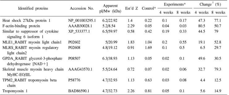
Table 1. The up- and down-regulated proteins in the rabbit penile corpus cavernosum tissue following bilateral orchiectomy Identified proteins Accession No. Apparent
pI/Mw (kDa) Est'd Z Control* Experiments* Change†(%) 4 weeks 8 weeks 4 weeks 8 weeks Heat shock 27kDa protein 1
F-actin-binding protein
Similar to suppressor of cytokine signaling 6 isoform 1
MLE1_RABIT myosin light chain1 MLRS_RABIT myosin regulatory
light chain2
GPDA_RABIT glycerol-3-phosphate dehydrogenase [NAD+]
Skeletal muscle myosin heavy chain MyHC-EO/IIL
TPM2_RABIT tropomyosin beta chain
Tropomyosin 1
NP_001003295.1 AAAB30028.1 XP_533377.1
P02602 P02608
P08507
AAAG43570.1
P58776
BAD86590.1
6.2/22.92 5.2/8.54 6.5/59.97
5/20.99 4.8/19.12
6.3/38.93
5.5/24.64
4.7/32.93
4.7/32.73 1.4 2.29 0.58
1.83 0.91
1.13
0.72
1.13
2.26
0.22 0.05 0.42
1.04 1.69
0.05
0.07
0.63
0.81 0.1 0.04 0.19
0.2 0.1
0.02
0.02
0.03
0.05
0.17 0.03 0.33
0.55 0.5
0.1
0.06
0.08
0.1
47.3 80.5 44.5
19.1 6.5
49.6
32.7
4.4
5.6
77.1 50.7 79
52.8 29.7
30.5
79.3
12.5
14.9
*: volume precent (vol%), †: experiment vol%/control vol%
Fig. 1. Two-dimension electrophoresis image of the rabbit penile corpus cavernosum tissue. The expressed name indicates the proteins that were up-regulated or down-regulated and these were identified using matrix assisted laser desorption/ionization-the time of flight mass spectrometry (MALDI-TOF MS).
Xi cell), Trans-Blot Cell, CHAPS, DTT, SDS, iodoacetamide, ampholyte와 IPG strip 등은 Bio-Rad (Richmond, USA)에서 구입하였다. DNase와 protein pI marker는 Amersham- Pharmacia Korea (서울, 한국)에서 구입하였다. Acrylamide, N,N’-methylene-bis-acrylamide (Bis), TEMED, ammonium per- sulfate, trisma base, glycine, methanol, glacial acetic acid, glycerol, potassium chloride, urea, thiourea, phenylmethyl- sulfonylfluoride (PMSF), ethylenedinitrilotetra acetic acid diso- dium salt (EDTA), benzamidine와 brilliant blue R-250들은 Sigma-Aldrich (St. Louis, USA)에서 구입하였다.
결 과
대조군과 실험군과의 비교에서 총 9가지의 단백질의 변 화가 관찰되었다. 대조군에 비해 실험군에서 증가된 단백질 은 없었다. Glycerol-3-phosphate dehydrogenase, F-actin bind- ing protein 등 2개의 단백질은 대조군에 비해 group 1과 group 2에서 점차 감소되는 양상을 보였다. 나머지 단백질 은 대조군에 비해 group 1에서 감소된 후 group 2에서 조금 씩 증가하는 양상을 보였는데 그중 myosin regulatory light chain 2, tropomyosin β chain, tropomyosin 1 등 3가지 단백
질의 변화가 크게 나타났다. 또한 skeletal muscle myosin heavy chain MyHC-EO/III, similar to suppressor of cytokine signaling 6 isoform, myosin light chain 1, heat shock 27kDa protein 1 등이 대조군에 비해 실험군에서 저발현되었다 (Table 1) (Fig. 1).
고 찰
평균 수명이 증가함에 따라 40-70대 남성의 52%에서 발 기부전이 발생하고,7 전립선암의 유병률이 높아지면서 약 물적 치료 혹은 고환절제술 후 발기부전이나 성욕 감소 등 의 합병증으로 삶의 질이 감소할 수 있다.8 음경발기는 복합 적인 신경-혈류의 역동학적 과정이므로 이러한 일련의 과 정 중 어느 한 요인의 변화가 나타나는 경우 음경발기조직 에 영향을 주어 발기부전이 유발된다. 하지만 발기부전의 원인을 밝혀내는 과정에서 남성 호르몬의 역할은 매우 중 요한 부분이며 이는 많은 거세 동물 시험을 통하여 그 결과 를 도출할 수 있었다. Carrier 등9은 남성 호르몬이 감소되어 있는 거세 쥐에서 산화질소 합성효소의 활성도가 감소하는 것으로 보고하였다. Dahiya 등3은 거세 쥐의 조직학적 소견 으로 TGF-β1의 증가 등에 의한 해면체 섬유화와 음경 평 활근의 위축으로 발기부전이 유발된다고 하였다. 또한 Bakircioglu 등10은 거세 쥐에서 해면체 내압의 의미 있는 감 소를 나타낸다고 보고했다. 하지만 이러한 일련의 모든 연 구들은 고환 절제한 쥐들의 음경해면체에서 밝혀낸 병리 및 조직학적 혹은 유전체학적 접근으로 도출해낸 결과가 대부분이며 자세한 발생기전을 분석하기에는 한계를 보이 는 부분이라고 할 수 있다.
최근 이러한 관점에서 단백체학적인 접근이 적용되고 있 다. 단백체학은 발현 단백질 정보 외에도 번역 후 수정 (post-translational modification, acetylation, ubiquitination, phos- phorylation과 glycosylation을 포함)에 대한 정보를 제공한 다. 따라서 단백체학은 표현된 유전자의 기능적 단위를 발 견하는 것이기 때문에 생물학적 표지자를 발견하는 데 가 장 적절한 방법이다. 따라서 단백질연구에 의한 표지 단백 질의 발굴은 발기부전으로 고통받는 환자들의 정확한 원인 분석과 추후, 치료단계까지의 실마리를 제공할 수 있을 것 으로 기대되는 바이다.
본 연구는 기존의 동물 실험의 목적과 설계와 맥락을 같 이 하여 고환절제술을 시행한 토끼의 음경해면체를 분석하 기 위하여 단백체학적 접근을 시도하였다. 대조군에 비해 변화가 크게 나타난 myosin regulatory light chain 2, tro- pomyosin β chain, tropomyosin 1 등 3가지 단백질들은 Group 1에서 감소된 후 Group 2에서 조금씩 증가하는 양상
을 보였는데, 이들 단백질은 음경해면체 평활근을 구성하 는 단백질로 간주가 된다. 정확한 단백질들의 기능과 단백 질간의 조절 기전을 알 수 없지만, tropomyosin 관련 단백질 들의 발현량의 변화와 음경 평활근의 상관관계를 생각했을 때 이 세가지 단백질은 음경 평활근의 위축과 연관됨을 시 사한다. 특히 tropomyosin 1 (TM-1)은 tropomyosin 분자의 비 근육 세포에서 발현되는 여러 가지 isoform 중 한가지로 tumor suppression protein으로 알려져 있다.11 TM-1은 근골격 계 단백질로 microfilament를 안정화시키고, stress fiber의 모 임을 유도하여 세포의 악성 전환을 억제한다. 또한 cell anoikis (detatchment induced apoptosis)를 유도한다고도 알려 져 있다. TM-1의 항암 작용은 주로 유방암에서의 연구가 많이 진행되어 있는데 이는 TM-1이 유방내피 악성 전환을 억제하기 때문이며 TM-1의 N말단은 TM-1의 기능을 결정 하는 중요한 인자로서 N말단 기능을 수정하면 항암작용과 섬유 기질화 등의 기능들이 없어진다는 보고도 있다.12 Zhang 등13은 거세된 백서의 음경해면체 세포고사의 원인으 로 남성호르몬이 음경해면체에 미치는 영향과 그와 관련된 분자학적 변화를 제시했다. 거세 후 쥐의 음경해면체에서 세포고사가 일어났고, 48시간 후 남성호르몬을 보충했을 때 DNA합성 일어남을 보고한 연구도 있다.14 TM-1은 남성 호르몬 박탈 후 세포 고사에 관련된 유전체학적 또는 단백 체학적 단계에서 의문점을 해소할 수 있는 중요한 역할을 하는 것으로 추측되며 이에 관련된 많은 연구가 필요할 것 으로 생각한다.
이 외에도 TM-1에 비해 상대적으로 변화의 양이 적었던 단백질에는 heat shock 27KDa protein 1, skeletal muscle myosin heavy chain MyHC-EO/III, similar to suppressor of cytokine signaling 6 isoform, myosin light chain 1 등이 있어 조직손상15 및 암의 발생 기전16과 관련 있는 것으로 이들 단백질에 대해서도 많은 연구가 필요할 것으로 생각한다.
결 론
양측 고환절제술을 시행한 토끼의 음경에서 9개의 단백 질의 변화가 있었고 이 중 두드러진 변화를 보인 myosin regulatory light chain 2, tropomyosin β chain, tropomyosin 1 은 근육을 구성하는 단백질이다. 그중에서도 tropomyosin 1 은 세포의 악성변화 및 세포고사에도 관련된 것으로도 알 려져 있어 이러한 단백질의 변화가 발기부전에 기여하는 역할에 대한 인체연구가 필요하다.
REFERENCES
1. Eri LM, Tveter KJ. Safety, side effects and patient acceptance of the luteinizing hormone releasing hormone agonist leu- prolide in treatment of benign prostatic hyperplasia. J Urol 1994;152:448-52
2. Wespes E. The ageing penis. World J Urol 2002;20:36-9 3. Dahiya R, Chui R, Perinchery G, Nakajima K, Oh BR, Lue
TF. Differential gene expression of growth factors in young and old rat penile tissues is associated with erectile dys- function. Int J Impot Res 1999;11:201-6
4. Traish AM, Munarriz R, O'Connell L, Choi S, Kim SW, Kim NN, et al. Effects of medical or surgical castration on erectile function in an animal model. J Androl 2003;24:381-7 5. Reilly CM, Lewis RW, Stopper VS, Mills TM. Androgenic
maintenance of the rat erectile response via a non-nitric- oxide-dependent pathway. J Androl 1997;18:588-94
6. Liu X, Gao X, Pang J, Zhang Y, Wang K, Fang Y, et al.
Proteomic analysis of rat penile tissue in a model of erectile dysfunction after radical prostatectomy. BJU Int 2007;99:
1500-5
7. Feldman HA, Goldstein I, Hatzichristou DG, Krane RJ, McKinlay JB. Construction of a surrogate variable for impotence in the Massachusetts Male Aging Study. J Clin Epidemiol 1994;47:457-67
8. Bacon CG, Giovannucci E, Testa M, Glass TA, Kawachi I. The association of treatment-related symptoms with quality-of-life outcomes for localized prostate carcinoma patients. Cancer
2002;94:862-71
9. Carrier S, Nagaraju P, Morgan DM, Baba K, Nunes L, Lue TF. Age decreases nitric oxide synthase-containing nerve fibers in the rat penis. J Urol 1997;157:1088-92
10. Bakircioglu ME, Sievert KD, Nunes L, Lau A, Lin CS, Lue TF. Decreased trabecular smooth muscle and caveolin-1 ex- pression in the penile tissue of aged rats. J Urol 2001;166:
734-8
11. Bharadwaj S, Shah V, Tariq F, Damartoski B, Prasad GL.
Amino terminal, but not the carboxy terminal, sequences of tropomyosin-1 are essential for the induction of stress fiber assembly in neoplastic cells. Cancer Lett 2005;229:253-60 12. Bharadwaj S, Hitchcock-DeGregori S, Thorburn A, Prasad GL.
N terminus is essential for tropomyosin functions: N-terminal modification disrupts stress fiber organization and abolishes anti-oncogenic effects of tropomyosin-1. J Biol Chem 2004;
279:14039-48
13. Zhang XH, Li-Quan HU, Zheng XM, Shi-Wen LI. Apoptosis in rat erectile tissue induced by castration. Asian J Androl 1999;1:181-5
14. Shabsigh R, Raymond JF, Olsson CA, O'Toole K, Buttyan R.
Androgen induction of DNA synthesis in the rat penis.
Urology 1998;52:723-8
15. Allen GV, Gerami D, Esser MJ. Conditioning effects of repetitive mild neurotrauma on motor function in an animal model of focal brain injury. Neuroscience 2000;99:93-105 16. Song H, Ethier SP, Dziubinski ML, Lin J. Stat3 modulates
heat shock 27kDa protein expression in breast epithelial cells.
Biochem Biophys Res Commun 2004;314:143-50