17-베타 에스트라디올이 OLETF 쥐의 지방세포 Lipin-1 발현에 미치는 영향
강은석1,2,3·김인숙4,5·고석진2,4·김철훈2,4·전성완6·안철우1,2,3·차봉수1,2,3·이현철1,2,3
연세대학교 의과대학 내과학교실1·BK21 사업단2·내분비연구소3·약리학교실4, 국립보건연구원5, 순천향대학교 의과대학
내분비내과학교실6
Received: 27 March 2010, Accepted: 20 July 2010 Corresponding author: Hyun Chul Lee
Department of Internal Medicine, Yonsei University College of Medicine, 250 Seongsan-ro, Seodaemun-gu, Seoul 120-752, Korea
Tel: +82-2-2228-1943, Fax: +82-2-393-6884, E-mail: [email protected] This work was supported by BumSuk Academic Research Fund of 2007.
Effect of 17-beta Estradiol on Adipocyte Lipin-1 Expression in OLETF Rat
Eun Seok Kang1,2,3, In Sook Kim4,5, Seok Jin Ko2,4, Chul Hoon Kim2,4, Sung Wan Chun6, Chul Woo Ahn1,2,3, Bong Soo Cha1,2,3, Hyun Chul Lee1,2,3
Department of Internal Medicine1 · BK21 Project for Medical Science2 · Institute of Endocrine Research3 · Department of Pharmacology4, Yonsei University College of Medicine, Seoul; National Institute of Health5, Seoul; Department of Endocrinology6, Soonchunhyang University College of Medicine, Seoul, Korea
Background: 17 beta-estradiol is known to play an important role in glucose homeostasis. Lipin-1 is a nuclear protein that is essen- tial in adipocyte differentiation and it is considered to play a role in ectopic fat deposition and the redistribution of fat. The aim of this study was to evaluate the effect of 17 beta-estradiol on the lipin-1 expression in the adipocytes of OLETF rats, which is an animal model of diabetes.
Methods: The OLETF rats were divided into 3 groups, 1) the sham-operation group (SHAM) 2) the castrated group (CAST) and 2) the castrated and estradiol treatment group (EST), and all the rats were at 6 weeks of age. LETO rats were used as a control group (LETO). 0.1 mg of estradiol valerate was injected subcutaneously every 4 weeks in the rats of the EST group. The visceral and subcu- taneous tissues were isolated to evaluate the lipin-1 protein expression. The lipin-1 expression was measured in human visceral and subcutaneous preadipocytes.
Results: Less body weight gain was observed in the EST group compared with that of the SHAM group. In addition, improvement in the glucose tolerance was observed in the EST group. The lipin-1 expression in visceral fat was decreased in the SHAM and CAST groups, but it was but recovered in the EST group. The lipin-1 expression in the subcutaneous fat was decreased in the SHAM, CAST, and EST groups.
Conclusion: Long term estradiol treatment in OLETF rats reduces the body weight gain and improves the glucose tolerance. Estra- diol enhances the lipin-1 protein expression in the visceral adipocytes, but not in the subcutaneous adipocytes. (Endocrinol Metab 25:199-205, 2010)
Key Words: Estradiol, Lipin-1, Visceral fat, Subcutaneous fat, Adipocyte, OLETF rat
서 론
제2형당뇨병은현재폭발적으로증가하고있으며 20년후에는당
뇨병인구가현재의두배가될것으로예측되고있다[1,2]. 현재당뇨
병의병태생리학적기전은명확하지않으나서구식식사와운동부
족으로인해지방이외의조직에포화지방산이축적되는것이하나
의기전으로제시되고있다[3]. 이렇게지방이외의조직에지방산이
축적되면골격근에서포도당섭취가감소하고간의포도당신생이억 제되지않으며췌장베타세포에서 포도당에의한인슐린분비에이
상이발생하는것으로알려져있다[3].
이러한 배경에서 17베타-에스트라디올(E2)이포도당 항상성유지 에중요한역할을한다는것은 30년전부터동물실험과임상시험을 통해꾸준히보고되고있으나아직명확한기전은밝혀지지않았다. 실제임상에서보면여성은남성에비해골격근의양은 2/3정도이며 지방의양은두배정도이지만폐경이될때까지는당뇨병의발생률
이남성과차이가없다[4]. 에스트로겐과당뇨병과의관계는많이보 고되었는데폐경후의건강한여성에서에스트로겐보충요법은인 슐린저항성을호전시키고혈당을낮추는효과가있으며[5-8] 폐경 후의당뇨병여성에서에스트로겐보충요법은인슐린저항성과혈당 에좋은효과를보여주었다[9-11]. 그러나에스트로겐의혈당강하 효과자체는크지않고당뇨병상태를정상혈당으로되돌릴수는없 으나당뇨병전단계에서당뇨병으로진행되는것을예방하는효과 가있으리라사료되고있다[12]. 당뇨병동물모델인 Otsuka Long Ev- ans Tokushima Fatty (OLETF) 쥐는자연적으로제2형당뇨병이발 생하는동물로인간의제2형당뇨병과유사하게만성합병증이발병
하는데[13-15] 특징적으로수컷에서만당뇨병이 발병한다[16]. 이러
한효과는에스트로겐의효과로추측되나정확한기전은아직탐색 되지않았다[16].
Lipin-1은핵내단백질로지방세포분화에있어서 PPAR-gamma
와같이필수적인인자로알려져있다[17-19]. 지방세포에서의 lipin
의결핍은지방이상증(lipodystrophy)을유발해인슐린저항성을일
으키며[19] 지방조직에서특이적으로 lipin을과발현시키면지방세포
는증가하나인슐린저항성은호전되며지방조직에서 lipin mRNA의
발현정도는인슐린감수성정도와비례한다[20,21]. 또한지방조직
의 lipin 발현정도는근육내지방함량과역비례한다고보고되어
[21] lipin은이소성지방침착과지방의재분포에관여할것으로사료
된다[22].
본연구에서는에스트로겐이당뇨병동물모델인 OLETF 쥐의지 방세포에서 lipin-1의발현에미치는영향을분석하고자하였다.
대상 및 방법
1. 실험동물
제2형당뇨병및인슐린저항성동물모델인수컷 OLETF 쥐 32마 리와 대조군으로 사용한 수컷 Long-Evans Tokushima Otsuka (LETO) 쥐 8마리를일본 Tokushima Research Institute (Otsuka Pharmaceutical, Tokushima, Japan)로부터구하였다. 생후 6주령된 OLETF 쥐와 LETO 쥐를우리당 1마리씩분리하여온도 23 ± 2°C, 습도 55 ± 5%에서 12시간밤낮의주기로사육한다. 모든쥐는정상 식이를투여하였고먹이와물의섭취를자유롭게하였으며, 본동물 실험프로토콜은본연구기관의동물실험지침을준수하여시행하 고기관의승인을얻었다.
2. Estradiol 투여
OLETF 쥐는세군으로나누어서각군은 8마리씩으로하고생후
6주령에한군은겉보기수술(sham-operation: SHAM)을시행하고 다른한군은거세(castration) 하였으며(CAST) 다른한군은거세후 에스트로겐투여(EST)를시작하였다. LETO 쥐는 대조군(LETO)으
로사용하였다. Estradiol valerate (일본 Schering社)는 sesame oil에 희석(Estradiol:sesame oil = 1 : 9)하여 1 mg/mL의농도가되도록하 였으며 SHAM군에서는매개체(vehicle)를 0.1 mL의양으로하여한 달에 1회 6주부터 42주까지피하주사를실시하였다[23].
3. 혈당, 체중, 식이섭취량 측정 및 포도당부하검사 및 지방조직채취 매주마다체중의변화를관찰하고공복혈당측정을실시하였으 며 38주령의 OLETF 쥐와 LETO 쥐를 16시간동안금식시킨후복
강내로포도당 2 g/kg을투여하고꼬리정맥에서혈액을채취하여
0, 30, 60, 90, 120분에혈당을측정(Roche-Diagnostic, Pleasanton,
CA, USA)하였다. 50주령의동물을희생하여피하지방인서혜부백
색지방조직과내장지방인후복막지방을분리하였다.
4. Lipin-1 polyclonal antibody의 제작 및 Western blot analysis 인간 lipin-1 단백질의 C-터미널쪽 12개의아미노산을선택해펩티 드를합성하여 Lipin-1 펩티드를 SuperInject 수송체에접합시킨다음, 애주번트와섞어서토끼에접종한후, 혈액을채취하였다. HeLa cell lysates와쥐 brain lysates를 SDS-PAGE 겔에서전개한후 Western blot을수행하여제작항체가 lipin-1을잘인식하는지조사하였다.
5. In vitro 실험
일차인간피하지방전구세포와내장지방전구세포(Lonza Walkers- ville Inc. Walkersville, MD, USA)를구입하여배양하였다[24]. 분화 는인슐린, 덱사메타존, IBMX, 인도메타신을포함한 EGMTM-2 Sin- gleQuots 보충액(Lonza)을넣어준 EBM-2 내피세포배양액(Lonza) 을인슐린, 덱사메타존, IBMX, 인도메타신이빠진배양액과교환해 줌으로써유도시켰다. 분화유도시, 에스트로겐을 0.1 μM, 1 μM을 처리한후 10일후 lipin-1에대한 Western blot을수행하였다.
6. 통계학적 분석
모든자료는평균값±표준오차로표시하여분석하였고실험결 과들의통계처리는 Students’ t-test와분산분석을 하였으며 P값은
0.05 미만을통계학적으로유의한것으로판단하였다. 모든통계분
석은 SPSS for Windows 프로그램(version 12.0; SPSS, Chicago, IL, USA)을이용하여시행하였다.
결 과
1. 체중 및 혈당에 대한 효과
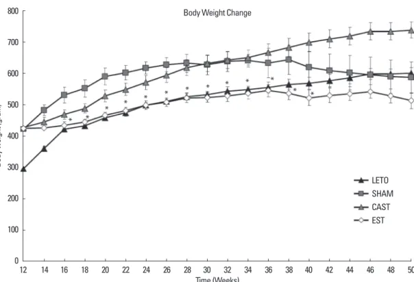
Figure 1은 LETO군, SHAM군, CAST군, EST군에서의 체중변화를 나타내었다. LETO군은꾸준히체중이증가하다체중이유지되었고
SHAM군은체중이증가하다고혈당이나타나는 38주이후체중이
감소하였다. CAST군은꾸준히체중이증가하였다. EST군은체중증가
500
400
300
200
100
00 30 60 90 120
Time (min)
Blood glucose (mg/dL)
LETO SHAM Castration Estrogen
*
*
* *
Fig. 2. Estradiol treated OLETF rats display improved glucose tolerance. *indi- cates P value < 0.05 when comparing estrogen treated OLETF rat group with sham operated OLETF rat group. Values are mean ± SE. n = 8 per group.
속도가현저히둔화되어 24주이후에는 LETO군과비슷하거나더낮
은체중을보였다. 38주에실시한복강내포도당부하검사결과 EST
군이 SHAM군보다유의하게당내성이호전됨을알수있었다(Fig. 2).
2. In vivo sample western blot analysis
본연구진이제작한 lipin-1 항체는 lipin-1이잘발현하지 않는
HeLa 세포에서는 lipin-1이검출되지않았고 lipin-1이상당량발현
하는것으로알려진뇌에서는 lipin-1이 100 kDa 근처에서잡히는
것을볼수있었다. LETO 쥐와 OLETF 쥐에서얻은피하지방과내장
지방에서단백질을분리한후 lipin-1 항체를이용하여 Western blot
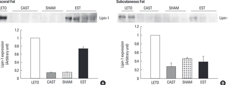
을시행한결과 Figure 3에서같이내장지방에서는 LETO 쥐에서발
현되는 lipin-1 단백이 SHAM군및 CAST군에서는 LETO군에비해 85% 감소하였고(P < 0.05) EST군에서 LETO군의 74%까지회복되는 것을알수있다(P < 0.05). 한편피하지방에서는 LETO군에 비해 SHAM군은 55% 감소(P < 0.05), CAST군은 72% 감소(P < 0.05), EST 군에서는 62% 감소(P < 0.05)되었다.
3. In vitro sample western blot analysis
일차인간지방전구세포를배양하면서에스트로겐을처리하여본 결과내장지방세포와피하지방세포모두에서지방세포로분화를시
작하면서 lipin-1의 발현이 증가하였다. 내장지방전구세포에서는
85% 증가하였고(P < 0.05) 피하지방전구세포에서는 30% 증가하였 다(P < 0.05). 내장지방세포에서 0.1 μM의에스트로겐을처리한경 우 lipin-1의발현이 10% 증가하였고(P < 0.05) 1 μM을처리한경우 에는 27% 감소되었다(P < 0.05). 피하지방세포에서 0.1 μM의에스트 로겐을처리한 경우 lipin-1의발현이 26% 감소하였고(P < 0.05) 1 μM을처리한경우에는 31% 감소되었다(P < 0.05)(Fig. 4).
Body weight (gram)
800
700
600
500
400
300
200
100
012 14 16 18 20 22 24 26 28 30 32 34 36 38 40 42 44 46 48 50 Time (Weeks)
Body Weight Change
* * * * * * * * * * *
* *
LETO SHAM CAST EST
Fig. 1. Body weight changes during experiment. LETO, CAST and EST group shows steadily increase in body weight while SHAM group shows decrease in body weight after 38 weeks. *indicates P value < 0.05 when comparing estrogen treated OLETF rat group with sham operated OLETF rat group. Values are mean ± SE. n=
8 per group.
고 찰
당뇨병동물모델인 OLETF 쥐는자연적으로제2형당뇨병이발생 하는동물로인간의제2형당뇨병과유사하게만성합병증이발생하
는데[13-15] 특징적으로 수컷에서만당뇨병이발병한다[16]. 이러한
효과는에스트로겐의효과로추측되나정확한기전은아직탐색되 지않았다. 본연구에서도에스트로겐투여군에서 SHAM군에비해 혈당이유의하게감소하였으나정상 LETO군수준으로는감소하지 않았다. 이러한결과는많은임상연구의 결과와도일치한다[9-11].
이러한효과는에스트로겐이직접근육에작용하여나타나는것은 아님이보고되었고[12,25] 간에서지방산대사를촉진[26,27], 간의포 도당생성을억제[25,28], 췌장베타세포기능보호[29,30] 및베타세포
사멸억제[31,32] 등의여러가지기전이복합적으로작용하여이루어
진것으로사료된다. 또한지방조직에서지방세포의분화를촉진시 키고[33-35] 지방분해(lipolysis)를억제[36]하는것역시에스트로겐
의혈당강하효과를설명해주는기전으로생각된다. 또한에스트 로겐은렙틴과유사한식욕억제효과가있어서[37] 체중을감소시키 며체중감소에따른이차적인효과로혈당이감소한다. 본연구에서 도에스트로겐투여에의해식이섭취량이감소되었으며이로인한 체중감소, 혈당강하가일어났을가능성을배제할수없다.
본연구에서는에스트로겐의지방세포의 lipin에대한영향에대 해연구하였는데 lipin은핵내단백질로지방세포분화에필수적인
인자로알려져있으며[17-19] 지방세포에서의 lipin의결핍은지방이
상증을유발해인슐린저항성을일으키며[19] 지방조직특이적으로
lipin을과발현시키면지방세포는증가하나인슐린저항성은호전되
는데이는지방의분화를촉진시켜서일어나는현상으로사료되고 있으며지방조직에서 lipin mRNA의발현정도는인슐린감수성정 도와비례한다고보고되었다[20,21]. Lipin-1은 mRNA의선택적스플 라이싱에의하여두가지형태로존재하는데다른기능을갖는다
[38]. 세포질에존재하는 lipin-1은효소역할을하여중성지방합성을
촉진시키는역할을하며[39,40] 핵내에위치한 lipin-1은전사과정에 부요소로작용하는데지방산산화에관여하는유전자의활성에관 여한다[41]. 즉 lipin-1은 peroxisome proliferator-activated receptor alpha (PPAR α)와 PPARγ coactivator 1α (PGC-1 α)에직접작용해지 방산산화유전자의활성을조절하는것으로알려져있다. 본연구에 서는지방조직에서 lipin-1 발현을보았다. 내장지방에서는 LETO 쥐 에서발현되는 lipin-1 단백이 SHAM군및 CAST군에서는 LETO군에 비해 85% 감소하였고(P < 0.05) EST군에서 LETO군의 74%까지회복 되는것을알수있다(P < 0.05). 그러나 피하지방에서는 EST군도 lipin-1 발현이 LETO군에비해유의하게감소되었다. 이러한결과는 에스트로겐이내장지방에서만 lipin-1의발현을증가시키는것을제 시하나정확한분자생물학적기전은향후연구되어야할것이다. Fig. 4. Lipin-1 expression in human primary visceral and subcutaneous adipo-
cytes. In visceral adipocyte, lipin-1 expression was increased by 10% with 0.1 μM estrogen and decreased 27% with 1 μM estrogen. In subcutaneous adipo- cyte, lipin-1 expression was decreased by 26% with 0.1 μM estrogen and de- creased 31% with 1 μM estrogen (n = 4).
Lipin-1
Actin
0 0.1 1 0 0.1 1
E2 (μM) E2 (μM)
Visceral Subcutaneous
No differentiation
No differentiation
Fig. 3. Protein expression of lipin-1 in visceral and subcutaneous adipose tissues of rats. A. In visceral fat: Lipin-1 expression was decreased by 85% in SHAM and CAST group comparing with LETO group. In EST group, lipin-1 level was restored to 74% of that of LETO group. B. In subcutaneous fat: Lipin-1 expression showed 55% decrease in SHAM group, 72% decrease in CAST group, and 62% decrease in EST group (n = 3-4 for each group).
Lipin-1 expression (Arbitrary unit) 1.2
1 0.8 0.6 0.4 0.2
0 LETO CAST SHAM EST
Lipin-1 LETO
Subcutaneous Fat
CAST SHAM EST
Lipin-1 expression (Arbitrary unit) 1.2
1 0.8 0.6 0.4 0.2
0 LETO CAST SHAM EST
Lipin-1 LETO
Visceral Fat
CAST SHAM EST
A B
SHAM군및 CAST군의내장지방세포에서 lipin-1의발현이감소 되어있는것은지방세포분화가덜되고늦게되어지방의저장이제 대로일어나지않아이소성지방침착을일으키는하나의기전이되 리라사료된다. 또한이소성지방침착및미성숙된지방세포에서나 오는아디포카인등은인슐린저항성증가, 혈당상승을 유발하는데 기여했을것으로사료된다. 에스트로겐을투여한 EST군의지방조직
에서는 lipin-1의발현이증가되었다. 이는에스트로겐을 투여하면
지방의분화가촉진되어이소성지방침착을감소시켜인슐린저항성 의감소, 혈당감소에역할을했을가능성을제시한다.
인간지방전구세포를이용한실험에서는내장지방세포에서는 0.1 μM 에스트로겐처리시 lipin-1의발현이증가하다 1 μM 처리시는 감소하는 양상을보였고피하지방세포에서는 에스트로겐처리 시 lipin-1의발현이감소하였다. In vivo 실험과 in vitro 실험의차이점
은 in vivo 실험에서는조직을얻은시점이비만과당뇨병과연관된
어떤병리기전에의해이미 lipin-1이감소되어있는또는제때증가되 지못하는상황에서에스트로겐이투여되었고, in vitro에서는정상 적으로지방세포로분화가일어나고있는상황에서에스트로겐이 추가로투여되었기때문에, 상대적으로 in vitro 실험에서는에스트
로겐의 lipin-1 매개효과가나타날수있는여지가상당히줄어들어
있다고 볼수있다. 또한, 내장지방세포에서 lipin-1을감소시킨 1 μM의농도는일반적인호르몬의유효 농도보다훨씬높기때문에 나타난역자U 약물반응으로유추가능하며, 향후실험에서는다양 한농도를시험함으로써유효농도를적정하는것이필요함을시사 한다고하겠다.
본연구의제한점으로는에스트로겐투여동물에서에스트로겐 의혈중농도는측정하지않아효과적으로에스트로겐의투여가이 루어졌는지판단하기어려운점이있으나본연구와동일한조건및 방법으로실험하여혈중농도를측정한다른연구가있다[42]. 또한 실험방법상의문제로 mRNA 발현검사에모든샘플을사용하지못 한한계점이있다.
결론적으로본연구에서는 OLETF 쥐에서 17-베타에스트라디올 을장기간투여한결과체중증가정도가감소되었고포도당부하검 사에서혈당상승정도가유의하게적었으며지방세포에서 lipin-1의 발현이증가됨을보였다. 이는향후에스트로겐의지방세포분화및 이소성지방침착에대한연구를위한기초적자료가될것으로기대 한다.
요 약
배경: 17베타-에스트라디올이포도당 항상성유지에중요한역할
을한다고사료되고있으나명확한기전은밝혀지지않았다. Lipin은
핵내단백질로지방세포분화에있어서필수적인인자로알려져있 으며이소성지방침착과지방의재분포에관여할것으로사료된다.
본연구에서는 17-베타에스트라디올이당뇨병동물모델인 OLETF 쥐에서지방세포의 lipin 발현에미치는영향을분석하고자하였다.
방법:생후 6주에 OLETF 쥐를 3군으로나누어한군은겉보기수
술을시행하고(SHAM), 다른한군에서는거세하고(CAST) 나머지
한군에서는거세한후에스트로겐치료를하였다(EST). LETO 쥐 8 마리를대조군(LETO)으로사용하였다. EST군에서는 estradiol val- erate를 0.1 mg을한달에 1회 6주부터 42주까지피하주사를실시 하였다.
결과: SHAM군에서는체중이증가하다고혈당증이나타나는 30
주이후체중증가정도가감소되었고 CAST군에서는지속적으로체 중이증가하였다. EST군에서는체중증가속도가느려져서대조군인 LETO군과비슷한수준으로체중이유지되었다. EST군에서포도당 부하검사상혈당상승정도가유의하게적었다. 내장지방세포에서의 lipin-1의발현은 SHAM군, CAST군에서는감소하였으나 EST군에서 는 회복됨이관찰되었다. 피하지방세포에서의 lipin-1의발현은 SHAM군, CAST군, EST군모두에서감소하였다.
결론: OLETF 쥐에서 17-베타에스트라디올을장기간투여한결과
체중증가정도가감소되었고포도당부하검사에서혈당상승정도가 유의하게적었으며내장지방세포에서 lipin-1의발현을회복시켜주 었으나이러한효과는피하지방세포에서는관찰되지않았다.
참고문헌
1. King H, Aubert RE, Herman WH: Global burden of diabetes, 1995-2025:
prevalence, numerical estimates, and projections. Diabetes Care 21:1414- 1431, 1998
2. Wild S, Roglic G, Green A, Sicree R, King H: Global prevalence of diabe- tes: estimates for the year 2000 and projections for 2030. Diabetes Care 27:
1047-1053, 2004
3. McGarry JD: Banting lecture 2001: dysregulation of fatty acid metabolism in the etiology of type 2 diabetes. Diabetes 51:7-18, 2002
4. Gale EA, Gillespie KM: Diabetes and gender. Diabetologia 44:3-15, 2001 5. Espeland MA, Hogan PE, Fineberg SE, Howard G, Schrott H, Waclawiw
MA, Bush TL: Effect of postmenopausal hormone therapy on glucose and insulin concentrations. PEPI Investigators. Postmenopausal Estrogen/Pro- gestin Interventions. Diabetes Care 21:1589-1595, 1998
6. Crespo CJ, Smit E, Snelling A, Sempos CT, Andersen RE; NHANES III:
Hormone replacement therapy and its relationship to lipid and glucose me- tabolism in diabetic and nondiabetic postmenopausal women: results from the Third National Health and Nutrition Examination Survey (NHANES III). Diabetes Care 25:1675-1680, 2002
7. Saglam K, Polat Z, Yilmaz MI, Gulec M, Akinci SB: Effects of postmeno- pausal hormone replacement therapy on insulin resistance. Endocrine 18:
211-214, 2002
8. Li C, Samsioe G, Borgfeldt C, Bendahl PO, Wilawan K, Aberg A: Low- dose hormone therapy and carbohydrate metabolism. Fertil Steril 79:550- 555, 2003
9. Andersson B, Mattsson LA, Hahn L, Mårin P, Lapidus L, Holm G, Bengts-
son BA, Björntorp P: Estrogen replacement therapy decreases hyperandro- genicity and improves glucose homeostasis and plasma lipids in postmeno- pausal women with noninsulin-dependent diabetes mellitus. J Clin Endo- crinol Metab 82:638-643, 1997
10. Brussaard HE, Gevers Leuven JA, Frölich M, Kluft C, Krans HM: Short- term oestrogen replacement therapy improves insulin resistance, lipids and fibrinolysis in postmenopausal women with NIDDM. Diabetologia 40:
843-849, 1997
11. Friday KE, Dong C, Fontenot RU: Conjugated equine estrogen improves glycemic control and blood lipoproteins in postmenopausal women with type 2 diabetes. J Clin Endocrinol Metab 86:48-52, 2001
12. Louet JF, LeMay C, Mauvais-Jarvis F: Antidiabetic actions of estrogen: in- sight from human and genetic mouse models. Curr Atheroscler Rep 6:
180-185, 2004
13. Takiguchi S, Takata Y, Takahashi N, Kataoka K, Hirashima T, Kawano K, Miyasaka K, Funakoshi A, Kono A: A disrupted cholecystokinin A recep- tor gene induces diabetes in obese rats synergistically with ODB1 gene.
Am J Physiol 274:E265-E270, 1998
14. Kawano K, Hirashima T, Mori S, Natori T: OLETF (Otsuka Long-Evans Tokushima Fatty) rat: a new NIDDM rat strain. Diabetes Res Clin Pract 24:S317-S320, 1994
15. Kawano K, Hirashima T, Mori S, Saitoh Y, Kurosumi M, Natori T: Spon- taneous long-term hyperglycemic rat with diabetic complications. Otsuka Long-Evans Tokushima Fatty (OLETF) strain. Diabetes 41:1422-1428, 1992
16. Shi K, Mizuno A, Sano T, Ishida K, Shima K: Sexual difference in the in- cidence of diabetes mellitus in Otsuka-Long-Evans-Tokushima-Fatty rats:
effects of castration and sex hormone replacement on its incidence. Metab- olism 43:1214-1220, 1994
17. Phan J, Peterfy M, Reue K: Lipin expression preceding peroxisome prolif- erator-activated receptor-gamma is critical for adipogenesis in vivo and in vitro. J Biol Chem 279:29558-29564, 2004
18. Phan J, Peterfy M, Reue K: Biphasic expression of lipin suggests dual roles in adipocyte development. Drug News Perspect 18:5-11, 2005
19. Peterfy M, Phan J, Xu P, Reue K: Lipodystrophy in the fld mouse results from mutation of a new gene encoding a nuclear protein, lipin. Nat Genet 27:121-124, 2001
20. Xu J, Lee WN, Phan J, Saad MF, Reue K, Kurland IJ: Lipin deficiency impairs diurnal metabolic fuel switching. Diabetes 55:3429-3438, 2006 21. Yao-Borengasser A, Rasouli N, Varma V, Miles LM, Phanavanh B, Starks
TN, Phan J, Spencer HJ 3rd, McGehee RE Jr, Reue K, Kern PA: Lipin expression is attenuated in adipose tissue of insulin-resistant human sub- jects and increases with peroxisome proliferator-activated receptor gamma activation. Diabetes 55:2811-2818, 2006
22. van Harmelen V, Rydén M, Sjölin E, Hoffstedt J: A role of lipin in human obesity and insulin resistance: relation to adipocyte glucose transport and GLUT4 expression. J Lipid Res 48:201-206, 2007
23. Tomiyoshi Y, Sakemi T, Aoki S, Miyazono M: Different effects of castration and estrogen administration on glomerular injury in spontaneously hyper- glycemic Otsuka Long-Evans Tokushima Fatty (OLETF) rats. Nephron 92:
860-867, 2002
24. Ocaña A, Gómez-Asensio C, Arranz-Gutiérrez E, Torres C, Señorans FJ, Reglero G: In vitro study of the effect of diesterified alkoxyglycerols with conjugated linoleic acid on adipocyte inflammatory mediators. Lipids Health
Dis 9:36, 2010
25. Mauvais-Jarvis F, Kulkarni RN, Kahn CR: Knockout models are useful tools to dissect the pathophysiology and genetics of insulin resistance. Clin Endocrinol (Oxf) 57:1-9, 2002
26. Nemoto Y, Toda K, Ono M, Fujikawa-Adachi K, Saibara T, Onishi S, Enzan H, Okada T, Shizuta Y: Altered expression of fatty acid-metabolizing en- zymes in aromatase-deficient mice. J Clin Invest 105:1819-1825, 2000 27. Hewitt KN, Pratis K, Jones ME, Simpson ER: Estrogen replacement re-
verses the hepatic steatosis phenotype in the male aromatase knockout mouse. Endocrinology 145:1842-1848, 2004
28. Cagnacci A, Soldani R, Carriero PL, Paoletti AM, Fioretti P, Melis GB: Ef- fects of low doses of transdermal 17 beta-estradiol on carbohydrate metab- olism in postmenopausal women. J Clin Endocrinol Metab 74:1396-1400, 1992
29. Paik SG, Michelis MA, Kim YT, Shin S: Induction of insulin-dependent diabetes by streptozotocin. Inhibition by estrogens and potentiation by androgens. Diabetes 31:724-729, 1982
30. Puah JA, Bailey CJ: Insulinotropic effect of ovarian steroid hormones in streptozotocin diabetic female mice. Horm Metab Res 17:216-218, 1985 31. Contreras JL, Smyth CA, Bilbao G, Young CJ, Thompson JA, Eckhoff DE:
17beta-Estradiol protects isolated human pancreatic islets against proin- flammatory cytokine-induced cell death: molecular mechanisms and islet functionality. Transplantation 74:1252-1259, 2002
32. Zhu M, Mizuno A, Kuwajima M, Ogino T, Murakami T, Noma Y, Sano T, Shima K: Ovarian hormone-induced beta-cell hypertrophy contributes to the homeostatic control of beta-cell mass in OLETF female rat, a model of Type II diabetes. Diabetologia 41:799-805, 1998
33. Heine PA, Taylor JA, Iwamoto GA, Lubahn DB, Cooke PS: Increased adi- pose tissue in male and female estrogen receptor-alpha knockout mice. Proc Natl Acad Sci U S A 97:12729-12734, 2000
34. Jones ME, Thorburn AW, Britt KL, Hewitt KN, Wreford NG, Proietto J, Oz OK, Leury BH, Robertson KM, Yao S, Simpson ER: Aromatase-defi- cient (ArKO) mice have a phenotype of increased adiposity. Proc Natl Acad Sci U S A 97:12735-12740, 2000
35. Misso ML, Murata Y, Boon WC, Jones ME, Britt KL, Simpson ER: Cellu- lar and molecular characterization of the adipose phenotype of the aroma- tase-deficient mouse. Endocrinology 144:1474-1480, 2003
36. O’Sullivan AJ, Ho KK: A comparison of the effects of oral and transder- mal estrogen replacement on insulin sensitivity in postmenopausal women.
J Clin Endocrinol Metab 80:1783-1788, 1995
37. Gao Q, Mezei G, Nie Y, Rao Y, Choi CS, Bechmann I, Leranth C, Toran- Allerand D, Priest CA, Roberts JL, Gao XB, Mobbs C, Shulman GI, Dia- no S, Horvath TL: Anorectic estrogen mimics leptin’s effect on the rewir- ing of melanocortin cells and Stat3 signaling in obese animals. Nat Med 13:89-94, 2007
38. Peterfy M, Phan J, Reue K: Alternatively spliced lipin isoforms exhibit distinct expression pattern, subcellular localization, and role in adipogene- sis. J Biol Chem 280:32883-32889, 2005
39. Carman GM, Han GS: Phosphatidic acid phosphatase, a key enzyme in the regulation of lipid synthesis. J Biol Chem 284:2593-2597, 2009 40. Reue K, Brindley DN: Thematic Review Series: Glycerolipids. Multiple
roles for lipins/phosphatidate phosphatase enzymes in lipid metabolism. J Lipid Res 49:2493-2503, 2008
41. Finck BN, Gropler MC, Chen Z, Leone TC, Croce MA, Harris TE, Law-
rence JC Jr, Kelly DP: Lipin 1 is an inducible amplifier of the hepatic PGC- 1alpha/PPARalpha regulatory pathway. Cell Metab 4:199-210, 2006 42. Ohtsuka N, Sakemi T, Tomiyoshi Y, Morito F: Attenuating effect of castra-
tion or estrogen administration on glomerular injury in adriamycin-induced nephropathy. Nephrology 2:45-52, 1996