pISSN 2093-596X · eISSN 2093-5978
Article
The Relationship of Body Composition and Coronary Artery Calcification in Apparently Healthy Korean Adults
Jung-Hee Yu, Seo Hyoung Yim, Su Hyeon Yu, Ji Yong Lee, Jong Dae Kim, Mi Hae Seo, Won Seon Jeon, Se-Eun Park, Cheol-Young Park, Won-Young Lee, Ki-Won Oh, Sung-Woo Park, Eun-Jung Rhee
Division of Endocrinology, Department of Internal Medicine, Kangbuk Samsung Hospital, Sungkyunkwan University School of Medicine, Seoul, Korea
Background: We investigated the association of coronary artery calcium score (CACS) with body composition and insulin resis- tance in apparently healthy Korean adults.
Methods: Nine hundred forty-five participants (mean age, 48.9 years; 628 men) in a medical check-up program were selected for analysis. Body composition was assessed by bioelectrical impedance analysis (BIA). Insulin resistance was evaluated using the ho- meostasis model assessment of insulin resistance (HOMA-IR). The CACS was assessed by multidetector computed tomography.
Results: One hundred forty-six subjects (15.4%) showed coronary artery calcification and 148 subjects (15.7%) had metabolic syndrome. CACS showed a significant positive correlation with age, fasting glucose level, waist circumference (WC), blood pressure, hemoglobin A1c, HOMA-IR, and waist-hip ratio (WHR) assessed by BIA. CACS had a negative correlation with high density lipoprotein cholesterol (HDL-C). Subjects with high CACS showed significantly higher mean WHRs and lower mean values for lean body mass compared with subjects without coronary artery calcification. In logistic regression analyses with coro- nary artery calcification as the dependent variable, the highest quartile of WHR showed a 3.125-fold increased odds ratio for cor- onary artery calcification compared with the lowest quartile after adjustment for confounding variables. When receiver operating characteristics analyses were performed with coronary artery calcification as the result variable, WHR showed the largest area under the curve (AUC) value among other variables except for age and WC in women (AUC=0.696 for WHR, 0.790 for age, and 0.719 for WC in women).
Conclusion: In our study population of apparently healthy Korean adults, WHR was the most significant predictor for coronary artery calcification among other confounding factors, suggesting that it may have implication as a marker for early atherosclerosis.
Keywords: Coronary artery calcium score; Waist-hip ratio; Obesity, abdominal
INTRODUCTION
Adipose tissue is not simply a mass of fat, but an active endo- crine organ that modifies the metabolic status of the human body [1]. Recent studies emphasize the importance of depots
where fat is accumulated rather than the simple fat mass, and visceral obesity is known to be a strong risk factor for meta- bolic and subsequent cardiovascular disease (CVD) [2].
Early detection of subclinical atherosclerosis is important to prevent overt atherosclerosis [3]. Coronary artery calcium
Received: 27 November 2012, Accepted: 4 February 2013 Corresponding author: Eun-Jung Rhee
Division of Endocrinology, Department of Internal Medicine,
Kangbuk Samsung Hospital, Sungkyunkwan University School of Medicine, 29 Saemunan-ro, Jongno-gu, Seoul 110-746, Korea
Tel: +82-2-2001-2485, Fax: +82-2-2001-1588, E-mail: hongsiri@hanmail.net
Copyright © 2013 Korean Endocrine Society
This is an Open Access article distributed under the terms of the Creative Com- mons Attribution Non-Commercial License (http://creativecommons.org/
licenses/by-nc/3.0/) which permits unrestricted non-commercial use, distribu- tion, and reproduction in any medium, provided the original work is properly cited.
score (CACS) is a risk marker for atherosclerosis and is posi- tively associated with CVD events [4]. The development of multislice computed tomography enabled the reliable detec- tion and quantification of CAC that correlates with the overall atherosclerotic plaque burden [5]. There are studies reporting that visceral adiposity and insulin resistance are significant risk factors for the development of subclinical atherosclerosis, as assessed by CACS, in apparently healthy subjects without previous history of CVD [6-9]. In addition, a few studies have reported an association between body composition and the de- velopment of metabolic and CVDs [10,11]. The importance of strategies to lessen abdominal adiposity, modify the body composition, and improve insulin resistance would be of great benefit to prevent atherosclerosis even in healthy subjects without documented CVD.
The aim of this study was to analyze the relationship be- tween body composition assessed by bioelectric impedance analysis (BIA) and coronary artery calcification assessed by multidetector computed tomography (MDCT) in asymptomat- ic healthy Korean adults.
METHODS
Study participants
Among the subjects who participated in the medical check-up program at Kangbuk Samsung Hospital’s Health Promotion Center from 2007 through 2009, 945 subjects (mean age, 48.9 years; range, 28 to 82 years; 628 men, 66.5%) with CACS data were selected for analysis. The study protocol conforms to ethical guidelines of the 1975 Declaration of Helsinki, and accordingly the Kangbuk Samsung Hospital Human Research Committee approved it. The informed consent requirement for this study was deemed exempt by the Institutional Review Board at the time the study was in the planning phase because researchers only accessed the database, which was free of identifying personal information, for analysis purposes.
Clinical and laboratory measurement
Height, weight, systolic and diastolic blood pressures were measured in duplicate and the results were averaged. The blood pressures were taken with a standardized sphygmomanometer after at least 5 minutes of rest, according to the Hypertension Detection and Follow-up Program protocol [12]. The body mass index (BMI) was calculated by dividing the patient’s weight (kg) by the height (m) squared. The waist circumfer- ence (WC) was measured in the standing position, at the mid-
point between the anterior iliac crest and lower border of the last palpable rib by a single examiner.
After 12 hours of fasting, fasting blood glucose, total cho- lesterol, triglyceride, high density lipoprotein cholesterol (HDL-C), and low density lipoprotein cholesterol (LDL-C) levels were checked. The hexokinase method (Advia 1650 Autoanalyzer, Bayer Diagnostics, Leverkusen, Germany) was used to measure blood glucose levels and an enzymatic colori- metric test was used to measure total cholesterol and triglycer- ide levels. The selective inhibition method was used to mea- sure the level of HDL-C and a homogeneous enzymatic calo- rimetric test was used to measure the level of LDL-C. Serum insulin concentrations were measured with an immunoradio- metric assay (INS-Irma, Biosource, Nivelles, Belgium), with intra and interassay coefficients of variation of 1.6% to 2.2%
and 6.1% to 6.5%, respectively.
Hemoglobin A1c (HbA1c) was measured by immunoturbi- dimetric assay with a Cobra Integra 800 automatic analyzer (Roche Diagnostics, Basel, Switzerland) with a reference val- ue of 4.4% to 6.4%. The methodology was aligned with the Diabetes Control and Complications Trial and National Gly- cohemoglobin Standardization Program (NGSP) standards [13]. The intra-assay coefficient of variation (CV) was 2.3%
and interassay CV was 2.4%, both within the NGSP accept- able limits [14]. The glycemic statuses of the participants were determined according to the self-questionnaire of the partici- pants and the American Diabetes Association’s diagnostic cri- teria [15].
Assessment of insulin resistance was calculated by homeo- stasis model assessment of insulin resistance (HOMA-IR) [16]: HOMA-IR=fasting insulin (μU/mL)×fasting glucose (mmol/L)/22.5.
Body composition analyses by bioelectrical impedance analyses
Body composition measurements were carried out by segmen- tal bioelectric impedance, using eight tractile electrodes ac- cording to the manufacturer’s instructions (InBody 3.0, Bio- space, Seoul, Korea). Lean mass (kg), fat mass (kg), percent fat mass (%), and waist-hip ratio (WHR) as a marker of ab- dominal obesity, were measured.
Measurement of CACS
The CAC imaging was performed using MDCT (Philips Bril- liance 40 slice, Amsterdam, The Netherlands). The 40-slice MDCT was performed using the following protocol: 0.625 mm
slice thickness, 120 kVp, 800 effective mAs, and a 0.4-second gantry rotation speed. CACS were expressed in Agatston units and CACS [17] as significant or CAC positive status. Having coronary artery calcification was defined as having CACS>0.
Subjects were divided into three groups according to CACS;
group with CACS=0, 1<CACS<100, and CACS≥100. The reason for dividing the subjects into three groups instead or four or five groups as with previous studies [17], was because there were too few subjects with CACS higher than 1 (n=146, 15.4%).
Statistical analyses
All data are presented as the mean and standard deviation, and were analyzed using IBM SPSS version 18.0 for Windows (IBM, Armonk, NY, USA). Bivariate correlation analyses be- tween coronary calcium score and the other variables were performed using Pearson’s correlation analysis. Comparison of the parameters among three groups divided by CACS was analyzed by one-way analysis of variance, and post-hoc analy- ses were performed with Tukey’s method. Multiple logistic re- gression analyses were performed with coronary calcification as the dependent variable with other confounding variables in- cluded in the model. The variables included in the model were selected according to the results of bivariate correlation analy- ses. Receiver operating characteristics (ROC) curve analyses were performed to calculate the area under the curve (AUC) and the cutoff of each variable for coronary artery calcifica- tion. A P value <0.05 was considered statistically significant.
RESULTS
Baseline characteristics of the participants are listed in Table 1.
Mean age of the study sample was 48.9 years; 66.5% were men. One hundred forty-six subjects (15.4%) showed signifi- cant coronary artery calcification with a CACS greater than 0.
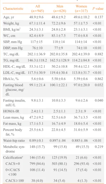
Seven hundred ninety-nine subjects (84.6%) were normal, 108 subjects (11.4%) had a CACS less than 100, and 38 subjects (4.0%) had a CACS larger than or equal to 100 (Table 1). As the subjects got older, the mean CACS linearly increased (Supplemental Table S1 online, Fig. 1). Six hundred and four- teen subjects (65%) showed normoglycemia, 274 subjects (29.0%) showed impaired fasting glucose, and 57 subjects (6.0%) had diabetes. Men were more obese than women, but showed a higher lean body mass and less body fat compared with women. Women showed a lower prevalence for coronary artery calcification compared with men (Table 1).
In bivariate correlation analysis, CACS showed a signifi- cant positive correlation with age, WC, blood pressure, HbA1c, fasting blood sugar (FBS), HOMA-IR, and WHR, and a negative correlation with HDL-C (Table 2). In women, CACS also showed a positive correlation with body fat mass.
When the subjects were divided into three groups according to the CACS, subjects were older as the mean CACS increased
Table 1. General Characteristics of the Participants Characteristic All
(n=945) Men
(n=628) Women
(n=317) P value Age, yr 48.9±9.6 48.6±9.2 49.6±10.2 0.137 Weight, kg 67.1±11.4 72.2±9.6 57.1±7.5 <0.01 BMI, kg/m2 24.3±3.1 24.8±2.8 23.1±3.1 <0.01 WC, cm 82.4±8.9 85.1±7.5 77.0±8.8 <0.01 SBP, mm Hg 117±15 118±14 114±14 <0.01
DBP, mm Hg 76±10 77±9 74±10 <0.01
TC, mg/dL 202.1±36.9 202.0±35.8 202.4±39.0 0.882 TG, mg/dL 146.3±118.2 162.5±128.9 114.2±84.8 <0.01 HDL-C, mg/dL 53.3±12.1 50.2±10.8 59.4±12.1 <0.01 LDL-C, mg/dL 117.5±30.9 119.4±30.4 113.8±31.7 <0.01
HbA1c, % 5.6±0.6 5.58±0.6 5.59±0.6 0.862
Fasting blood glucose, mg/
dL
99.1±21.4 100.1±22.1 97.0±20.0 0.032
Fasting insulin,
mIU/L 9.8±3.1 10.0±3.3 9.6±2.6 0.040
HOMA-IR 2.4±1.1 2.5±1.1 2.3±.9 <0.01 Lean mass, kg 47.2±9.2 52.5±6.0 36.7±3.5 <0.01 Fat mass, kg 17.1±5.1 16.7±4.9 18.0±5.4 <0.01 Percent body
fat, % 25.5±6.3 22.8±4.5 31.0±5.9 <0.01 Waist-hip ratio 0.89±0.1 0.897±.04 0.885±.06 <0.01 Metabolic syn-
drome 148 (15.7) 99 (15.8) 49 (15.5) 0.219 Calcificationa 146 (15.4) 125 (19.9) 21 (6.6) <0.01
CACS=0 799 (84.6) 503 (80.1) 296 (93.4) <0.01 0<CACS
<100 108 (11.4) 91 (14.5) 17 (5.4) <0.01 CACS≥100 38 (4.0) 34 (5.4) 4 (1.3) <0.01 Values are expressed as mean±SD or number (%).
BMI, body mass index; WC, waist circumference; SBP, systolic blood pressure; DBP, diastolic blood pressure; TC, total cholesterol; TG, tri- glyceride; HDL-C, high density lipoprotein cholesterol; LDL-C, low density lipoprotein cholesterol; HbA1c, hemoglobin A1c; HOMA-IR, homeostasis model assessment of insulin resistance; CACS, coronary artery calcium score.
aHaving coronary artery calcification was defined by CACS>0.
from 0 to greater than 100 (Table 2). Mean values for body weight, BMI, WC, blood pressure, HbA1c, FBS, HOMA-IR, and WHR became higher and mean values for HDL-C became lower as the mean CACS increased from 0 to that greater than 100 (Table 3). In all groups, the mean value for lean body mass became lower as the mean CACS increased from 0 to that greater than 100; however, when the analysis was per- formed only in men, the mean value for lean body mass was significantly lower in subjects with a CACS higher than 0 and lower than 100 compared with subjects without coronary ar- tery calcification (Supplemental Table S2 online). In women, mean lean body mass did not show any significant differences among the three groups (Supplemental Table S3 online). In contrast, for total body fat and percent body fat, subjects with coronary artery calcification showed significantly increased mean values as the CACS increased. Men did not show any differences among the three groups regarding total fat mass or percent body fat (Supplemental Table S2 online). Mean WHR significantly increased as the CACS increased from 0 to great- er than 100 even when the analyses were performed separately for different genders (Supplemental Tables S2, S3 online).
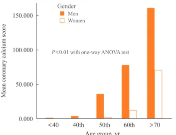
When the subjects were divided into four groups according to WHR, mean CACS was significantly higher in the 4th quar- tile group compared to the 1st group in men (Supplemental Table S4 online, Fig. 2A). In women, mean values of CACS increased linearly from 1st to 4th quartile groups with signifi- cant differences between that of 1st quartile group and those of the other three quartile groups in post-hoc analyses (Sup- plemental Table S4 online, Fig. 2B).
In logistic regression analyses with coronary artery calcifi-
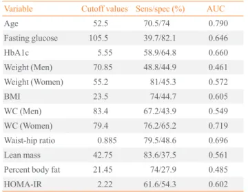
cation as the dependent variable, the 4th quartile group of WHR showed a 3.125-fold increased risk for coronary artery calcification compared to the lowest quartile group after ad- justment for other confounding variables (Table 4). When ROC curve analyses were performed with coronary artery cal- cification as the result variable, WHR showed the largest AUC value among other variables except for age and WC in women (AUC=0.696 for WHR, 0.790 for age and 0.719 for WC in women) (Table 5).
DISCUSSION
In this study, abdominal obesity assessed by WHR showed a significant positive correlation with CACS. The subjects with high CACS showed significantly worse metabolic parameters Table 2. Bivariate Correlation of Coronary Calcium Score with Various Parameters
Parameter
Correlation coefficients All subjects
(n=945) Men
(n=628) Women (n=317)
Age, yr 0.277a 0.320a 0.262a
Weight, kg 0.026 -0.069 0.057
BMI, kg/m2 0.019 -0.045 0.151a
WC, cm 0.068a 0.008 0.115a
SBP, mm Hg 0.126a 0.117a 0.139a
DBP, mm Hg 0.097a 0.089a 0.083
TC, mg/dL 0.008 -0.002 0.061
TG, mg/dL 0.036 0.011 0.058
HDL-C, mg/dL -0.073a -0.047 -0.017
LDL-C, mg/dL 0.028 0.012 0.063
HbA1c, % 0.146a 0.168a 0.097
FBS, mg/dL 0.140a 0.147a 0.102
Fasting insulin, mIU/L 0.002 -0.015 0.064
HOMA-IR 0.075a 0.066 0.093
Lean mass, kg 0.045 -0.074 -0.052
Fat mass, kg -0.024 -0.040 0.118a
Percent body fat, % -0.050 -0.012 0.147a
Waist-hip ratio 0.107a 0.076 0.244a
BMI, body mass index; WC, waist circumference; SBP, systolic blood pressure; DBP, diastolic blood pressure; TC, total cholesterol; TG, tri- glyceride; HDL-C, high density lipoprotein cholesterol; LDL-C, low density lipoprotein cholesterol; HbA1c, hemoglobin A1c; FBS, fast- ing blood sugar; HOMA-IR, homeostasis model assessment of insulin resistance.
aP<0.05 in bivariate correlation analyses with Pearson’s correlation analyses.
Fig. 1. The distribution of coronary calcium score according to age group.
150.000
100.000
50.000
0.000
<40 40th 50th 60th >70 Age group, yr
Mean coronary calcium score
P<0.01 with one-way ANOVA test Gender
MenWomen
compared with those with low CACS, supporting the role of metabolic parameters in the development of subclinical ath- erosclerosis. In men, mean lean mass was significantly higher in subjects without coronary artery calcification compared to those with significant coronary artery calcification. In con- trast, women showed a significant positive correlation be- tween CACS and fat mass. In both genders, higher WHR
showed an increased risk for coronary artery calcification, and WHR showed the highest AUC for the prediction of coronary calcification except for age and WC in women, suggesting that WHR plays a significant role as a marker for early atheroscle- rosis. This is the first study performed in Koreans regarding the relationship between body composition and coronary ar- tery calcification.
In this study, abdominal obesity assessed by WHR showed the most significant correlation with coronary artery calcifica- tion among the measured components of body composition analysis. This is in line with the result of a previous study [11].
The Dallas Heart Study analyzed the relationship between CACS and differing methods of measuring obesity in 2,744 multi-ethnic adults. They discovered that WHR was indepen- Table 3. Comparisons of the Mean Values of Parameters Ac-
cording to Calcification Status in all subjects Parameter CACS=0
(n=799)
0<CACS
<100 (n=108)
CACS≥100
(n=38) P
valuea Age, yr 47.4±8.7b 56.0±9.3c 61.8±8.5d <0.01 Weight, kg 66.7±11.6 68.9±9.7 70.8±10.4 0.025 BMI, kg/m2 24.1±3.1 25.0±2.6b 25.4±3.0b 0.001 WC, cm 81.8±9.0 85.2±7.0b 87.4±8.0b <0.01 SBP, mm Hg 115.7±14.3 120.6±15.9b 125.0±12.1b <0.01 DBP, mm Hg 75.2±9.5b 77.5±9.4 80.0±8.3c 0.001 TC, mg/dL 201.5±36.8 206.6±37.2 202.5±37.4 0.402 TG, mg/dL 144.7±121.5 145.5±74.4 183.9±143.5 0.135 HDL-C, mg/
dL 53.8±12.2 51.3±11.4 47.8±9.7 0.002
LDL-C, mg/dL 116.6±30.8 123.7±30.8 118.6±33.8 0.083 HbA1c, % 5.5±0.5 5.9±0.9b 6.1±1.1b <0.01 Fasting glu-
cose, mg/dL 97.1±17.4b 106.6±28.6c 118.7±46.5d <0.01 Fasting insu-
lin, IU/L 9.8±3.1 10.2±3.1 10.3±3.1 0.301 HOMA-IR 2.4±1.0 2.7±1.2b 3.0±1.7b <0.01 Lean body
mass, kg 46.9±9.4 48.6±7.5 50.0±8.0 0.029 Total body fat,
kg 17.0±5.1 17.4±4.9 17.9±4.8 0.505
Percent body
fat, % 25.6±6.4 25.2±6.1 25.2±6.0 0.795 Waist-hip ratio 0.89±0.05 0.92±0.04b 0.93±0.04b <0.01 Values are expressed as mean±standard deviation.
CACS, coronary artery calcification score; BMI, body mass index;
WC, waist circumference; SBP, systolic blood pressure; DBP, diastol- ic blood pressure; TC, total cholesterol; TG, triglyceride; HDL-C, high density lipoprotein cholesterol; LDL-C, low density lipoprotein cholesterol; HbA1c, hemoglobin A1c; HOMA-IR, homeostasis model assessment of insulin resistance.
aP values for the comparison of the mean values among the three groups divided according to the CACS analyzed by one-way analysis of variance test; b,c,dSame or different letters denote significant or no differences between the designated groups in post-hoc analysis. If not designated, they are the opposite of the designated groups.
Fig. 2. Mean coronary calcium score in groups according to quar- tiles of waist-hip ratio in (A) men and (B) women. (A) Different letters denote significant differences between the groups in post- hoc analysis, and (B) different letters denote significant differenc- es from 1st quartile group in post-hoc analysis.
30.000
20.000
10.000
0.000
1 2 3 4 Quartile groups of waist-hip ratio
Mean coronary calcium score
P=0.012 in one-way ANOVA test
a
b
A 20.000
15.000 10.000 5.000
0.000
1 2 3 4 Quartile groups of waist-hip ratio
Mean coronary calcium score
P=0.006 in one-way ANOVA test
a
b
c
B
dently associated with prevalent coronary artery calcification and that WHR was a more accurate predictor than either BMI or WC. In the Jackson Heart Study performed in 2,884 sub- jects who underwent noncontrast CT, visceral adipose tissue was positively associated with CAC, but this association was diminished with multivariable adjustment [8]. Additionally, in a study performed in 321 Japanese subjects, Ohashi et al. [7]
showed that visceral adiposity was significantly associated with CAC as a marker of subclinical atherosclerosis. From these previous studies and the results of our study, we can con- clude that abdominal adiposity, especially assessed by WHR rather than overall fat mass, contributes more to the develop- ment of subclinical atherosclerosis in apparently healthy adults.
The mechanistic reason for the association of visceral fat with atherosclerosis is explained fully in the previous litera- ture. Although obesity as defined by BMI is undoubtedly as- sociated with an increased cardiovascular risk, recent studies have emphasized on the metabolic differences between differ- ent types of obese subjects. For example, there are suggestions for a subset of obese subjects who are obese but metabolically healthy [18]. The main differences between the “metabolically healthy obese” and “metabolically unhealthy obese” subjects are in their fat depots. “Metabolically healthy obese” subjects have BMI levels similar to their metabolically unhealthy
peers, but they tend to accumulate fat less in the abdomen, and more subcutaneously [19]. Therefore, in a sense, abdominal obesity in the absence of increased BMI, could negatively af- fect metabolic status and thus, vascular health. There is al- ready strong consensus on the deleterious effects of visceral fat accumulation on the development of subclinical athero- sclerosis and subsequently increased CVD risk [2].
In this study, the effect of body composition on CACS was different between genders. Male subjects showed a negative correlation between lean body mass and coronary artery calci- fication, and women showed a positive correlation between body fat mass and coronary artery calcification. In the study by Alexandersen et al. [10], peripheral lean mass showed a strong and independent inverse association with aortic calcifi- cation assessed by lateral radiograph. Although the significant association of sarcopenia with CVD is not clear yet, recent studies strongly suggest the possible role of lack of muscle mass in the development of metabolic diseases [20]. The rea- son for the different correlation of CACS with different com- ponents of body composition between genders might be due to the role of sex hormones. In addition, since most of the female participants in this study were premenopausal, the degree of atherosclerosis might be very low, reflected in the significantly lower mean CACS in women compared with men. Also, the vasculoprotective effects of estrogen might still be present in these women, suggesting the level of estrogen might correlate Table 4. Logistic Regression Analyses with Coronary Artery
Calcification as the Dependent Variable
Model P value Exp (B) 95% CI
Lower Upper
Age <0.01 1.135 1.108 1.163
Gender <0.01 0.169 0.095 0.302
Fasting blood glucose 0.001 1.013 1.005 1.021 Systolic blood pressure 0.184 1.010 0.995 1.024 Percent body fat 0.541 0.976 0.905 1.054
Lean body mass 0.699 0.992 0.950 1.035
Waist-hip ratio 1st Q 0.163 1.000 - -
Waist-hip ratio 2nd Q 0.073 2.546 0.917 7.070 Waist-hip ratio 3rd Q 0.115 2.306 0.815 6.524 Waist-hip ratio 4th Q 0.030 3.125 1.119 8.728
HOMA-IR 0.712 1.046 0.823 1.331
Total cholesterol 0.185 1.004 0.998 1.010
Triglyceride 0.128 0.999 0.996 1.001
CI, confidence interval; HOMA-IR, homeostasis model assessment of insulin resistance.
Table 5. AUROC Curve of the Variables for the Prediction of Coronary Calcification
Variable Cutoff values Sens/spec (%) AUC
Age 52.5 70.5/74 0.790
Fasting glucose 105.5 39.7/82.1 0.646
HbA1c 5.55 58.9/64.8 0.660
Weight (Men) 70.85 48.8/44.9 0.461
Weight (Women) 55.2 81/45.3 0.572
BMI 23.5 74/44.7 0.605
WC (Men) 83.4 67.2/43.9 0.549
WC (Women) 79.4 76.2/65.2 0.719
Waist-hip ratio 0.885 79.5/48.6 0.696
Lean mass 42.75 83.6/37.5 0.561
Percent body fat 21.45 74/27.9 0.485
HOMA-IR 2.22 61.6/54.3 0.602
AUROC, area under the receiver operating characteristic curve; Sens, sensitivity; spec, specificity; AUC, area under the curve; HbA1c, he- moglobin A1c; BMI, body mass index; WC, waist circumference;
HOMA-IR, homeostasis model assessment of insulin resistance.
with subclinical atherosclerosis, which is significantly corre- lated with body fat mass. This could explain why the positive correlation of CACS with fat mass was observed only in women.
This study has several limitations. First, the study popula- tion was not representative of the Korean population. There- fore, the results of our study cannot be extrapolated to whole Korean population. Second, specific history of the participants such as smoking, past medical history and medication were not available for analysis. Third, since this was a cross-sec- tional study, a cause-and-effect relationship cannot be deter- mined from the study. Lastly, the BIA method used for the measurement of body composition analyses was not as accu- rate as dual X-ray absorptiometry; thus the assessment of spe- cific components of body composition could have had some biases and limitations. In spite of these limitations, this study is meaningful in that it is the first study performed in Koreans regarding the relationship between body composition and CACS.
In conclusion, we found that WHR showed a significantly higher correlation with coronary artery calcification compared with other metabolic parameters in Korean adults. This result suggests the superiority of WHR as the marker for early ath- erosclerosis compared to other parameters that assess obesity status. In addition, our study adds to the body of knowledge suggesting that abdominal obesity may have significant dele- terious effects, particularly the development of atherosclerosis with the potential for future cardiovascular risk. Further, pro- spective studies are needed to determine whether WHR is as- sociated with presence of CAC across different ethnic groups in apparently healthy subjects.
CONFLICTS OF INTEREST
No potential conflict of interest relevant to this article was re- ported.
ACKNOWLEDGMENT
This work was supported by grant from Sungkyunkwan Uni- versity Industry-Academy Cooperation Group.
REFERENCES
1. Fortuno A, Rodriguez A, Gomez-Ambrosi J, Fruhbeck G, Diez J. Adipose tissue as an endocrine organ: role of leptin
and adiponectin in the pathogenesis of cardiovascular dis- eases. J Physiol Biochem 2003;59:51-60.
2. Mathieu P, Pibarot P, Larose E, Poirier P, Marette A, De- spres JP. Visceral obesity and the heart. Int J Biochem Cell Biol 2008;40:821-36.
3. Simon A, Levenson J. May subclinical arterial disease help to better detect and treat high-risk asymptomatic individu- als? J Hypertens 2005;23:1939-45.
4. Greenland P, LaBree L, Azen SP, Doherty TM, Detrano RC. Coronary artery calcium score combined with Fram- ingham score for risk prediction in asymptomatic individu- als. JAMA 2004;291:210-5.
5. Nasir K, Clouse M. Role of nonenhanced multidetector CT coronary artery calcium testing in asymptomatic and symptomatic individuals. Radiology 2012;264:637-49.
6. Lee CD, Jacobs DR Jr, Schreiner PJ, Iribarren C, Hankin- son A. Abdominal obesity and coronary artery calcification in young adults: the Coronary Artery Risk Development in Young Adults (CARDIA) Study. Am J Clin Nutr 2007;86:
48-54.
7. Ohashi N, Yamamoto H, Horiguchi J, Kitagawa T, Hirai N, Ito K, Kohno N. Visceral fat accumulation as a predictor of coronary artery calcium as assessed by multislice comput- ed tomography in Japanese patients. Atherosclerosis 2009;
202:192-9.
8. Liu J, Musani SK, Bidulescu A, Carr JJ, Wilson JG, Taylor HA, Fox CS. Fatty liver, abdominal adipose tissue and ath- erosclerotic calcification in African Americans: the Jack- son Heart Study. Atherosclerosis 2012;224:521-5.
9. Arad Y, Newstein D, Cadet F, Roth M, Guerci AD. Associ- ation of multiple risk factors and insulin resistance with in- creased prevalence of asymptomatic coronary artery dis- ease by an electron-beam computed tomographic study.
Arterioscler Thromb Vasc Biol 2001;21:2051-8.
10. Alexandersen P, Tanko LB, Bagger YZ, Jespersen J, Skou- by SO, Christiansen C. Associations between aortic calci- fication and components of body composition in elderly men. Obesity (Silver Spring) 2006;14:1571-8.
11. See R, Abdullah SM, McGuire DK, Khera A, Patel MJ, Lindsey JB, Grundy SM, de Lemos JA. The association of differing measures of overweight and obesity with preva- lent atherosclerosis: the Dallas Heart Study. J Am Coll Cardiol 2007;50:752-9.
12. Curb JD, Ford C, Hawkins CM, Smith EO, Zimbaldi N, Carter B, Cooper C. A coordinating center in a clinical tri- al: the Hypertension Detection and Followup Program.
Control Clin Trials 1983;4:171-86.
13. NGSP. List of NGSP certified methods [Internet]. [place unkown]: NSGP; c2010 [updated 2013 Feb 1; cited 2012 Nov 26]. Available from: http://www.ngsp.org/docs/meth- ods.pdf.
14. Schwartz KL, Monsur JC, Bartoces MG, West PA, Neale AV. Correlation of same-visit HbA1c test with laboratory- based measurements: a MetroNet study. BMC Fam Pract 2005;6:28.
15. American Diabetes Association. Standards of medical care in diabetes: 2011. Diabetes Care 2011;34 Suppl 1:S11-61.
16. Matthews DR, Hosker JP, Rudenski AS, Naylor BA, Treacher DF, Turner RC. Homeostasis model assessment:
insulin resistance and beta-cell function from fasting plas- ma glucose and insulin concentrations in man. Diabetolo- gia 1985;28:412-9.
17. Rumberger JA, Brundage BH, Rader DJ, Kondos G. Elec- tron beam computed tomographic coronary calcium scan- ning: a review and guidelines for use in asymptomatic per- sons. Mayo Clin Proc 1999;74:243-52.
18. Bluher M. Are there still healthy obese patients? Curr Opin Endocrinol Diabetes Obes 2012;19:341-6.
19. Samocha-Bonet D, Chisholm DJ, Tonks K, Campbell LV, Greenfield JR. Insulin-sensitive obesity in humans: a ‘fa- vorable fat’ phenotype? Trends Endocrinol Metab 2012;23:
116-24.
20. Kim TN, Park MS, Lim KI, Yang SJ, Yoo HJ, Kang HJ, Song W, Seo JA, Kim SG, Kim NH, Baik SH, Choi DS, Choi KM. Skeletal muscle mass to visceral fat area ratio is associated with metabolic syndrome and arterial stiffness:
the Korean Sarcopenic Obesity Study (KSOS). Diabetes Res Clin Pract 2011;93:285-91.