하인두암의 보존적 수술
Conservation Surgery for Hypopharyngeal Cancer
김민식
가톨릭대학교 의과대학 이비인후과학교실
Min-Sik Kim, M.D., Ph.D.
Department of Otolaryngology-HNS, College of Medi- cine, The Catholic University of Korea, Seoul, Korea
책임저자 주소: 137-040, 서울시 서초구 반포동 505번지 가톨릭대학교 의과대학 이비인후과학교실
Tel: 02-2258-6211, Fax: 02-595-1354 E-mail: entkms@catholic.ac.kr
투고일자: 2009년 6월 8일, 심사일자: 2009년 7월 26일, 게재확정일자: 2009년 8월 10일
Abstract
Hypopharyngeal carcinoma is a very aggressive cancer that is generally diagnosed at advanced stages and consequently has a poor prognosis and low survival rate. The treatment of these tumors is designed to cure the patient with the cancer and to preserve or restore physiologic function of the laryngo-pharyngeal complex as well. However, the need for extensive ablative surgery often coupled with radiation therapy renders many patients incap- able of performing the basic human functions of swallowing and speech. Loss of such integral func- tions has a dramatically negative influence on the patients’ quality of life, which has already been threatened by the aggressive nature of this disease.
Therefore, it is imperative to use a reliable surgical strategy with low morbidity that will allow expedient restoration of speech and swallowing. Consequently, conservation surgery for hypopharynx carcinoma offers a wider resection with promising functional
results in hypopharyngeal cancer patients. Recently several procedures were introduced for this purpose and we review the introduction and indications of those conservation methods in hypopharyngeal cancer surgery.
Key Words: Hypopharynx, Cancer, Conservation surgery
서 론
하인두암은 상부호흡소화기계(upper aerodigestive tract) 에 발생하는 암 중 약 7%를 차지하며 이 중 약 95%는 편평 상피암종이다.1 하인두의 범위는 설골의 상방으로부터 윤 상연골의 하연에 이르며, 이상와, 하인두후벽, 후윤상부로 세분된다. 이 중 하인두암은 60%가 이상와에서 발생하며, 다음으로 하인두후벽에 발생하고 약 5% 미만에서 후윤상 부에 발생한다.
하인두암은 두경부 암 중 가장 예후가 나쁜 것으로 알려 져 있으며, 이는 증상이 늦게 나타나 발견 당시 진행된 병 기가 대부분이며 점막하 전파(submucosal spread)를 잘 하기 때문이다.2 또한 병소 주위의 정상 점막을 뛰어 넘어 해부학적으로 멀리 구분되는 다른 병변에 혼재하는 도약 병변이 빈발한다는 이유로 두경부 영역에서 치료하기 어려 운 종양의 하나이다. 경부림프절로의 전이가 60~80%의 환 자에게서 발견되며 다른 두경부 종양에 비해 원격전이를 잘하고 이차암의 발병율이 높아서 생존율이 높지 않다.1-4 하인두암의 치료에 대한 궁극적인 목표는 종양의 제거이지 만, 초기 병기에서도 후두와 하인두전체와 양쪽 경부청소 술을 포함하는 광범위한 수술 및 술 후 방사선치료의 병합 요법이 치료의 근간이 되고 있으나, 이러한 병합요법으로 도 5년 생존율이 30%정도로 매우 예후가 좋지 않다. 또한 수술적 치료도, 낮은 생존율과 광범위한 절제에 의한 기능 의 상실 및 절제술과 재건술에 의한 높은 이환율을 등의 제
본 론
초기 치료에 관한 결정에 중요한 영향을 미치는 인자는 원발 부위, T-병기, 국소 침범부위, 종양의 다발성, 후두 침 범 범위, 조직학적 성상, 국소 림프절의 침범 등이다. 환자 와 관련된 요인 중에 일반적인 건강 상태, 기도 침범, 폐 기 능 등이 매우 중요하다.10-13 만성 폐쇄성 질환을 가진 환자 는 술 후 흡인을 해결할 수 있는 능력이 부족하므로 부분 인후두 적출술을 시행하기에 부적절하다.14 또한 하인두암 환자에 대한 초기 치료의 성공적인 결과에 영향을 미치는 가장 중요한 환자 외적인 인자로 다양한 두경부 외과 및 종 양내과, 방사선 종양학과 등의 두경부암 전문 의사들로 이 루어진 두경부 종양팀 구성 및 운영이다.15-17 이들은 수술 기술, 특히 내시경 진단 기능, 보존적 수술 경험과 미세 수 술을 포함한 재건 능력과 수술 후 연하 및 음성 재활을 담 당하는 팀을 포함한다.
1. 부분 인두절제술(Partial Pharyngectomy) 부분 인두절제술의 적응증으로 종양의 병기가 T1 또는 T2이고 이상와의 후벽이나 외측벽에 국한되어 있을 때이며 금기로는 종양이 이상와의 한쪽 측벽 이상을 침범했을 때, 이상와첨(pyriform sinus apex)을 침범했을 때, 혹은 후두 를 침범했을 때이다.
1) 표준 측방 인두절개술(Standard lateral pharyngoto- my)
술식은 다음과 같다. 우선 적응이 될 경우 먼저 경부 청소 술을 시행한다. 다음으로 경동맥초(carotid sheath)와 그 안의 구조물을 확인 후 갑상연골의 후부에서 조심스럽게
ryngotomy)
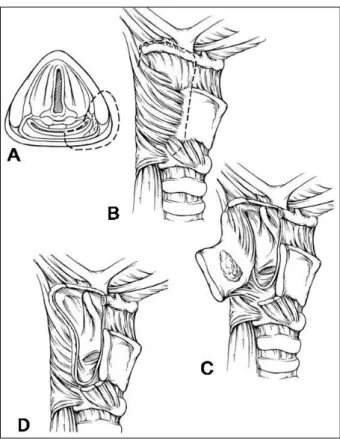
종양이 좀 더 광범위하게 하인두의 측벽을 침범한 경우 갑상 연골의 후부와 설골을 포함하는 광범위한 절제가 필 요하다. 종양으로의 접근은 이상와를 통해서가 아니라 후 두개곡(vallecula)을 통해서 이루어진다. 술식은 다음과 같 다. 일단 경부 청소술을 시행 후 갑상연골의 하부 기반 연 골막 피판(inferiorly based perichondrial flap)을 거상한 다. 설골을 lesser cornu에서 측방으로 노출시킨다. 이후 설골과 갑상연골을 lesser cornu부근에서 수직으로 절개한 다. 다음으로 하인두는 후두개곡을 통해 관찰이 가능하며, 이 때 견인기를 이용하면 쉽게 진입이 가능하다. 연골 절개 부위를 통해 외측, 아래측으로 절개선을 연장하며 종양의 안전 영역을 확인할 수 있다. 병변을 확인하며 환상 절재를 시행 후 동결 절편 검사를 시행한다. 마지막으로 점막을 재 봉합 후 연골막에 강화 봉합을 시행한다(Fig. 1, 2).
3) 전방 경설골 인두절개술(Anterior transhyoid pha- ryngotomy)
병변이 주로 하인두 후벽을 포함하고 있을 때 시행되지 만 이 술식의 제한점은 전체 하인두의 관찰에 장애가 있다 는 점이다. 따라서 적절한 종양의 선택이 이 술식의 성패를 좌우한다. 술식은 다음과 같다. 먼저 기관 절개술을 시행한 다. 설골은 제거되거나 혹은 접근법이 설골의 위, 아래로 진행할 수 있다. 후두개곡을 식별 후 점막의 절개를 시행한 다. 이후 종양을 관찰하며 전척추근막을 심부 안전 영역으 로 하여 종양을 절제한다. 동결 절편검사로 안전 영역을 평 가 후 split thickeness 피부 이식을 시행한다. 후두개곡의 점막을 봉합한다.
Fig. 1. Partial pharyngectomy via the lateral pharyngotomy. (A) Area removed, including the posterolateral hypopharyngeal wall and a portion of the thyroid ala. (B) Dotted lines show cuts on the thyroid cartilage, hyoid, and vallecula. (C) After entry into the hypopharynx, the specimen is reflected laterally and final cuts are made. (D) Defect following excision of the posterolateral hypopharyngeal tumor.
Fig. 2-1. Case of partial pharyngectomy via lateral pharyngo- tomy. Right lateral wall of the hypopharynx cancer (star).
Fig. 2-2. Intraoperative complete excision of the tumor with safe margin.
Fig. 2-3. Postoperative finding shows clearly removed tumor and healing.
2. 부분 인후두절제술(Partial Laryngopharyngec- tomy)
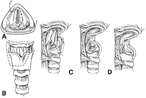
수직 후두 부분 절제술과 부분 인두절제술을 결합하여 실시하는 술식이다. 남아있는 후두 일부분은 발성, 연하 및 기도 보호 작용을 할 수 있는 장점이 있다. 하인두 이상와 의 내측벽을 침범하고 후두개와 피열 연골로 암종이 닿아 있는 병변에 대해서 절제 범위는 이상와의 내측벽, 후두개 의 일부, 병변측의 피열 연골과 설골 그리고 갑상연골의 상 부 2/3가 포함된다. 이 술식은 이상와의 내측벽을 침범하는 종양에 대해서 주로 적용할 수 있다. 설기저부, 외측 이상 와, 후두 계곡으로 절개를 연장함으로써 잠재적으로 이 부
Fig. 3. Partial Laryngopharyngectomy. (A) The resection margin after a partial laryngo- pharyngectomy for a medial pyriform tumor is shown in the superior view. (B) This view demonstrates the bone and cartilage cuts for a partial laryngopharyngectomy. (C) The completed resection of the medial pyriform tumor is shown. The epiglottis may be divided (as described in the text) or removed completely as shown here. (D) With resection of the arytenoid, the ipsilateral true vocal cord must be sutured in the midline to the cricoid to prevent aspiration. The laryngeal mucosa is reapproximated as much as possible as shown.
위가 절제된 표본에 포함될 수 있다. 그러나 이 술식은 암 종이 이상와 첨부를 침범한 경우, 윤상연골 후부로 확장, 동측의 성대 마비, 윤상인두근으로 침범이 있을 경우에는 금기증이 된다. 술식은 다음과 같다. 우선 기관절개술을 시 행하고 경부 림프절 청소술을 시행하게 된다. 상설골근을 절제한 후 상부기저의 연골막피판을 갑상연골로부터 거상 한다. 갑상연골은 중앙에서 수직으로 절개하고 높이는 약 2/3까지 연장할 수 있다. 수평절개는 갑상연골의 후방경계 까지 연장하고 동측의 설골을 박리하여 후두계곡으로 들어 간다. 후두개를 잡아서 밖으로 당김으로써 시야를 좋게 하 며 종양 주위의 후두개는 안전 거리를 확보하고 절제하면 서 전연합부까지 이르게 된다. 수술 가위의 한 날은 후두실 에 위치하고 다른 날은 수평으로 잘려진 갑상연골에 근접 시킨 후 절제하여 피열연골까지 이르게 되며 종양이 피열 연골을 침범했을 경우에는 이를 함께 절제한다. 후두계곡 의 절개는 아래로 종양과 안전역을 확보하여 연장하고 만 일 종양이 침범되었으면 외측 이상와벽이 절제시에 포함되 어야 하며 외측과 내측 절개는 피열연골에서 만난다. 종양 이 성문상부 이상을 침범했을 경우는 고식적인 성문상 후 두절제술을 시행하여 후두개 전체, 후두개 전공간 혹은 설
기저부 일부를 절제 할 수 있다. 안전역을 동결절편 조직검 사로 확인하고 피열연골이 절제된 경우에는 동측의 성대를 윤상연골의 중앙에 고정하여 술후 흡인을 방지할 수 있다.
윤상인두근 절개술을 시행하며 결손부위는 근피판이나 유 리피판을 이용하여 재건을 한다(Fig. 3).
3. 상윤상 인후두반절제술(Supracricoid Hemilary- gopharyngectomy)
이 술식은 부분 후인두절제술을 확장하여 이상와와 동측 의 전체 후윤상 후두 절반을 절제하는 것을 말한다. 이 술 식의 금기증으로는 암종이 이상와의 첨부를 침범하였거나 동측 성대 고정이 있는 경우, 후윤상부위를 침범했을 경우, 또는 후인두부위를 침범했을 때이다. 흡인의 위험성이 높 기 때문에 수술 후 적극적인 객담배출을 하도록 하고 수술 전에 적절한 폐기능을 유지하고 있는 환자를 선택하는 것 이 중요하다. 다른 수술 방법과 비슷하게 기관주위림프절 을 포함하는 적절한 림프절 청소술 후 일측의 갑상선 반절 제술을 하게된다. 갑상연골의 정중앙 바로 옆에서 근연골 피판을 거상하며 동측 설골의 반을 소각 부위에서 절제하 고 윤상갑상관절을 확인하여 탈골시킨다. 기관절개술을 시
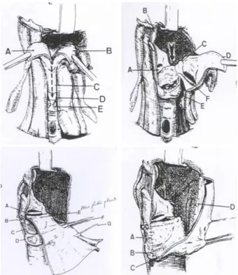
Fig. 4. Wide Vertical Hemipharyngolaryngectomy. Top left.
Vertical section of the epiglottis. A, B=two parts of epiglottis;
C=thyroid cartilage; D=cricothyroid membrane; E=cricoids car- tilage. Top right. Section through the inetrarytenoid space and subperichondrial dissection of the cricoids cartilage until the cricoarytenoid joint. A=right vocal cord; B=right hemiepiflottis;
C=left arytenoid cartilage; D=left hemiglottis; E=cricoids cartil- age; F=left vocal cord. Bottom left. Fixation of the tendon through the two drilled holes and the suture of the skin around the tendon to create a new glottis plane. A=remaining vocal cord; B=anterior fixation of tendon; C=anterior suture between the flap and the subglottic mucosa; D=cricoid cartilage; E=new glottic plane; F=
radial vascular pedicle; G=cutaneous sensitive branch. Bottom right. Progressive suture between the borders of the flap and the neighboring structures, creating a new pyriform sinus. A=
anterior fixation with the perichondrium of the thyroid cartilage;
B, C=inferior fixation to the cricoid cartilage; D=posterior fixation to the pharyngeal mucosa.
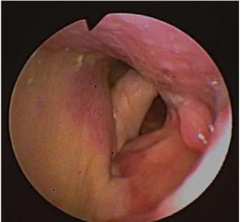
Fig. 5-1. Case of wide vertical laryngopharyngectomy. Tumor involving medial wall of the right side pyriform sinus.
행하여 경구기관내 튜브를 옮기고 윤상갑상막을 수직으로 중앙에서 절제하여 하인두로 들어가게 되는데 중앙의 수직 갑상연골절제부와 연결을 시킨다. 절개를 상부로 연장하여 후두개와 전후두개의 지방층까지 확장하여 후두계곡에서 절개를 마치게 된다. 절제할 부위를 회전시켜 개방하여 하 인두내의 종양을 확인하게 된다. 윤상 연골 위에서 수평 절개를 중앙에서 윤상연골 관절로 연결하여 끝내게 된다.
후두계곡으로부터의 절개는 아래로 내려와서 피열간 절개 와 연결하여 마침내 절제된 부위를 일괴로 빼내게 된다. 반
대측 피열연골은 주위 점막으로 덮어 주며 동측의 성대를 버팀목으로 만들어 반대측의 남아있는 성대와의 접촉을 원 활하게 한다. 근연골막 피판으로 외측 인두벽을 봉합한다.
4. CO2 레이저를 이용한 내시경적 절제
이 술식의 장점은 기관지 절개술이 필요하지 않으며 설 골상근을 보존하여 술후 정상 연하를 가능하게 하고 재건 술을 필요로 하지 않아 재원기간을 줄인다는 점이다. 미국 에서 전향적으로 내시경 절제술을 시행한 530명 환자에 대 한 예비 보고에서 42명(8%)이 하인두암이었다. 평균 21개 월의 추적 관찰 후 무병생존률이 1기/ 2기에서 75%, 3기/4 기에서 67%였으며 재원기간이 유의하게 감소하는 것이 관 찰되었다. 고식적인 개방술과 달리 종양은 내시경 수술시 에 잘리거나 축소되어 종양의 깊이나 연골 침범여부를 확 인할 수 있고 10 mm 정도의 경계를 두고 종양을 완전히 절 제할 수 있다.
5. 광범위 수직 인두후두부분절제술(Wide Vertical Hemipharyngolaryngectomy)
수직인두후두부분절제술(WVHLP)은 인후두 국소 진행 암종에서 후두 및 연하기능을 보존하면서 인두후두종양을 절제한 후 생긴 커다란 인후두 결손과 성대를 장장근의 건 을 포함한 요전박유리 피판을 이용하여 재건하는 술식이다 (Fig. 4, 5). 수직인두후두부분절제술의 개념은 1991년 Chantrain에 의해 최초로 도입되었다.18, 19 이 술식은 결손
Fig. 5-2. Intraoperative finding and tumor.
Fig. 5-3. Reconstruction of defect by radial forearm free flap with palmaris longus tendon.
Fig. 5-4. Postoperative finding.
결 론
하인두암에 대하여 종양의 완전 절제와 동시에 기능을 보존하는 수술은 현재 진행형이며 앞으로도 계속되는 도전 의 연속이다. 종양적 혹은 기능적 측면에서 모두 만족스러 운 결과를 가져올 수 있는 이상적인 술식은 아직 확립되어 있지 않다. 하지만 위에서 저자들이 소개한 광범위 수직인 두후두부분절제술을 포함한 다양한 술식을 종양의 크기와 위치에 따라 다양하게 적용한다면 이전에 후두전적출술을 시행하여 음성 소실을 초래한 환자들보다 좀 더 나은 삶의 질을 제공할 수 있을 것이다. 향후 조직 재생 공학이나 이 상적인 피판이 개발 된다면 하인두암의 수술적 치료는 더 욱 진일보 할 것이다.
References
1. Muir C, Weiland L. Upper aerodigestive tract cancers.
Cancer 1995;75(suppl):147-53.
2. Hussey DH, Latourette HB, Panje WR. Head and neck cancer: an analysis of the incidence, patterns of treat- ment, and survival at the University of Iowa. Ann Otol Rhinol Laryngol Suppl 1991;100(suppl 152):2-16.
3. Driscoll WG, Nagorsky MJ, Cantrell RW, Johns ME.
Carcinoma of the pyriform sinus: analysis of 102 cases.
Laryngoscope 1983;93:556-60.
4. Bataini JP, Bernier J, Brugere J, Jaulerry C, Picco C, Brunin F. Natural history of neck disease in patients with squamous cell carcinoma of oropharynx and pha-
ryngolarynx. Radiother Oncol 1985;3:245-55.
5. Scharpf J, Esclamado RM. Reconstruction with radial forearm flaps after ablative surgery for hypopharyngeal cancer. Head Neck 2003;25:261-6.
6. Seidenberg B, Rosenak S, Hurwitt ES, Som ML. Im- mediate reconstruction of the cervical esophagus by a revascularized isolated jejunal segment. Ann Surg 1959;149:162-71.
7. Harii K, Ebihara S, Ono I, Saito H, Terui S, Takato T.
Pharyngoesophageal reconstruction using a fabricated forearm free flap. Plast Reconstr Surg 1985;75:463-74.
8. Gilbert RW, Neligan PC. Microsurgical laryngotracheal reconstruction. Clin Plastic Surg 2005;32:293-301.
9. Plouin-Gaudon I, Lengel ˊe B, Desuter G, Rombaux P, Ledeghen S, Gr ˊegoire V, Hamoir M. Conservation la- ryngeal surgery for selected pyriform sinus cancer. Eur J Surg Oncol 2004;30:1123-30.
10. Kelly KE, Anthony JP, Singer M. Pharyngoesophageal reconstruction using the radial forearm fasciocutaneous free flap: preliminary results. Otolaryngol Head Neck Surg 1994;111:16-24.
11. Disa JJ, Pusic AL, Hidalgo DA, Cordeiro PG. Microvas- cular reconstructionof the hypopharynx: defect classi- fication, treatment algorithm, and functional outcome based on 165 consecutive cases. Plast Reconstr Surg 2003;111:652-60.
12. Stark B, Nathanson A. The free radial forearm flap: a reliable method for reconstruction of the laryngohypo- pharynx after in-continuity resection. Acta Otolaryngol 1998;118:419-22.
13. Urken ML, Blackwell K, Biller HF. Reconstruction of the laryngopharynx after hemicricoid/hemithyroid cartilage resection. Preliminary functional results. Arch Otolaryn- gol Head Neck Surg 1997;123:1213-22.
14. Delaere PR, Liu Z, Feenstra L. Tracheal autograft reva- scularization and transplantation. Arch Otolaryngol Head Neck Surg 1994;120:1130-6.
15. Delaere PR, Blondeel PN, Hermans R, Guelinckx PJ, Feenstra L. Use of a composite fascial carrier for laryn- gotracheal reconstruction. Ann Otol Rhinol Laryngol 1997;106:175-81.
16. Nakatsuka T, Harii K, Ueda K, et al. Preservation of the larynx after resection of a carcinoma of the posterior wall of the hypopharynx, versatility of a free flap patch graft. Head Neck 1997;19:137-42.
17. Scharpf J, Esclamado RM. Analysis of recurrence and survival after hypopharyngeal ablative surgery with radial forearm free flap reconstruction. Laryngoscope 2005;115:429-32.
18. Chantrain G, Deraemaecker R, Andry G, Dor P. Wide vertical hemipharyngolaryngectomy with immediate glottic and pharyngeal reconstruction using a radial forearm free flap: preliminary results. Laryngoscope 1991;101:869-75.
19. Chantrain G, Deraemaecker R. Glottic reconstruction after cricohyoidoepiglottopexy with a radial forearm free flap that includes the tendon of the palmaris longus:
preliminary results and perspectives. Laryngoscope 1994;104:1035-40.