90
90 THE EWHA MEDICAL JOURNALTHE EWHA MEDICAL JOURNAL
Stress-induced Cardiomyopathy Associated with Non-Small Cell Lung Cancer Presenting as Hyponatremia
Jong Taek Kim, Seok Ho Seo, Seung Hyun Lee, Dae Won Park, Dong Goo Kang, Seung Uk Lee
Division of Cardiology, Department of Internal Medicine, Kwangju Christian Hospital, Gwangju, Korea
Introduction
Stress-induced cardiomyopathy was first introduced in Japan under the name Takotsubo cardiomyopathy [1]. Since then, several cases with similar clinical features have been reported [2,3]. Stress-induced cardiomyopathy is accompanied by apical akinesia, along with apical ballooning, and is characterized by transient left ventricular dysfunction. In addition, it has clinical features that are similar to acute myocardial infarction but has normal coronary angiographic findings.
Clinical features of stress-induced cardiomyopathy include acute chest pain, ischemic echocardiographic change, and in- creased cardiac enzyme. Because these clinical features are similar to those of acute myocardial infarction, stress-induced cardiomyopathy can be mistaken for acute myocardial infarction in clinical settings. In most cases, coronary angiography is per- formed, thereby differential diagnosis can be made from acute myocardial infarction. Unlike acute myocardial infarction, most
patients with stress-induced cardiomyopathy show improvements in their symptoms within 1 week and a pattern of return to near-normal cardiac function within 1 month [4].
Authors encountered a patient with stress-induced cardiomy- opathy that presented clinical features of hyponatremia, who was also diagnosed with lung adenocarcinoma, and hereby report this case along with a literature review.
Case
A 72-year-old woman visited the Kwangju Christian Hospi- tal with a chief complaint of dizziness, nausea, and vomiting for the previous 3 days. She was being treated for 2 weeks before admission at a nearby hospital for cystitis with fever and dys- uria, during which time she was transferred to our hospital with a chief complaint of dizziness, nausea, and vomiting.
According to her medical history, she was diagnosed with hypertension 15 years prior and had been treated with medica-
Case Report
Ewha Med J 2015;38(2):90-93
http://dx.doi.org/10.12771/emj.2015.38.2.90 pISSN 2234-3180 • eISSN 2234-2591
Stress-induced cardiomyopathy, so-called Takotsubo cardiomyopathy, has recently been reported in Japan. Stress-induced cardiomyopathy is characterized by transient left ventricular apical dysfunction and ballooning, with normal coronary angiographic findings. We describe a rare case of stress-induced cardiomyopathy associated with lung adenocarcinoma presenting as hyponatremia. (Ewha Med J 2015;38(2):90-93)
Received April 6, 2015 Accepted June 11, 2015 Corresponding author Dong Goo Kang
Division of Cardiology, Department of Internal Medicine, Kwangju Christian Hospital, 37 Yangnim-ro, Nam-gu, Gwangju 503-715, Korea
Tel: 82-62-650-5230, Fax: 82-62-650-5116 E-mail: [email protected]
Key Words
Takotsubo cardiomyopathy; Acute coronary syndrome; Hyponatremia; Inappropriate ADH syndrome; Lung neoplasms
91
THE EWHA MEDICAL JOURNAL Stress-induced Cardiomyopathy Associated with NSCLC as Hyponatremia
tion since then. She had no history of diabetes, tuberculosis, or liver disease. She had a right knee joint replacement surgery for degenerative arthritis 8 years earlier. Her family medical his- tory indicated no particular findings. Upon admission, her blood pressure was 120/70 mmHg; pulse rate, 64 bpm, and body temperature, 36.2oC. She showed signs of acute conditions such as dizziness, nausea, and vomiting, but no signs of dyspnea or chest pain. Her pulse rate was regular, while no prominent rale was heard in the lung sound examination. She did not complain of tenderness or rebound tenderness during the abdominal ex- amination.
Electrocardiography revealed normal sinus rhythm, and a T inversion of ≥9 mm was observed in the aVL, V1, V2, V3, V4, V5, and V6 leads (Fig. 1).
Chest radiography revealed cardiomegaly, along with enlarged right hilum. The result of the blood test performed at admission showed the following values: Na, 120 mEq/L and K, 3.8 mEq/
L, indicating hyponatremia. BUN (13.0 mg/dL) was normal range, Cr (0.8 mg/dL) was normal range. Serum Osm (244 mOsmo/kg) decreased, and urine Osm (530 mOsmo/kg), urine sodium (150 mEq/L), creatine kinase (CK; 114 IU/L), creatine
kinase isoenzyme MB (CK-MB; 8.46 ng/mL), troponin I (0.96 ng/mL), troponin T (0.071 ng/mL), and D-dimer (11.1 µg/mL) increased. The urine analysis values were white blood cell count of 0–1/HPF and red blood cell count of 30–49/HPF.
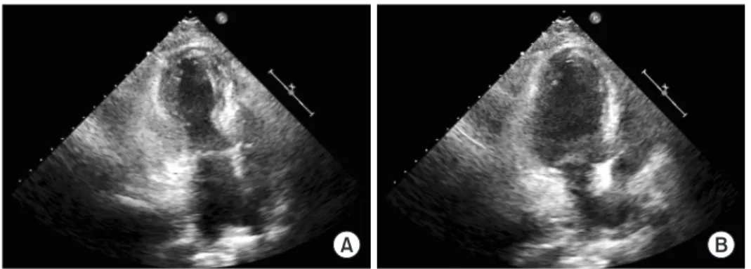
Thyroid function test was within normal limits and rapid adre- nocorticotropic (ACTH) stimulation test was unremarkable. On the transthoracic two-dimensional echocardiography performed on the first day of admission, the left ventricular ejection frac- tion was 56%, which indicated good left ventricular systolic function. However, left ventricular apical wall motion abnormal- ity and ballooning were observed. Moreover, mild aortic valve regurgitation due to calcified aortic valve was also observed (Fig. 2).
Because the patient complained primarily of nausea and vom- iting rather than chest pain, 3% hypertonic saline was admin- istered for hyponatremia until serum sodium level 131 mEq/L, and the symptoms improved following conservative therapy. The patient was maintained on conservative management, including aspirin 100 mg, clopidogrel 75 mg orally. Three days after, car- diac enzyme were significant for CK 190 IU/L, CK-MB 11.25 ng/mL, troponin I 0.83 ng/mL, troponin T 0.065 ng/mL.
A B
Fig. 2. Echocardiography during systole (A) and diastole (B), on the first day of ad- mission. Initial echocardiography reveals left ventricular apical wall motion abnor- mality and ballooning.
Fig. 1. Electrocardiogram (ECG). (A) ECG obtained two years ago shows sinus rhythm with left ventricular hypertrophy. (B) ECG obtained on ar- rival shows normal sinus rhythm with T inversion in all the leads.
A B
92 THE EWHA MEDICAL JOURNAL Kim JT, et al
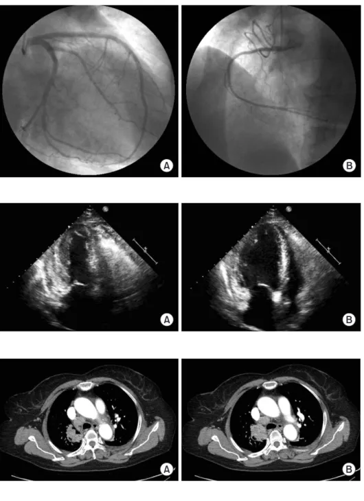
Seven days after, patient remained stable and cardiac enzymes were decreased (CK 65 IU/L, CK-MB 2.09 ng/mL, troponin I 0.26 ng/mL, troponin T 0.037 ng/mL). On the coronary angiography performed on the seventh day after admission, no significant stenosis was found (Fig. 3). On the transthoracic two-dimensional echocardiography performed again on the seventh day after admission, improvements were observed in the left ventricular apical dyskinesia that was exhibited previously (Fig. 4). On the chest computed tomography, an indication of primary lung malignancy was found in the right upper lobe of lung (Fig. 5). Endobronchial ultrasonography-guided fine-
needle aspiration biopsy results indicated a poorly differentiated lung adenocarcinoma (Fig. 6). On positron emission tomo- graphic computed tomography, indications of metastasis in both the pelvic bones and right femur were found, for which pallia- tive systemic chemotherapy was to be administered. However, in consideration of the systemic condition, conservative therapy was administered instead.
Discussion
In this case study, we report a case of stress-induced car-
A B
Fig. 5. Chest computed tomography image obtained on admission. Chest computed tomography shows an approxi- mately 4.4-cm long, enhancing mass-like consolidation in the right upper lobe of the lung at serial axial image (A) and (B).
A B
Fig. 4. Echocardiogram obtained during systole (A) and diastole (B) on the seventh day after admission. Echocardiogram shows the recovery of the previous wall motion abnormalities.
A B
Fig. 3. Coronary angiogram. Coronary an- giogram shows normal left (A) and right (B) coronary arteries.
93
THE EWHA MEDICAL JOURNAL Stress-induced Cardiomyopathy Associated with NSCLC as Hyponatremia
diomyopathy accompanied with lung adenocarcinoma that pre- sented with clinical symptoms of hyponatremia. Stress-induced cardiomyopathy occurs in association with various conditions. Its causes include emotional stress, surgical disposition, and diseases such as septicemia and malignant tumors [5]. Most clinical cases of stress-induced cardiomyopathy show good prognosis.
However, some cases are accompanied by various complications.
Known complications include cardiogenic shock, thrombosis, heart failure, and cerebral infarction [6]. In lung cancer patients, hyponatremia caused by the syndrome of inappropriate antidi- uretic hormone (SIADH) secretion is often known to develop into paraneoplastic syndrome. In particular, hyponatremia as- sociated with the SIADH secretion occurs commonly in patients with small cell lung cancer [7,8]. Hyponatremia associated with the SIADH secretion is known to be rare in patients with non- small cell lung cancer, accounting only for <1% of patients, according to previous studies [9,10]. In this case, the patient was admitted owing to atypical symptoms such as nausea and vomiting, and the laboratory test results showed indications of T inversion and increased cardiac enzyme, besides hyponatremia.
Based on these results, coronary angiography was performed to eliminate the possibility of acute myocardial infarction. The coronary angiography did not reveal significant indications of stenosis, and an indication of primary lung malignancy was dis- covered during the evaluation for hyponatremia. We considered
SIADH induced by lung cancer as a cause of hyponatremia.
There was no other reason to cause of hyponatremia in this case. Although hyponatremia associated with the SIADH secre- tion is known to rarely occur in non-small cell lung cancer, an indication of lung adenocarcinoma was found on the endobron- chial ultrasonography-guided fine-needle aspiration biopsy.
Stress-induced cardiomyopathy is a heart disease with various causes. Its prognosis may vary depending on the cause. The au- thors experienced a case of stress-induced cardiomyopathy with similar features as acute myocardial infarction in a patient with non-small cell lung cancer that presented with hyponatremia and hereby report the case.
References
1. Dote K, Sato H, Tateishi H, Uchida T, Ishihara M. Myocardial stunning due to simultaneous multivessel coronary spasms: a review of 5 cases. J Cardiol 1991;21:203-214.
2. Wittstein IS, Thiemann DR, Lima JA, Baughman KL, Schulman SP, Gerstenblith G, et al. Neurohumoral features of myocardial stunning due to sudden emotional stress. N Engl J Med 2005;352:
539-548.
3. Kawai S, Suzuki H, Yamaguchi H, Tanaka K, Sawada H, Aizawa T, et al. Ampulla cardiomyopathy (‘Takotusbo’ cardiomyopathy):
reversible left ventricular dysfunction: with ST segment eleva- tion. Jpn Circ J 2000;64:156-159.
4. Lee HH, Gwon HC, Kim BJ, Lee KJ, Im ES, Won KH, et al. Clinical manifestation of novel stress-induced cardiomyopathy mimick- ing acute myocardial infarction: single center prospective regis- try. Korean Circ J 2002;32:1054-1063.
5. Andrade AA, Stainback RF. Takotsubo cardiomyopathy. Tex Heart Inst J 2014;41:299-303.
6. Donohue D, Movahed MR. Clinical characteristics, demograph- ics and prognosis of transient left ventricular apical ballooning syndrome. Heart Fail Rev 2005;10:311-316.
7. McDonald P, Lane C, Rojas GE, Masood A. Syndrome of inap- propriate anti-diuretic hormone in non-small cell lung carci- noma: a case report. Ecancermedicalscience 2012;6:279.
8. Vanhees SL, Paridaens R, Vansteenkiste JF. Syndrome of inap- propriate antidiuretic hormone associated with chemotherapy- induced tumour lysis in small-cell lung cancer: case report and literature review. Ann Oncol 2000;11:1061-1065.
9. Sorensen JB, Andersen MK, Hansen HH. Syndrome of inappro- priate secretion of antidiuretic hormone (SIADH) in malignant disease. J Intern Med 1995;238:97-110.
10. Bose CK, Dey S, Mukhopadhyay A. Hyponatremia of non-small cell lung cancer: Indian experience. Indian J Med Paediatr Oncol 2011;32:139-142.
Fig. 6. Biopsy in the right upper lobe with endobronchial ultrasonog- raphy guidance. The adenocarcinoma is poorly differentiated. Histo- logic examination of the lung shows atypical cell proliferation with indistinct glandular structures (H&E, ×200).