대 한 골 절 학 회 지 제16권, 제4호, 2003년 10월 The Journal of the Korean Society of Fractures
Vol. 16, No. 4, October, 2003
종골 골절 치료에 있어서의 전산화 단층 촬영의 유용성
김학준・하권익・윤정로・심재익・김택선・김영배・이우승・최재혁
한국보훈병원 정형외과[국문 초록]
목 적: 종골 골절은 해부학적 특성상 골절된 양상에 따른 정확한 분류가 어렵고 분류에 따른 치료 방법이 확립되어 있지 않다. 이에 저자들은 단순 방사선 사진상의 Essex-Lopresti에 의한 분류 및 전산화 단층 촬영상의 Sanders 분류에 따른 차이점과 치료 결과에 대한 분석을 하고 자 하였다.
대상 및 방법: 1993년 1월부터 2002년 1월까지 종골 골절로 인해 도수 정복 후 축성 핀 고정술 또는 관혈적 정복술 후 내고정술을 시행 받은 24명 (26례) 중 전산화 단층 촬영이 가능하였던 16명 (17례)을 대상으로 하여 단순 방사선 사진상 Essex-Lopresti 분류를 시행하였으며 전산화 단층 촬영을 시행할 수 있었던 17례에서 Sanders 분류를 시행하였고, 술전 및 술후의 측면 단순 방사선 사진을 이용하여 Böhler 각을 측정하였으며, 임상적 결과는 동통, 환자의 만족도, 보행 능 력, 보조기 사용 여부에 중점을 둔 Salama 평가 기준에 의거하여 분석하였다.
결 과: 평균 추시기간은 6년 (12개월~8년 8개월)이었으며, 단순 방사선 사진상 설상형은 9례, 관절 함몰형은 17례이었고, 전산화 단층 촬영을 할 수 있었던 예에서는 설상형이 8례, 관절 함몰 형이 9례이었다. Sanders 분류상 IIA형이 5례, IIC형이 1례, IIIA형이 4례, IIIB형이 4례, IV형이 3 례이었다. 도수 정복 후 축성 핀 고정술을 시행한 예는 12례, 관혈적 정복술후 금속판 고정술을 시행한 예는 5례이었다. 지연 유합 및 불유합의 예는 관찰되지 않았다. 관절 함몰형 1례에서 외상성 관절염의 소견을 보였다. 수술 방법에 따른 임상적 결과에는 차이가 없었으나, 술후 Böhler 각 의 회복 여부와 임상적 결과 사이의 통계학적으로 유의한 연관성이 있었으며 (Linear Regression, correlation coefficient=0.40), 단순 방사선 사진상에서 설상형으로 분류되었던 골절 7례에서 종골 조면과 종골 후관절과의 연결이 유지되어 있는 Sanders IIC형은 1례만이 관찰되었으며 나 머지 예에서는 종골 관절면이 침범되었다.
결 론: 종골의 관절내 골절의 치료는 아직 논란의 여지가 있으며, 단순 방사선 사진상 종골 골 절의 정확한 분류를 위해서는 전산화 단층 촬영이 필요하며 종골 관절면의 침범 정도에 따른 수술 술기의 선택이 필요할 것으로 사료된다.
색인단어: 종골 골절, 관절내 골절, 전산화 단층 촬영
526
※통신저자: 윤 정 로
134-060, 서울특별시 강동구 둔촌동 6-2 한국보훈병원 정형외과학교실
전화: +82-2-2225-1352, Fax: +82-2-487-0754 e-mail: [email protected]
*본 논문의 요지는 2003년 대한골절학회 춘계학술대회에서 구연됨.
서 론
종골 골절은 족부 골절의 약 60%를 차지하는 가장 흔한 족부 골절이며, 체중 부하 골의 관절을 형성함으 로서 종골 관절의 정확한 해부학적 정복이 술후 기능 에 중요한 영향을 미치는 것으로 보고되고 있다1).
종골 골절이 상대적으로 빈도가 많음에도 불구하고 치료 방법에 대한 확립이 되어 있지 않으나 단순 방 사선 사진에 의한 Essex-Lopresti 분류상 관절내 설상 형 골절에서는 도수 정복 및 축성 핀 고정술이 시행 되고 있으며 관절 함몰형에서는 관혈적 정복술 및 내 고정술이 일반적으로 시행되고 있다1,9). 전산화 단층 촬영의 발달과 더불어 종골 후관절면의 침범 여부를 기준으로 한 Sanders19) 분류가 보고되어 종골 후관절 침범에 대한 비교적 정확한 진단과 함께 종골 후관절 면의 정확한 해부학적 정복과 견고한 내고정이 시도 되어 지고 있다.
이에 저자들은 전산화 단층 촬영 사진상 종골 후관 절의 침범 여부가 수술적 술기의 선택에 중요한 요건 으로 생각되어 후향적으로 단순 방사선 사진 및 전산 화 단층 촬영 사진상의 종골 골절에 대한 분류 및 수 술 방법에 따른 방사선학적, 임상적 결과를 비교 분석 하였다.
연구 대상 및 방법
1993년 1월부터 2002년 1월까지 본원에서 관절내 종골 골절로 인해 도수 정복 후 축성 핀 고정술 및 관 혈적 정복술후 내고정술을 시행받은 후 1년 이상 추 시 가능하였던 24명 (26례)을 대상으로 하여 단순 방 사성 사진을 기준으로 Essex-Lopresti 분류법을 이용하 여 관절내 골절을 설상형 및 관절 함몰형으로 분류하 였다. 이중 전산화 단층 촬영을 시행하였던 17례 (16 명)에서, 서로 다른 2명의 관찰자가 Sanders 분류를 다시 시행하였으며, 방사선학적 결과는 술전 및 술후 의 단순 측면 방사선 사진을 이용하여 Böhler 각을 측 정하여 분석하였고, 임상적 결과는 동통, 환자의 만족 도, 보행 능력, 보조기 사용 여부에 중점을 둔 Sal- ama18)의 평가 기준에 의거하여 분석하였다 (Table 1).
환자의 평균 연령은 52.5세 (27세~74세)이었고 남 자는 15명이었으며, 여자는 2명이었다. 평균 추시기간 은 6년 (1년~8년 8개월)이었다.
술전 Böhler 각은 평균 9.1° (-23°~32°)이었으며, 도 수 정복 후 축성 핀 고정술을 시행한 례는 12례이었 고 Böhler 각은 평균 9.5° (-23°~32°)이었으며, 관혈적 정복술 후 금속판 고정술을 시행한 례는 5례이었고 Böhler 각은 평균 7.8° (0°~22°)이었다. 단순 방사선 사진상 설상형에서 도수 정복 후 축성 핀 고정을 시행 하였던 예는 6례이었으며 관혈적 정복술후 내고정술 을 시행하였던 예는 2례이었고, 관절 함몰형에서 도수 정복 후 축성 핀 고정을 시행하였던 예는 6례이었으며 관혈적 정복술후 내고정술을 시행하였던 예는 3례이 었다.
도수 정복 후 핀 고정술은 전례에서 Essex-Lopresti9) 술식을 이용하였으며, 관혈적 정복술 및 내고정술은 Zwipp 등21)과 Benirschke와 Sangeorzan2)이 기술한 광 범위 외측 절개술 (extensile right-angle lateral incision)을 Table 1. Salama's Criteria of assessment
Patient satisfied, Normal mobility of joint Asymptomatic broadening of the heel Excellent:
No pain
Patient satisfied but occasional pain Walking ability unaffected Slight limitation of inversion Good :
Mild flat foot
Patient not entirely satisfied (reserved) Pain after exertion
Walking ability markedly reduced Limitation of tarsal movements Fair :
Special shoes Patient not satisfied Pain even on slight effort Walking ability markedly reduced Severely limitation of joint movement Poor :
Change of occupation
이용하여 종골에 도달한 후 종골 관절의 관절면의 해 부학적 정복을 한 후 3.5 mm 유관나사 (cannulated screw)와 3.5 mm H-plate (Mathys®, Bettlach, Switzerland) 를 이용하여 견고한 고정을 시도하였다.
통계학적 분석은 Epi info 2002 (CDC®)와 Win SAS 6.11 (Microsoft®) 프로그램을 이용하여 분석하였다.
결 과
1. 방사선학적 결과
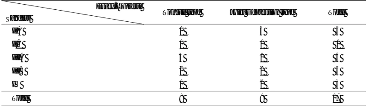
단순방사선 사진상 설상형으로 분류되었던 7례에서 전산화 단층 촬영 사진에 의한 Sanders 분류를 시행한 결과 IIA형이 1례, IIC형이 1례, IIIA형이 3례, IIIB형 이 2례이었으며, 단순 방사선 사진상 관절 함몰형으로 분류되었던 9례에서는 Sanders 분류상 IIA형이 4례, IIIA형이 1례, IIIB형이 2례, IV형이 2례이었다. 단순 방사선 사진상으로 설상형으로 분류되었던 7례 중 6 례 (85.7%)에서 2 mm 이상의 전위를 보인 종골 후관 절의 침범이 관찰되었으며 관절이 함몰된 Sanders III 형이 5례 (71.4%)나 관찰되었다 (Table 2).
술후 Böhler 각은 평균 25.6° (0°~40°)로 측정되었
으며, 단순 방사선 사진상 설상형에서 도수 정복 후 축성 핀 고정술을 시행하였던 6례에서의 술후 평균 Böhler 각은 22.2° (10°~32°)이었으며, 관혈적 정복술 을 시행한 1례는 5°이었다. 관절 함몰형에서 도수 정 복 후 축성 핀 고정을 시행하였던 6례에서의 술후 평 균 Böhler 각은 26° (0°~39°)이었고, 관혈적 정복술을 시행한 3례의 술후 평균 Böhler 각은 38° (35°~40°) 이었으며 통계학적으로는 의미 있는 차이를 보이지는 않았다 (Kruskal-Wallis test, p>0.05) (Table 3).
Table 3. Postoperative mean after Treatment modalities according to Essex-Lopresti classification Fx type
Böhler angle Tongue type Joint depression type Mean angle after C/R & axial pinning* 22.2° (10°~32°) 26° ( 0°~39°) Mean angle after O/R & I/F† 5° 38° (35°~40°)
(Kruskal-Wallis Test, p>0.05)
*Closed reduction and axial pin fixation by Essex-Lopresti method, †Open reduction and plate fixation by Zwipp method Table 2. Essex-Lopresti classification and Sanders classification
Essex-Lopresti
Sanders Tongue type Joint Depression type Total
IIA 1 4 5
IIC 1 0 1
IIIA 3 1 4
IIIB 2 2 4
IV 1 2 3
Total 8 9 17
Table 4. Results according to Böhler angle Result
Angle Excellent Good Fair Poor Total
0°~10° 0 0 2 1 3
11°~20° 0 1 1 0 2
21°~30° 2 0 1 0 3
31°~40° 3 4 1 1 9
Total 5 5 5 2 17
(Linear Regression, correlation coefficient = 0.04)
2. 임상적 결과
설상형에서 도수 정복 후 핀 고정술을 시행한 후의 임상적 결과는 우수가 2례, 보통이 4례이었으며, 관혈 적 정복술 및 내고정술을 시행한 후의 임상적 결과는 우수가 1례, 불량이 1례이었다. 관절 함몰형에서 도수 정복 후 핀 고정술을 시행한 후의 임상적 결과는 우수 가 2례, 양호가 2례, 불량이 2례이었고 관혈적 정복술 및 내고정술을 시행한 후의 임상적 결과는 우수가 1 례, 양호가 2례이었다.
임상적 결과는 수술 방법에 따른 차이는 없었으나
(Kruskal-Wallis test, p>0.05), 술후 Böhler 각의 회복과 밀접한 연관 관계 (Linear Regression, correlation coeffi- cient = 0.40)가 있었다 (Table 4).
단순 방사선 사진상 설상형으로 분류되며 전산화 단층 촬영 사진에서 Sanders IV형인 종골 골절의 예에 서는 관혈적 정복 및 내고정술을 시행하였으며 술후 Böhler 각이 34°로 회복되었으며 임상적 결과는 우수 로 분류되었다 (Fig. 1). 단순 방사선 사진상 관절 함몰 형으로 분류되었고 전산화 단층 촬영 사진상 Sander IIIA형으로 분류되었던 예에서는 도수 정복 및 축성 핀 고정을 시행하였으며 술후 Böhler 각이 32°로 측
A B
C D Fig. 1. 51 years olds female suffered fall down injury
A. Preoperative radiography: Essex-Lopresti tongue type with -16° in Böhler angle.
B. Preoperative CT scan: classified to Sanders type IV
C. Immediate postoperative plain radiography after open reduction and internal fixation: Böhler angle was restored to 34°
D. After 1 year 6 months, Böhler angle was not depressed and clinical outcome was good grade.
정되었으나 최종 추시 관찰하시에 Böhler 각이 8°로 감소하면서 거골하 관절의 외상성 관절염이 발생하여 임상적 결과는 불량으로 분류할 수 있었다 (Fig. 2).
고 찰
종골 골절은 족부 골절 중에서 가장 흔한 골절이며, 특히 거골하 관절면을 침범하는 종골 골절은 Cave7)에 의하면 모든 종골 골절의 75%를 차지한다고 하였고,
Kang 등11)은 90%를 차지한다고 발표하였다. 이는 관 절면의 정확한 해부학적 정복이 술후 환자의 예후에 많은 영향을 미칠 것으로 생각된다.
종골 골절의 분류는 1952년 발표된 Essex-Lopresti9) 가 발표한 단순 방사선 사진을 통한 분류법이 널리 쓰여 왔으나 Burkley와 Meek4)은 관절면의 정복의 중 요성을 역설하며 전산화 단층 촬영으로 관절면의 전 위 정도를 측정하여 관절면의 전위를 평가하였으며, Sanders19)는 전산화 단층 촬영을 통하여 종골의 후관
A B
C D Fig. 2. 27 years olds male suffered fall down injury.
A. Preoperative radiography: Essex-Lopresti tongue type with 5° in Böhler angle.
B. Preoperative CT scan: classified to Sanders type IIA
C. Immediate postoperative plain radiography after Closed reduction and axial pin fixation: Böhler angle was restored to 32°
D. After 2 years Böhler angle was decreased to 8° and clinical outcome was poor grade.
절면을 침범하여 전위된 관절면을 중심으로 새로운 분류법을 제시하였다.
종골의 관절내 골절은 거골하 관절을 침범한 골절 이지만, 그 해부학적 복잡성 및 빈약한 연부 조직에 쌓여 있어 다양한 치료법이 제시되고 있으며, 보통 설 상형에서는 Essex-Lopresti 술식에 의한 도수 정복 후 축성 핀 고정술이 널리 쓰이고 있고, 관절 함몰형에서 는 관혈적 정복술후 내고정술이 이용되어 지고 있다.
Koh와 Kim13)은 27례의 설상형 및 14례의 종골 골절에 서 Essex-Lopresti 술식에 의한 수술적 치료 후 77.7%
에서 임상적으로 만족할 만한 결과를 얻었음을 보고 하였고, Park 등15)은 103례의 설상형 및 관절 함몰형, 분쇄형 종골 골절에서 제한적 후방 절개술을 통한 거 골 후관절의 정복으로 70.9%의 우수한 임상적 결과를 얻었음을 보고하였다. 또한 Tornetta17)는 핀 대신에 나 사못을 이용하여 Essex-Lopresti 술식을, 설상형이고 Sanders IIC형 종골 골절에 시행한 결과 우수한 결과 를 얻었으며, Essex-Lopresti 술식은 설상형 Sanders IIC 형이 가장 이상적인 적응증이라고 보고하였다. 또한 Kang 등11)은 Sanders II형과 II형에서 관혈적 정복 및 내고정을 시행한 군이 Essex-Lopresti 술식 또는 보존 적 치료를 시행한 군 보다 우수한 임상적 결과를 보 였음을 보고하였다. 그러나 Burckley와 Meek4)은 전위 된 종골 관절내 골절에서 전체적으로 보존적 치료와 수술적 치료와의 술후 기능적 차이는 환자의 만족도 를 제외하고는 차이가 없지만 종골 관절면의 해부학 적 정복이 이루어진 경우는 장기적 예후가 우수하다고 보고하여 관절면 정복의 중요성을 보고하였다.
관혈적 정복술은 내측, 외측 또는 내외측 병합 도
달법1~3,20)이 있으며 Burdeaux3)는 종골 골절시 종골
내측이 외측보다 덜 분쇄되고 종골 조면을 거골 지주 돌기에 해부학적 정복을 가능하게 함으로서 발뒤꿈치 의 폭을 좁게 할 수 있는 장점이 있다고 보고하였으나, Carr6)는 내측 도달법은 함몰된 종골 후관절면의 관찰 이 불가능한 단점이 있으므로 외측 도달법을 시행하여 함몰된 관절면을 직접 관찰하여 정복을 시행하는 것 이 견고한 내고정에 적당한 방법이며 전통적인 외측 도달법보다는 광범위 외측 도달법이 비골건의 합병증 을 줄일 수 있다고 보고하였다. 이에 저자들은 관혈적 정복시 광범위 외측 도달법을 이용하여 종골 후관절
면의 해부학적 정복과 견고한 내고정을 이루려고 시도 하였다.
술후 임상적 결과에 대해서 Kim 등12)은 술후 Böhler 각의 회복과 만족할 만한 임상적 결과와의 연관성이 있음을 보고하였으며, Loucks와 Burckely14)는 전향적 코호트 조사를 통해 수상 당시의 Böhler 각 감소 정 도가 더욱 나쁜 임상적 결과와 연관성이 있고 술후 Böhler 각의 회복 및 유지가 임상적 결과에 중요한 영 향을 미친다고 보고하였다. 그러나 Hutchinson과 Hue- bner10)는 술후 Böhler 각의 회복이 임상적 결과와 통 계학적으로 의미가 없음 보고하였다.
저자들의 연구에서도 술후 Böhler 각과 임상적 결과 가 통계학적으로 의미 있는 연관성을 가졌으나 수술 적 방법에 따른 Böhler 각의 변화는 통계학적으로 차 이가 없었다. 단순 방사선 사진상에서 설상형으로 분 류되었던 예서, 전산화 단층 촬영 사진에서 후관절면 과 종골 조면의 연결이 유지되어져 있는 상태인 San- ders IIC형은 14.3%에서만 관찰되었고 종골 후관절의 심각한 침범이 있는 Sanders III형은 71.4%에서 관찰되 었다. 수술적 방법과 임상적 결과와의 통계학적 차이 가 없게 나타난 것은 초기의 수술 술기의 부족으로 인해 관혈적 정복시 종골 관절면의 정확한 해부학적 정복이 이루어지지 않은 예가 다수 포함되었기 때문으 로 생각되어지며 좀더 정확한 연구 결과를 위해 많은 추가적인 증례 연구가 필요할 것으로 사료된다.
결 론
종골 골절의 관절내 골절은 해부학적 특성 및 수상 기전에 의해 관절의 침범 정도가 다양하고 치료 방법 또한 다양하므로, 종골 관절면 침범의 정확한 분류를 위해서는 단순 방사선 사진 뿐만이 아니라 반드시 전 산화 단층 촬영을 실시하여 수술적 술기를 선택하고 종골 관절면의 해부학적 정복과 견고한 고정을 시행 함으로서 좀더 나은 임상적 결과를 얻을 수 있을 것 으로 사료된다.
REFERENCES
1) Barei DV, Sangeorzan BJ and Benirschke SK: Frac-
ture of calcaneus. Ortho Clin North Am, 33: 263-285, 2002.
2) Benirschke SK and Sangeorzan BJ: Extensive intra- articular fractures of the foot: surgical management of calcaneal fractures. Clin Orthop, 292: 128-134, 1993.
3) Burdeaux BD: Reduction of calcaneal fractures by McReynolds medial approach techinque and its experi- mental basis. Clin Orthop, 177: 87-103, 1983.
4) Buckley RE and Meek RN: Comparison of open versus closed redction of intraarticular calcaneal frac- tures: A matched cohort in workmen. J Orthop Trauma, 6: 216-222, 1992.
5) Buckly R, Tough S and McCormack R, et al: Opera- tive compared with nonoperartive treatment of dis- placed intraarticular calcaneal fracture; A prospective, randomized, controlled multicenter trial. J Bone Joint Surg, 84-A: 1733-1744, 2002.
6) Carr JB: Surgical treatment of the intraarticular cal- caneal fracture. Orthop Clin North Am, 25: 665-675, 1994.
7) Cave EF: Fractures of the os calcis: the problem in general. Clin Orthop, 30: 64-66, 1963.
8) Crosby LA and Fitzgibbons TC: Intraarticular calca- neal fractures. results of closed treatment. Clin Orthop, 290: 47-54, 1993
9) Essex-Lopresti P: Mechanism, reduction technique, and results in fractures of the os calcis. Br J Surg, 39:
395-419, 1952.
10) Hutchinson F III and Huebner MK: Treatment of os calcis fractures by open reduction and intrenal fixation.
Foot Ankle Int, 15: 225-232, 1994.
11) Kang CS, Pyun YS and Kwak YC: Clinical study of calcaneal fractures. J Kor Soc Orthop, 10: 71-75, 1975.
12) Kim JO, Kim DW, Koh YD, Yoo JD and Lee DW:
The prognostic factors in tongue shaped calcaneal fractures treated by Essex-Lopresti method. J Kor Soc Fractures, 12: 328-334, 1999.
13) Koh YD and KIm JO: Essex-Lopresti axial fixation for intra-articular calcaneal fractures. J Kor Soc Frac- tures, 11: 371-377, 1998.
14) Loucks C and Buckley R: Böhler Angle: Correlation with outcome in displaced intra-articular calcaneal fractures. J Orthop Trauma, 13: 554-558, 1999.
15) Park IH, Song KW, Shin SI, Lee JY, Kim TG and Park RS: Displaced intra-articular calcaneal fracture treated surgically with limited posterior incision. Foot Ankle Int, 21: 195-205, 2000.
16) Park SR, Kim HS, Kang JS, Lee WH and Park SJ:
Treatments of intraarticular alcaneal fracture-Based on CT classification and comparison of treatments. J Kor Soc Fractures, 12: 103-334,1999.
17) Tornetta III P: Percutaneous treatment of calcaneal fractures. Clin Orthop, 375: 91-96, 2000.
18) Salama R, Bernamara A and Weissman SL: Fuc- tional treatment of intraarticular ractures of the cal- caneus. Clin Orthop, 115: 236-240, 1976.
19) Sanders R, Fortin P and DiPasquale T, et al: Opera- tive treatment in 120 displaced ntraarticular calcaneal fractures: Results using a prognostic computed tomo- graphy can classification. Clin Orthop, 290: 87-95, 1993.
20) Stephenson JR: Surgical treatment of displaced inra- articular fractures of the calcaneus: A combined lateral and medial approach. Clin Orthop, 290: 68-75, 1993.
21) Zwipp H, Tscherne H, Thermann H and Weber T:
Osteosynthesis of displaced intraarticular fracture of calcaneus. Results in 123 cases. Clin Orthop, 290: 76- 86, 1993.
Usefulness of CT Scan in Treatment of Calcaneal Fracture
Hak-Jun Kim, M.D., Kwon-Ick Ha, M.D., Jeong-Ro Yoon, M.D., Jae-Ik Shim, M.D., Taik-Seon Kim, M.D., Young-Bae Kim, M.D., Woo-Seung Lee M.D., Jae-Hyuck Choi, M.D.
Department of Orthopedic Surgery, Korea Veterans Hospital, Seoul, Korea
Purpose: Accurate fracture classification is difficult because of its complex bony architecture and there is no definitive treatment modality according to fracture pattern or classification. We evaluated the fracture pattern using simple radiography and CT scan simultaneously and then the result according to treatment modalities and fracture classification.
Materials and Methods: We evaluated 24 patients (26 cases) who suffered intraarticular calcaneal fracture were treated with closed reduction and axial pinning or open reduction and plate fixation. There were had taken CT scan. Fractures were classified with two different manner using both plain radiography and CT scan. Essex-Lopresti classification was made with plain radiography and Sanders classification was made with CT scan. Radiographic results were measured of preoperative and postoperative Böhler angle on plain x-ray. 16 patients (17 cases) were analyzed for clinical outcomes by Salama's criteria focused on pain, patients' satisfaction, walking abilities and usage of orthosis.
Results: Mean followed-up period was 6 years (range 1~8.8 years). 8 cases were classified into tongue type intraarticular calcaneal fractures by plain radiography and 9 cases into joint depression type. According to Sanders classification by CT scan, 5 cases were classified into type IIA, 1 case IIC, 4 cases type IIIA, 4 cases IIIB and 3 case type IV. 12 fractures were reduced by closed reduction and axial fixation and 5 cases by open reduction and internal fixation. There was no correlation between clinical outcomes and treatment modality but there was linear correlation between clinical outcomes and postoperative Böhler angle (correlation coefficient = 0.04). In 8 cases of tongue type by plain radiography, sanders type IIC was only 1 case which calcaneal tuberosity connected to articular facet but articular involvements were observed in remaining cases.
Conclusion: Accurate eavaluation of articular facet in calcaneal fracture by CT scan is necessary to determining to select the treatment option.
Key Words: Calcaneal fracture, Intraarticular fracture, CT scan
Address reprint requests to Jeong-Ro Yoon
Department of Orthopaedic Surgery, Korea Veterans Hospital 6-2, Dunchon-dong, Gangdong-gu, Seoul, Korea
Tel : +82-2-2225-1352, Fax : +82-2-487-0754 E-mail : [email protected]