ORIGINAL ARTICLE
직장 카르시노이드 종양에서 Ki-67 발현의 예후적 중요성
홍수민, 김유선, 문정섭, 김진남, 오명기, 권선옥, 정성연, 홍성우1, 강윤경2
인제대학교 의과대학 내과학교실, 외과학교실1, 병리학교실2
Prognostic Significance of Ki-67 Expression in Rectal Carcinoid Tumors
Su Min Hong, You Sun Kim, Jung Seop Moon, Jin Nam Kim, Myoung Ki Oh, Sun Ok Kwon, Seong Yeon Jeong, Seong Woo Hong1 and Yun Kyung Kang2
Departments of Internal Medicine, Surgery1, and Pathology2, Inje University College of Medicine, Seoul, Korea
Background/Aims: Rectal carcinoid tumors can be resected with endoscopy, and it is important to assess their prognostic factors. We evaluated the potential of Ki-67 expression as a prognostic factor in rectal carcinoid tumors.
Methods: We retrospectively reviewed the medical records of 37 patients with rectal carcinoid tumors who got endoscopic resection from January 2001 to January 2011 at Inje University Seoul Paik Hospital. We analyzed their endoscopic and histologic findings, Ki-67 expression, clinical outcome, and prognosis.
Results: The mean age (±SD) of the patients was 56.3±10.7 years, and the male : female ratio was 3.6:1. The mean tumor size was 0.5±0.4 cm, 33 patients showed grade 1 tumors (89.2%) and the average Ki-67 expression was 0.7±1.2%.
Thirty five patients underwent endoscopic mucosal resection, and two required endoscopic submucosal dissection. Eight patients had positive margins after resection, but no cases of lymphovascular invasion were identified. The median follow-up duration was 21.4±25.4 months, and no recurrences were observed.
Conclusions: In low grade rectal carcinoid tumors which are lack of central depression on colonoscopy, the expression of a molecular marker of malignant potential, Ki-67, was low. Therefore, endoscopic resection seemed to be a safe and effective treatment for these tumors. (Korean J Gastroenterol 2013;61:82-87)
Key Words: Rectum; Carcinoid tumor; Ki-67; Endoscopy; Therapeutics
Received August 6, 2012. Revised October 18, 2012. Accepted October 23, 2012.
CC This is an open access article distributed under the terms of the Creative Commons Attribution Non-Commercial License (http://creativecommons.org/licenses/
by-nc/3.0) which permits unrestricted non-commercial use, distribution, and reproduction in any medium, provided the original work is properly cited.
교신저자: 김유선, 100-032, 서울시 중구 마른내로 9, 인제대학교 서울백병원 내과
Correspondence to: You Sun Kim, Department of Internal Medicine, Inje University Seoul Paik Hospital, 9 Mareunnae-ro, Jung-gu, Seoul 100-032, Korea. Tel: +82-2- 2270-0015, Fax: +82-2-2270-0579, E-mail: [email protected]
Financial support: This study was supported by the grants of Inje University (2011). Conflict of interest: None.
INTRODUCTION
Carcinoid tumors can occur in nearly any gastrointestinal (GI) tissue, and historically they have been reported most fre- quently in the appendix. However, the bronchus/lung (27.9%), ileum (14.9%) and rectum (13.6%) are now the most commonly reported sites.1 Rectal carcinoid tumors account for 1.1-1.3% of all rectal tumors, their metastatic incidence is 3.9%, and compared to other tumors they are less invasive and less progressive.2 Rectal carcinoid tumors are often in-
cidentally diagnosed during screening lower endoscopy for colorectal cancer or other unrelated indications.3 They are most commonly found in the mid-rectum and can have a wide range of sizes. Rectal carcinoid tumors that are less than 1 cm in diameter rarely metastasize (3-5%), and they can be managed with local endoscopic resection.4-7 If the carcinoid tumor is over 2 cm, a full oncologic resection should be performed.2 The treatment of rectal carcinoid tumors be- tween 1-2 cm in diameter is controversial. In these cases it is critical to assess prognostic factors such as World Health
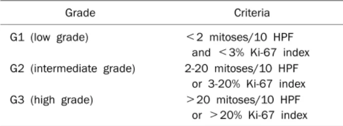
Table 1. Grading of Gastroenteropancreatic NET according to Proliferative Activity by WHO Classification
Grade Criteria
G1 (low grade) <2 mitoses/10 HPF
and <3% Ki-67 index G2 (intermediate grade) 2-20 mitoses/10 HPF
or 3-20% Ki-67 index G3 (high grade) >20 mitoses/10 HPF or >20% Ki-67 index NET, neuroendocrine tumor; WHO, World Health Organization; G, grade.
Organization (WHO) classification, tumor/nodes/metasta- sis (TNM) stage, histologic grade, size of the tumor, depth of invasion, lymphovascular invasion, and mitotic bodies on his- tologic examination.
Ki-67, a marker of cellular proliferation, has recently been introduced as a promising prognostic factor for rectal carci- noid tumors.8 In this study, we evaluated the role of Ki-67 ex- pression as a prognostic factor and predictive biomarker in low grade rectal carcinoid tumors.
SUBJECTS AND METHODS
1. Patients
We conducted cross sectional study on human partic- ipants with the approval of the Institution Ethics Committee of Seoul Paik Hospital. We retrospectively reviewed the medi- cal records of 37 patients with rectal carcinoid tumors who were treated with endoscopic resection between January 1, 2001 and January 1, 2011 at Inje University Seoul Paik Hospital. For each patient we analyzed the endoscopic and histologic findings, clinical outcome, and prognosis. In re- gards to treatment strategy, 35 patients underwent endo- scopic mucosal resection (EMR), and two patients required endoscopic submucosal dissection (ESD). Follow-up colono- scopic endoscopic examinations were performed 3-6 months after therapeutic resection and every 1-2 years thereafter to monitor for local recurrence.
2. Immunohistochemical staining
All rectal carcinoid tumor specimens were reviewed by one expert pathologist and classified based on WHO criteria (Table 1).9,10 The tissue specimens were then stained for Ki-67 expression by the following method. Paraffin blocks
were cut into 4 μm thick sections and peeled off by xylene.
Each paraffin block and section was mounted on a silanized slide. Blocking was performed by incubation with citrate buf- fer (10 mM, pH 6.0) for 10 minutes, and the buffer was wash- ed off with cold water. On dewaxed and rehydrated slides, we performed endogenous peroxidase blocking by incubation with 3% hydrogen peroxide for five minutes. After incubation with Ki-67 monoclonal antibodies (NCL-L-Ki-67-MMI dilution 1:250; Novocastra, Newcastle Upon Tyne, UK) for 50 mi- nutes at room temperature, we washed the slides with Tris buffered saline (TBS). We then incubated the slides with Dextran coupled with peroxidase molecules and goat secon- dary antibodies against rabbit and mouse immunoglobulins (REAL envision/HRP, Rabbit/Mouse envision; Dako, Glostrup, Denmark), which minimized the effect of endogenous biotin interruption by excluding avidin-biotin. The secondary anti- body solution was washed off with TBS. Sections were then stained with diamino-benzidine for five minutes, followed by hematoxyline staining immediately before viewing.11
3. Determination of staining results
Tumor cells were observed by light microscopy at 200×
magnification. Ki-67 positivity was calculated as the percent- age of the total neoplastic cells within 1 mm2 that had pos- itive nuclear staining. Stromal and endothelial cells were ex- cluded from the analysis.
4. Statistical analysis
All statistical calculations were performed with PASW Statistics version 18.0 (IBM, Armonk, NY, USA). Patient age, gender, tumor size, and Ki-67 expression were reported as means±SD. Variables were grouped by grade 1 or grade 2 proliferative activity and Ki-67 expression <0.7% or ≥0.7%.
Analysis was performed with an independent samples t-test.
RESULTS
1. Patient characteristics and outcomes
The mean age of the patients was 56.3±10.7 years (range 37-79 years), and there were 29 males (78.4%) and 8 fe- males (21.6%). Mean rectal carcinoid tumor size was 0.5±0.4 cm (range 0.2-1.5 cm). Tumor size of 31 patients was <1 cm, of 6 patients was ≥1 cm and ≤1.5 cm. The base- line characteristics of 37 lesions resected by endoscopy are
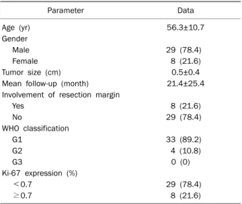
Table 2. Baseline Characteristics of Patients and Tumors
Parameter Data
Age (yr) 56.3±10.7
Gender
Male 29 (78.4)
Female 8 (21.6)
Tumor size (cm) 0.5±0.4
Mean follow-up (month) 21.4±25.4
Involvement of resection margin
Yes 8 (21.6)
No 29 (78.4)
WHO classification
G1 33 (89.2)
G2 4 (10.8)
G3 0 (0)
Ki-67 expression (%)
<0.7 29 (78.4)
≥0.7 8 (21.6)
Values are presented as mean±SD or n (%).
WHO, World Health Organization; G, grade.
Fig. 1. (A) Endoscopic findings. Co- lonoscopy showed a 0.6 cm sized mild yellowish elevated lesion with intact mucosa at anal verge 7.0 cm site. (B) EUS findings. EUS showed a 0.7×0.6 cm sized hypoechoic lesion in the submucosal layer of the rectum.
Fig. 2. Ki-67 staining in tumors resected by endoscopic mucosal resection (×200). On light microscopic examination, moderately uniform, small, round tumor cells with minimal cellular atypia were seen. The arrows indicate Ki-67 labeled nuclei, and the Ki-67 labeling index was 4% in this sample.
presented in Table 2.
Rectal carcinoid tumors had a smooth, round, mobile, sub- mucosal, and nodular appearance. There were no tumors which showed central depression on colonoscopy (Fig. 1A).
EUS was performed before definitive treatment in 19 pa- tients, including 6 patients who have the tumor size ≥1 cm (Fig. 1B). EUS findings typically showed a hypoechoic mass in the submucosal layer of the rectum. Through the work-up including abdominopelvic CT or EUS, no lymph node enlarge- ment or distant metastasis were found in any patients.
Therefore, endoscopic excision was tried for these patients.
In regards to margin positivity after endoscopic resection, 29 patients (78.4%) had clear margins, and 8 patients (21.6%)
had tumor involvement at the resected margins. In patient who showed tumor involvement at the resected margins, short-term follow-up (within 3 months) was done and re- vealed no residual tumors in all cases. Endoscopic examina- tions were performed periodically to monitor for local re- currence during the follow-up period. The median follow-up duration was 21.4±25.4 months (range 0-87 months). Six patients were followed up by more than 3 years, and no recur- rences were identified.
2. Ki-67 expression
The average Ki-67 expression was 0.7±1.2% (Fig. 2).
Subgroups of Ki-67 expression based on tumor size, patient age, gender, and margin positivity are presented in Table 3.
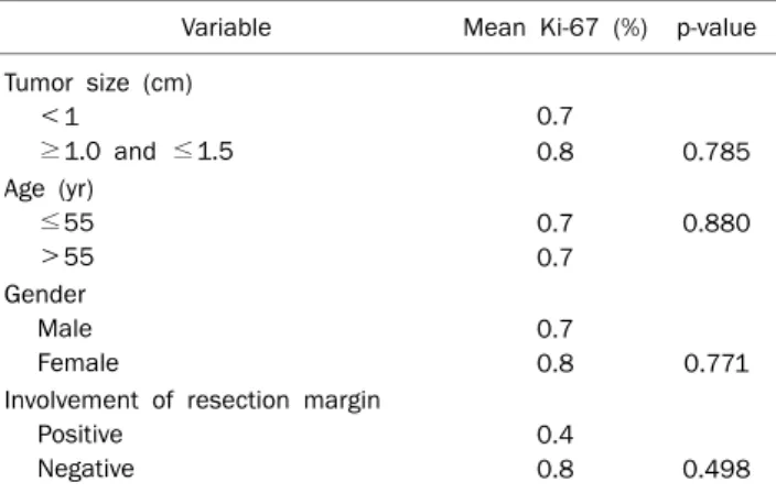
Table 3. Comparative Results of Ki-67 Expression with Size, Age, Gender and Involvement of Resection Margin
Variable Mean Ki-67 (%) p-value Tumor size (cm)
<1
≥1.0 and ≤1.5
0.7
0.8 0.785 Age (yr)
≤55
>55
0.7 0.7
0.880
Gender Male Female
0.7
0.8 0.771 Involvement of resection margin
Positive Negative
0.4
0.8 0.498 p<0.05 accepted as statistically significant.
Table 4. Correlation of WHO Classification Grade with Size, Age, Gender and Involvement of Resection Margin
Variable G1 (n=33) G2 (n=4) p-value Tumor size (cm) 0.5±0.4 0.6±0.4 0.776
Age (yr) 56.6±10.5 53.5±13.2 0.589
Gender
Male 26/33 (78.8) 3/4 (75.0) 0.867
Resection margin
Positive 8/33 (24.2) 0/4 (0.0) 0.279 Values are presented as mean±SD or n (%).
p<0.05 accepted as statistically significant.
WHO, World Health Organization; G, grade.
Table 5. Correlation of Ki-67 Expression with Size, Age, Gender and Involvement of Resection Margin
Ki-67 <0.7%
(n=29)
Ki-67 ≥0.7%
(n=8) p-value
Tumor size (cm) 0.5±0.4 0.4±0.3 0.460
Age (yr) 56.1±10.5 56.8±12.0 0.888
Gender
Male 22/29 (75.9) 7/8 (87.5) 0.493
Resection margin
Positive 6/29 (20.7) 2/8 (25.0) 0.800 Values are presented as mean±SD or n (%).
p<0.05 accepted as statistically significant.
In regards to tumor grading by WHO criteria, 33 patients had grade 1 tumors, and four patients had grade 2 tumors.
In aspect of mitosis, no mitosis was seen in all cases. Tumor size, patient age, gender, and margin positivity were not re- lated to tumor grade (Table 4).
When rectal carcinoid tumor specimens were divided into two groups based on mean value of Ki-67 expression, that was <0.7% or ≥0.7%, there was no correlation between Ki-67 expression and tumor size, patient age, gender, or mar- gin positivity (Table 5).
DISCUSSION
GI carcinoid tumors arise from subepithelial neuroen- docrine cells, penetrate the muscularis mucosa, and invade the submucosal layer at an early stage. Therefore, they ap- pear as submucosal lesions on endoscopic finding. Since the widespread implementation of colorectal cancer screening,
GI carcinoid tumors have begun to be discovered at an early stage.12 Well-differentiated, grade 1 carcinoid tumors less than 10 mm in diameter that are confined to the submucosal layer can be treated with endoscopic resection. A variety of endoscopic techniques are used, including conventional pol- ypectomy, band-snare resection, cap-assisted EMR and ESD.13-15 Previous reports have shown that ESD results in a higher proportion of histologically complete resections with a similar complication rate to EMR.16,17 In our study, most pa- tients received EMR as treatment for their rectal carcinoid tu- mor; only two patients underwent ESD.
EUS has a high sensitivity (87%) in the diagnosis of rectal carcinoid tumors, which are usually well-demarcated iso- echoic or hypoechoic masses.13 The main applications of EUS in rectal carcinoid tumors are for the evaluation of tumor size, depth of invasion, and perirectal lymphadenopathy.18 In our study, EUS was performed in 19 patients, and all tumors were confined to the submucosa without invasion of the mus- cularis propria. Especially, in 6 patients who had tumor sized
≥1 cm, EUS revealed that tumor was confined to the submucosa.
In this study, we evaluated the role of Ki-67 expression as a prognostic factor in low grade rectal carcinoid tumors. Ki-67 expression is widely used as a marker of proliferation in vari- ous types of tumors.19-21 However, the role of Ki-67 ex- pression as a prognostic factor in rectal carcinoid tumors has not been fully established.21-24 Kawahara et al.24 investigated 41 patients with GI carcinoid tumors. Fifty percents were Ki-67 positive in metastatic samples, while 0% were Ki-67 postitive in non-metastatic samples. Tumor cells were con- sidered positive for Ki-67 when ≥10% of the cells were im- munoreactive in the study. Hotta et al.23 investigated 43 pa-
tients with rectal carcinoid tumors and found that the mean Ki-67 expression in metastatic samples was 3.9%, while in non-metastatic samples it was 1.0% (p<0.01). Based on these results they suggested that Ki-67 expression is a reli- able microscopic predictor of metastatic potential in rectal carcinoid tumors.23,24 However, there are no reports on Ki-67 expression in low grade rectal carcinoid tumors.
In our study the mean Ki-67 expression was 0.7±1.2%, suggesting low malignant potential among our 37 patients with small, low grade rectal carcinoid tumors. Other prog- nostic factors such as tumor size, depth of invasion, and lym- phovascular invasion were reviewed. There was no correla- tion between patient age, gender, tumor size, marginal in- volvement and Ki-67 expression.
The mean tumor size in our study was 0.52 cm, and most tumors had grade 1 histology. Therefore, low grade rectal car- cinoid tumors and low Ki-67 expression suggested a good prognosis. We conclude that endoscopic resection is an ef- fective therapeutic strategy for low grade rectal carcinoid tumors. EUS might be helpful to be performed prior to resection. If the tumor is less than 10 mm, without a central depression, with invasion restricted to the mid-submucosa, and no lymph node involvement or distant metastasis, endo- scopic resection is indicated.
Our study has several limitations. The number of sample size was small, and all data were retrospectively reviewed.
There were no comparable control groups, and all enrolled patients had non-metastatic, non-invasive tumors. These compositions of our study population may have contributed to the overall low levels of Ki-67 expression. The follow-up pe- riod of 10 years (with a median follow-up duration of 21.4±25.4 months) was relatively short.
Our study showed that Ki-67 expression was low in low grade rectal carcinoid tumors which are lack of central de- pression on colonoscopy. Therefore, we suggest that endo- scopic resection seemed to be a safe and effective treatment for these tumors.
REFERENCES
1. Jensen RT. Endocrine tumors of the gastrointestinal tract and pancreas. In: Longo D, Fauci A, Kasper D, Hauser S, Jameson J, Loscalzo J, eds. Harrison’s principles of internal medicine.
Volume 2. 18th ed. New York: McGraw Hill, 2011:3056-3065.
2. Pinchot SN, Holen K, Sippel RS, Chen H. Carcinoid tumors.
Oncologist 2008;13:1255-1269.
3. Modlin IM, Kidd M, Latich I, Zikusoka MN, Shapiro MD. Current status of gastrointestinal carcinoids. Gastroenterology 2005;
128:1717-1751.
4. Sun JM, Jung HC. Gastrointestinal carcinoid tumor. Korean J Gastroenterol 2004;44:59-65.
5. Soga J. Early-stage carcinoids of the gastrointestinal tract: an analysis of 1914 reported cases. Cancer 2005;103:1587- 1595.
6. Mashimo Y, Matsuda T, Uraoka T, et al. Endoscopic submucosal resection with a ligation device is an effective and safe treat- ment for carcinoid tumors in the lower rectum. J Gastroenterol Hepatol 2008;23:218-221.
7. Kinoshita T, Kanehira E, Omura K, Tomori T, Yamada H. Transanal endoscopic microsurgery in the treatment of rectal carcinoid tumor. Surg Endosc 2007;21:970-974.
8. Lin MX, Wen ZF, Feng ZY, He D. Expression and significance of Bmi-1 and Ki67 in colorectal carcinoma tissues. Ai Zheng 2008;27:1321-1326.
9. Klimstra DS, Modlin IR, Coppola D, Lloyd RV, Suster S. The patho- logic classification of neuroendocrine tumors: a review of no- menclature, grading, and staging systems. Pancreas 2010;39:
707-712.
10. Bosman F, Carneiro F, Hruban R, Theise N. WHO classification of tumours of the digestive system. Lyon, France: IARC Press, 2010.
11. McCormick D, Yu C, Hobbs C, Hall PA. The relevance of antibody concentration to the immunohistological quantification of cell proliferation-associated antigens. Histopathology 1993;22:543- 547.
12. Modlin IM, Sandor A. An analysis of 8305 cases of carcinoid tumors. Cancer 1997;79:813-829.
13. Kobayashi K, Katsumata T, Yoshizawa S, et al. Indications of en- doscopic polypectomy for rectal carcinoid tumors and clinical usefulness of endoscopic ultrasonography. Dis Colon Rectum 2005;48:285-291.
14. Okamoto Y, Fujii M, Tateiwa S, et al. Treatment of multiple rectal carcinoids by endoscopic mucosal resection using a device for esophageal variceal ligation. Endoscopy 2004;36:469-470.
15. Ahmad NA, Kochman ML, Long WB, Furth EE, Ginsberg GG.
Efficacy, safety, and clinical outcomes of endoscopic mucosal resection: a study of 101 cases. Gastrointest Endosc 2002;55:
390-396.
16. Moon SH, Hwang JH, Sohn DK, et al. Endoscopic submucosal dissection for rectal neuroendocrine (carcinoid) tumors. J Lapa- roendosc Adv Surg Tech A 2011;21:695-699.
17. Park HW, Byeon JS, Park YS, et al. Endoscopic submucosal dis- section for treatment of rectal carcinoid tumors. Gastrointest Endosc 2010;72:143-149.
18. Glancy DG, Pullyblank AM, Thomas MG. The role of colonoscopic endoanal ultrasound scanning (EUS) in selecting patients suit- able for resection by transanal endoscopic microsurgery (TEM).
Colorectal Dis 2005;7:148-150.
19. Costes V, Marty-Ané C, Picot MC, et al. Typical and atypical bron- chopulmonary carcinoid tumors: a clinicopathologic and KI-67- labeling study. Hum Pathol 1995;26:740-745.
20. Moyana TN, Xiang J, Senthilselvan A, Kulaga A. The spectrum of
neuroendocrine differentiation among gastrointestinal carci- noids: importance of histologic grading, MIB-1, p53, and bcl-2 immunoreactivity. Arch Pathol Lab Med 2000;124:570-576.
21. Sökmensüer C, Gedikoglu G, Uzunalimoglu B. Importance of proliferation markers in gastrointestinal carcinoid tumors: a clin- icopathologic study. Hepatogastroenterology 2001;48:720- 723.
22. Shimizu T, Tanaka S, Haruma K, et al. Growth characteristics of
rectal carcinoid tumors. Oncology 2000;59:229-237.
23. Hotta K, Shimoda T, Nakanishi Y, Saito D. Usefulness of Ki-67 for predicting the metastatic potential of rectal carcinoids. Pathol Int 2006;56:591-596.
24. Kawahara M, Kammori M, Kanauchi H, et al. Immunohistochem- ical prognostic indicators of gastrointestinal carcinoid tumours.
Eur J Surg Oncol 2002;28:140-146.