The Correlation between the Injury Patterns of the Medial Patellofemoral Ligament in an Acute First-Time Lateral Patellar Dislocation on MR Imaging and the Incidence of a Second-Time Lateral Patellar Dislocation
Guang-ying Zhang, MD
1, Hong-xia Zhu, MB
2, En-miao Li, MM
3, Hao Shi, MD
4, Wei Liu, MD
1, Lei Zheng, MM
5, Zheng-wu Bai, MD
6, Hong-yu Ding, MB
1Departments of 1Ultrasonography, 4Radiology, and 6Orthopedics, Shandong Provincial Qianfoshan Hospital of Shandong University, Jinan 250014, China; 2Department of Ultrasonography, The Second People’s Hospital of Liaocheng, Liaocheng 252601, China; 3Department of Ultrasonography, Jinan Third People’s Hospital, Jinan 250132, China; 5Department of Radiology, Shandong Provincial Corps Hospital of Chinese People’s Armed Police Force, Jinan 250014, China
Objective: To evaluate the correlation between the injury patterns of the medial patellofemoral ligament (MPFL) on magnetic resonance imaging in an acute first-time lateral patellar dislocation (LPD) and incidence of a second-time LPD.
Materials and Methods: Magnetic resonance images were prospectively analyzed in 147 patients after an acute first-time LPD with identical nonoperative management. The injury patterns of MPFL in acute first-time LPDs were grouped by location and severity for the analysis of the incidence of second-time LPD in a 5-year follow-up. Independent t tests, chi-square tests and Kruskal-Wallis tests were performed as appropriate.
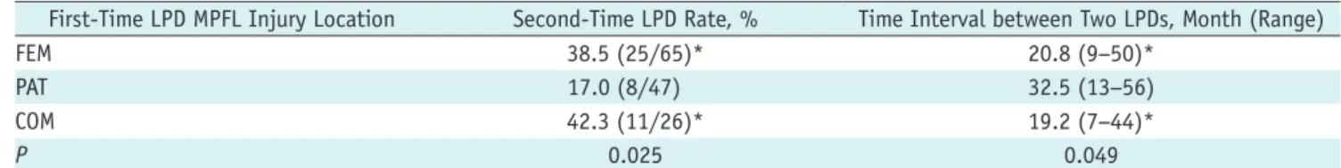
Results: Forty-six cases (46/147, 31.3%) of second-time LPD were present at the 5-year follow-up. Fourteen (14/62, 22.6%) and 31 cases (31/80, 38.8%) were present in the partial and complete MPFL tear subgroups, respectively. Twenty- five cases (25/65, 38.5%), 11 cases (11/26, 42.3%), and 8 cases (8/47, 17%) were present in the isolated femoral-side MPFL tear (FEM), combined MPFL tear (COM), and isolated patellar-side MPFL tear (PAT) subgroups, respectively. Compared with the partial MPFL tears, complete tears showed higher incidence of a second-time LPD (p = 0.04). The time interval between the two LPDs was shorter in the complete MPFL tear subgroup (24.2 months) than in the partial tear subgroup (36.9 months, p = 0.001). Compared with the PAT subgroup, the FEM and COM subgroups showed a higher incidence of a second-time LPD (p = 0.025). The time intervals between the two LPDs were shorter in the FEM and COM subgroups (20.8 months and 19.2 months) than in the PAT subgroup (32.5 months, p = 0.049).
Conclusion: A complete MPFL tear, isolated femoral-side tear and combined tear in a first-time LPD predispose a second- time LPD.
Keywords: Patellar dislocation; Knee; Medial patellofemoral ligament; Recurrence; MRI
Received May 27, 2017; accepted after revision August 10, 2017.
Corresponding author: Hong-yu Ding, MB, Department of Ultrasonography, Shandong Provincial Qianfoshan Hospital of Shandong University, 16766 Jingshi Rd, Li-Xia District, Jinan 250014, China.
• Tel: 86-18663733771 • Fax: (860531) 8296-3647
• E-mail: [email protected]
This is an Open Access article distributed under the terms of the Creative Commons Attribution Non-Commercial License (http://creativecommons.org/licenses/by-nc/4.0) which permits unrestricted non-commercial use, distribution, and reproduction in any medium, provided the original work is properly cited.
INTRODUCTION
Lateral patellar dislocation (LPD) is a common injury that typically occurs in young, active patients as a result of a variety of activities and accounts for approximately 2–3% of all knee injuries (1). Previous studies have shown that anatomic factors such as patella alta, increased tibial tubercle-trochlear groove (TT-TG) distance, rotational deformity and trochlear dysplasia are all associated with an increased risk of a recurrent LPD (2-10). Although injury Korean J Radiol 2018;19(2):292-300
pISSN 1229-6929 · eISSN 2005-8330
to the medial patellofemoral ligament (MPFL) may reduce passive stability and predict subsequent instability with non-operative treatment (11-16), the correlation between injury patterns of MPFL (injury severity and location) in acute first-time LPD and incidence of a recurrent (second- time) LPD has not been fully and clearly clarified (9, 15, 16). Thus, the purpose of this study was to use qualitative and quantitative methods to clarify the correlation between the injury patterns of MPFL in an acute first-time LPD and the incidence of second-time LPD at 5-year follow-up.
MATERIALS AND METHODS
Patients
This study was approved by our Institutional Ethics Committee, and informed consent was obtained from all patients. Patients were eligible for inclusion if they had suffered an acute, first-time LPD regardless of the injury mechanism. The inclusion criteria were as follows (17-21):
1) the presence of a locked acute dislocation or history of reduced dislocation within 15 days of the injury; 2) typical clinical findings including haemarthrosis, medial parapatellar structures, femoral epicondyle that is painful on palpation, and apprehension sign (except in locked dislocation) are present; and 3) a bone contusion involving both the lateral femoral condyle and medial patella as demonstrated on the MRI image.
The exclusion criteria included previous surgery on the injured knee, conditions associated with serious neuromuscular or congenital diseases, and a knee joint with a multiple-ligament injury. When no distinct fibers or remnants were identified at the expected MPFL course on the magnetic resonance (MR) image and there was no appreciable surrounding edema, the ligament was designated as absent and the case was excluded from the study. In addition, the purpose of this study was to investigate long-term nonoperative healing of the injured MPFL; thus, we excluded patients who had undergone patellofemoral surgery (MPFL repair or MPFL reconstruction) after the first-time LPD. However, the patients who only had undergone an arthroscopic operation to remove the osteochondral loose bodies were included in this study.
The selected patients were voluntarily participating in a five-year follow-up study at our hospital. All patients underwent a clinical examination in our orthopedics department six months after the first-time LPD, and then were interviewed by telephone every three months. The
follow-up was performed by an orthopedic surgeon in our study group. The final interview was conducted at 60 months after the first-time LPD. During the follow-up, patients who had experienced knee injuries were examined and cured in our hospital. If the LPD was a relapse (per the criteria mentioned above), the time interval between the two LPDs was recorded. The injury patterns of MPFL in the intraoperative findings were compared with the preoperative MRI examination in patients who were surgically treated (MPFL suture or reconstruction) in the follow-up because of a recurrent LPD. Moreover, during the follow-up, patients were excluded from the study because of other injuries or diseases that occurred between the two LPDs or occurred in the non-recurrent LPD subgroup that prevented the knee joint from returning to its optimal status.
Using the criteria mentioned above, the study enrolled 147 knees of 147 consecutive patients with a mean age of 20 years (range, 8−42 years; 80 women [mean age, 19 years; range, 8−41 years] and 67 men [mean age, 22 years;
range, 9−42 years]).
MRI Technique
The MRI was performed on a 1.5-tesla system (Magnetom Symphony Syngo MR A30; Siemens, Erlangen, Germany), and all patients underwent imaging with their knees positioned in full extension. The following sequences were performed:
a transverse fat-saturated proton-density weighted fast spin-echo imaging sequence (repetition time/echo time, 4500 ms/33 ms; flip angle, 150°; field of view [FOV], 160 x 160 mm; section thickness, 3.0 mm) was followed by a coronal fat-saturated proton density (2000/15; FOV, 150 mm; matrix, 320 x 224 pixels; slice thickness, 3.0 mm; skip, 0.3 mm), a sagittal proton density (1950−2766/14; FOV, 140−150 mm; matrix range, 320−384 x 192−224 pixels;
slice thickness, 3.0 mm; skip, 0.3 mm), a sagittal fat- saturated proton density (2650−4366/13−16; FOV, 140−150 mm; matrix range, 256−384 x 224−256; slice thickness, 3.0 mm; skip, 0.3 mm), and a sagittal T1-weighted sequence (450−600/10−20; FOV range, 140−150 mm; matrix range, 320−384 x 224; slice thickness, 3.0 mm; skip, 0.3 mm).
MRI Evaluation
The MR images were analyzed independently by two radiologists who had 13 years and 16 years of clinical experience in musculoskeletal radiology, respectively, and were both unaware of the results of the previous imaging interpretations. The conclusions of each radiologist were
initially recorded. In the event of disagreement, the images were then reviewed to reach consensus.
Using the diagnostic classification criteria from studies of Zhang et al. and Balcarek et al. (17-24), the degrees of MPFL injury were divided into partial and complete tears.
The manifestations of a partial MPFL tear were defined as thickening and irregularity of the contour, including discontinuity of normal fibers, and intraligamentous or extensive periligamentous edema. The manifestations of a complete MPFL tear were defined as completely discontinuous or apparently absent fibers in the expected region of the MPFL with extensive surrounding edema.
Partial or complete disruption of the MPFL was evaluated at 3 locations: the patellar insertion (PAT), the mid-substance (MID), and the femoral attachment (FEM). An evaluation was also performed on avulsion-type fractures at the PAT or FEM. Simultaneous injury at more than one location of the MPFL was classified as a combined injury (COM) (17-23).
Trochlear dysplasia, which is dysplasia of the femoral trochlea, was categorized as normal, low-grade trochlear dysplasia or high-grade trochlear dysplasia for further analysis (25). The patellar height was evaluated on the sagittal MR images according to the index of Insall and Salvati (i.e., the patellar tendon length to the longest sagittal dimension of the patella). Patella alta was considered with ratios of 1.3 or greater (2, 5, 26, 27). The TT-TG distance was assessed in conformity with Schoettle et al. (28). A TT-TG distance greater than 20 mm was classified as abnormal (4, 10, 27).
Statistical Analysis
Data analyses were performed using SPSS version 17.0 software (SPSS Inc., Chicago, IL, USA). The mean value, standard deviation, and range are presented. Independent samples t tests, chi-square tests and Kruskal-Wallis tests were used to compare the variables in the study subgroups (as described later). A p value of less than 0.05 was considered statistically significant.
RESULTS
Injury Patterns of MPFL in Acute First-Time LPD
An injury to the MPFL was found in 142 patients (96.6%) after an acute first-time LPD, including 62 cases with a partial tear (42.2%) and 80 cases with a complete tear (54.4%). For the remaining 5 patients (3.4%), no obvious MPFL injury was identified. Of the 80 cases with a complete
MPFL tear, 11 of them included an avulsion-type fracture at the patellar insertion and 3 of them included an avulsion- type fracture at the femoral attachment.
Based on the injury patterns of the MPFL (injury location and severity) of an acute first-time LPD, the study group was divided into 2 subgroups for further analyses: injury location (FEM, PAT, or COM) and injury severity (partial or complete tear). Patients with an isolated injury at the MID of the MPFL (4 patients) were excluded from the statistical analyses of the injury location subgroup because of the small sample size. The subgroups were considered parametric in relation to age and sex; there were no
Table 1. Statistical Analysis between Subgroups of MPFL Injury Severity in First-Time LPD and Anatomical Variants
Anatomic Variants MPFL Injury Severity Partial Complete P
Trochlear dysplasia 0.605
Normal 23 36
Low-grade 19 23
High-grade 20 21
Patella alta 0.969
No 37 48
Yes 25 32
TT-TG distance 0.878
Normal 51 65
Abnormal 11 15
Five patients without obvious MPFL injury in first-time LPD were excluded from statistical analysis. LPD = lateral patellar dislocation, MPFL = medial patellofemoral ligament; TT-TG = tibial tuberosity-trochlear groove
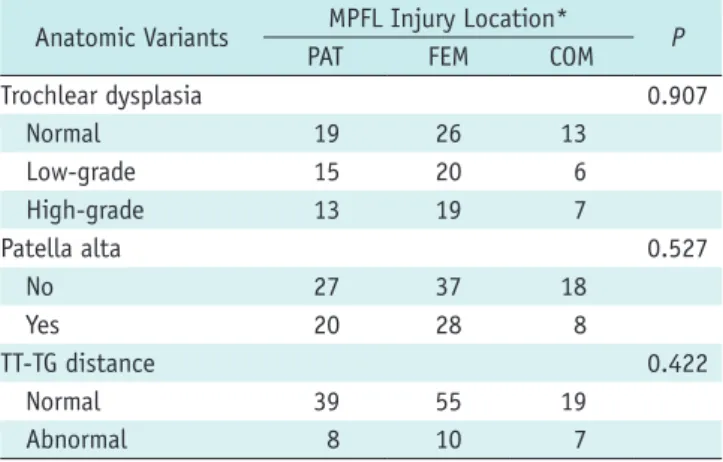
Table 2. Statistical Analysis between Subgroups of MPFL Injury Location in First-Time LPD and Anatomical Variants
Anatomic Variants MPFL Injury Location*
PAT FEM COM P
Trochlear dysplasia 0.907
Normal 19 26 13
Low-grade 15 20 6
High-grade 13 19 7
Patella alta 0.527
No 27 37 18
Yes 20 28 8
TT-TG distance 0.422
Normal 39 55 19
Abnormal 8 10 7
*Patients with isolated lesion at mid-substance (four patients) were excluded from statistical analysis. Five patients without obvious MPFL injury were excluded from statistical analysis. COM = injuries occurred simultaneously in more than one location, FEM = isolated femoral attachment, PAT = isolated patellar insertion
statistically significant differences between the groups.
The results of the correlation analyses between the injury patterns of MPFL in an acute first-time LPD and anatomic variants of the patellofemoral joint are shown in Tables 1 and 2.
Second-Time LPD
In the ensuing 5-year follow-up, the incidence rate of second-time LPD was 31.3% (46 of 147). The time interval between the two LPDs ranged from 7 to 56 months. The injury locations of MPFL in the second-time LPDs were exactly the same as those of the first-time LPD (100%).
Diagnostic Performance of Preoperative MRI
Of the 24 patients who underwent surgery (MPFL suture or reconstruction) after a second-time LPD in our study, 18 had a complete MPFL tear and 6 had a partial MPFL tear.
Overall, 28 localizations of MPFL injury were present in 24 patients. In a site-based analysis, the diagnostic accuracy of MRI was 100% for partial MPFL tears and 100% for complete MPFL tears.
Correlation Analyses between Injury Patterns of MPFL in Acute First-Time LPD and the Incidence of Second-Time LPD
The results of the correlation analyses of the injury patterns of MPFL in acute first-time LPD and the incidence of second-time LPD are shown in Tables 3 and 4.
The incidence rate of second-time LPD in the complete tear subgroup was statistically higher than that of the partial tear subgroup at the 5-year follow-up (38.8% vs.
22.6%, p = 0.04). The mean time interval between two LPDs in the complete tear subgroup was significantly
shorter than that of the partial tear subgroup (24.2 months vs. 36.9 months, p = 0.001) (Table 3). The incidence rates of second-time LPD in the FEM and COM subgroups were significantly higher than that of the PAT subgroup (38.5 and 42.3% vs. 17.0%, p = 0.025). In addition, the mean time intervals between two LPDs were significantly shorter in the FEM and COM subgroups than that of the PAT subgroup (20.8 and 19.2 months vs. 32.5 months, p = 0.049) (Table 4).
Representative cases are shown in Figures. 1-4.
DISCUSSION
As the most important passive restraint against LPD, the MPFL accounts for 50–60% of the total restraining force against a LPD (29-32). A MPFL injury occurred in up to 78.4−100% of the cases after an acute LPD (17-23, 33-38).
The results of the present study were consistent with those of previous studies that MPFL was more easily injured at the FEM, followed by the PAT (17, 20, 38). The distribution of the MPFL tears may be related to the anatomy and injury mechanism of the MPFL and the ossification process of the medial patella border and distal femoral epiphysis (18, 34, 39-41).
Trochlear dysplasia, patella alta and an increased TT-TG distance are primary anatomical variants for a first-time and recurrent LPD (2-10, 27). Trochlear dysplasia is the main risk factor for a LPD, while patellar alta and abnormal TT- TG distance act as additional risk factors for a LPD (27). Our study results were consistent with the findings of Zhang et al. and Weber-Spickschen et al. that demonstrated there were no correlations between the injury patterns of MPFL and trochlear dysplasia, and patellar height and TT-TG groove distance in a first-time LPD (19, 42).
Table 3. Statistical Analysis between Subgroups of MPFL Injury Severity in First-Time LPD and Incidence of Second-Time LPD First-Time LPD MPFL Injury Severity Second-Time LPD Rate, % Time Interval between Two LPDs, Month (Range)
Partial 22.6 (14/62) 36.9 (15−56)
Complete 38.8 (31/80) 24.2 (7−50)
P 0.04 0.001
Five patients without obvious MPFL injury in first-time LPD were excluded from statistical analysis.
Table 4. Statistical Analysis between Subgroups of MPFL Injury Location in First-Time LPD and Incidence of Second-Time LPD First-Time LPD MPFL Injury Location Second-Time LPD Rate, % Time Interval between Two LPDs, Month (Range)
FEM 38.5 (25/65)* 20.8 (9−50)*
PAT 17.0 (8/47) 32.5 (13−56)
COM 42.3 (11/26)* 19.2 (7−44)*
P 0.025 0.049
Patients with isolated lesion at mid-substance (four patients) in first-time LPD were excluded from statistical analysis. *Significant difference compared with isolated patellar insertion injury subgroup.
Fig. 1. 14-year-old girl was diagnosed with partial MPFL tear at its patellar insertion in acute first-time LPD. After 22 months, she had recurrent LPD. Axial fat-saturated proton density- weighted fast spin-echo imaging sequence shows irregularity with intraligamentous edema of MPFL at its patellar insertion (arrow) in first-time LPD. LPD = lateral patellar dislocation, MPFL = medial patellofemoral ligament
Fig. 3. 24-year-old man was diagnosed with complete MPFL tear at its femoral attachment in acute first-time LPD. After 16 months, he had recurrent LPD. Axial fat-saturated proton density-weighted fast spin-echo imaging sequence shows complete discontinuity of MPFL at its femoral attachment with retraction of fibers anteriorly and extensive surrounding edema (open arrow) in first-time LPD.
Fig. 2. 16-year-old male was diagnosed with partial MPFL tear at its femoral attachment in acute first-time LPD. After 23 months, he had recurrent LPD. Axial fat-saturated proton density- weighted fast spin-echo imaging sequence shows irregularity with intraligamentous and periligamentous edema of MPFL at its femoral attachment (arrow) in first-time LPD.
Fig. 4. 22-year-old woman was diagnosed with combined MPFL injury (complete MPFL tear at its femoral attachment and patellar insertion) in acute first-time LPD. After 7 months, she had recurrent LPD. Axial fat-saturated proton density-weighted fast spin-echo imaging sequence shows complete discontinuity of MPFL at its patellar insertion and femoral attachment with retraction of fibers and extensive surrounding edema (arrows) in first-time LPD. Note also small avulsion fracture from medial margin of patella.
The results of the present study showed that recurrent LPD was very common after conservative treatment of the first-time LPD. Moreover, the injury locations of MPFL in the first- and second-time LPDs were exactly the same.
This outcome may be an indicator of the poor ability to self-repair and self-heal a torn MPFL. On the other hand, the present study showed that there was a significant difference in the partial and complete MPFL injuries in a acute first-time LPD with respect to the incidence rate of second-time LPD in the ensuing 5-year follow-up. In addition, the mean interval of time between the two LPDs was shorter in the complete MPFL tear subgroup than that of the partial tear subgroup. This finding can be explained by the fact that MPFL is the primary passive restraint against LPD with a poor ability to self-repair and self-heal, as mentioned above. As the severity of the MPFL injury in first-time LPD (complete tear) increases, the stable force to the patellofemoral joint decreases, thereby becoming more predisposed to a recurrent LPD.
In addition, the present study also showed that the isolated femoral-side and combined MPFL tears in an acute first-time LPD also predisposes the patient to a recurrent LPD. This outcome also can be explained by the anatomy of MPFL. The femoral attachment is the weakest part of MPFL. On the contrary, the patellar insertion of MPFL is much thicker than the femoral attachment and is reinforced by the tendon of vastus medialis obliquus muscle, medial retinaculum, and medial patellotibial ligament at the patellar attachment (39-41). So compared with the patellar insertion of MPFL, the ability to self-repair and self- heal in the FEM subgroup may be even worse. Therefore, the FEM subgroup is more predisposed to re-dislocation with the same external force. We also surmise that the relevant factors are liable to cause re-dislocation in the COM subgroup. However, the anatomy of patellofemoral joint and the injury mechanisms in a LPD are not yet fully understood. Hence, further studies are needed to confirm these hypotheses.
The appropriate therapy for patients with first-time LPD remains a controversial issue (12, 26, 37, 43-48). According to the optimal available evidence, compared with non- operative treatment, operative treatment of acute LPD may result in a lower recurrence rate of LPD, higher health- related quality of life, and sporting function (43, 44, 49).
Although many anatomic variants of the patellofemoral joint have an impact on the incidence of a recurrent LPD, a MPFL repair or reconstruction remains the primary
surgical treatment method (2-10, 43, 44, 49). In fact, MPFL reconstruction has been broadly accepted as the primary surgical treatment for recurrent LPDs with good clinical results (50). Although still in dispute, most studies have suggested that trochlear dysplasia, patella alta and TT-TG distance did not result in a significant difference in the outcomes of an isolated MPFL reconstruction (8, 51-56).
Therefore, the injury patterns of MPFL in first-time LPD may be another important factor to consider when determining the optimal treatment. When the complete, isolated femoral-side or combined MPFL tears are identified after an acute first-time LPD, surgical treatment, including MPFL repair or reconstruction, may be considered.
This study had some limitations. First, the MPFL injury patterns in acute first-time LPDs were not evaluated surgically but based on the MRI findings only; therefore, it was impossible to verify the diagnostic accuracy of the MRI findings in the present study. Nonetheless, of the 24 patients who underwent surgical treatment after a second- time LPD, the accuracy of the preoperative MRI was 100%
in the diagnosis of MPFL injury patterns. In addition, previous studies also have shown that MRI is an accurate method for the diagnosis of the injury patterns of MPFL after an acute LPD (17, 19, 20, 22, 57, 58). Therefore, the present study could be carried out based on the MR images.
Second, anatomical factors of the patellofemoral joint are included only as considerations for the subgroups of injury patterns of MPFL in a first-time LPD and not incorporated as independent parameters in this follow-up study.
Nonetheless, previous studies have already investigated the correlations between the anatomical factors of the patellofemoral joint and incidence of a recurrent LPD (2-10).
The purpose of our study was just to analyze the correlation between the injury patterns of MPFL in a first-time LPD and incidence of a second-time LPD. Accordingly, the anatomical factors are not taken into consideration in a recurrent LPD.
To evaluate the risk factors of a recurrent LPD, further multivariate logistic regressions are required to assess the anatomic factors of patellofemoral joint and injury patterns of MPFL in a first-time LPD. Third, only the incidence of a second-time LPD was analyzed in the present study; the number of LPDs in the 5-year follow-up was not evaluated.
It deserves further attention in future studies.
In conclusion, second-time LPD is very common after conservative treatment following an acute first-time LPD.
Compared with partial MPFL tears, complete tears predispose recurrent LPD, including a higher incidence rate of second-
time LPD and a shorter time interval between dislocations.
Compared with the isolated patellar-side MPFL tears, the isolated femoral-side and combined MPFL tears predispose recurrent LPD, including a higher incidence rates of second- time LPD and shorter time intervals between dislocations.
Thus, the MPFL injury patterns in an acute first-time LPD may be another factor to consider in treatment selections.
REFERENCES
1. Casteleyn PP, Handelberg F. Arthroscopy in the diagnosis of occult dislocation of the patella. Acta Orthop Belg 1989;55:381-383
2. Balcarek P, Jung K, Ammon J, Walde TA, Frosch S, Schüttrumpf JP, et al. Anatomy of lateral patellar instability: trochlear dysplasia and tibial tubercle-trochlear groove distance is more pronounced in women who dislocate the patella. Am J Sports Med 2010;38:2320-2327
3. Balcarek P, Oberthür S, Hopfensitz S, Frosch S, Walde TA, Wachowski MM, et al. Which patellae are likely to redislocate?
Knee Surg Sports Traumatol Arthrosc 2014;22:2308-2314 4. Camp CL, Heidenreich MJ, Dahm DL, Stuart MJ, Levy BA,
Krych AJ. Individualizing the tibial tubercle-trochlear groove distance: patellar instability ratios that predict recurrent instability. Am J Sports Med 2016;44:393-399
5. Diederichs G, Issever AS, Scheffler S. MR imaging of patellar instability: injury patterns and assessment of risk factors.
Radiographics 2010;30:961-981
6. Jaquith BP, Parikh SN. Predictors of recurrent patellar instability in children and adolescents after first-time dislocation. J Pediatr Orthop 2017;37:484-490
7. Lewallen L, McIntosh A, Dahm D. First-time patellofemoral dislocation: risk factors for recurrent instability. J Knee Surg 2015;28:303-309
8. Lewallen LW, McIntosh AL, Dahm DL. Predictors of recurrent instability after acute patellofemoral dislocation in pediatric and adolescent patients. Am J Sports Med 2013;41:575-581 9. Seeley M, Bowman KF, Walsh C, Sabb BJ, Vanderhave KL.
Magnetic resonance imaging of acute patellar dislocation in children: patterns of injury and risk factors for recurrence. J Pediatr Orthop 2012;32:145-155
10. Steensen RN, Bentley JC, Trinh TQ, Backes JR, Wiltfong RE.
The prevalence and combined prevalences of anatomic factors associated with recurrent patellar dislocation: a magnetic resonance imaging study. Am J Sports Med 2015;43:921-927 11. Buckens CF, Saris DB. Reconstruction of the medial
patellofemoral ligament for treatment of patellofemoral instability: a systematic review. Am J Sports Med 2010;38:181- 188
12. Camanho GL, Viegas Ade C, Bitar AC, Demange MK, Hernandez AJ. Conservative versus surgical treatment for repair of the medial patellofemoral ligament in acute dislocations of the patella. Arthroscopy 2009;25:620-625
13. Giordano M, Falciglia F, Aulisa AG, Guzzanti V. Patellar dislocation in skeletally immature patients: semitendinosous and gracilis augmentation for combined medial patellofemoral and medial patellotibial ligament reconstruction. Knee Surg Sports Traumatol Arthrosc 2012;20:1594-1598
14. Petri M, Liodakis E, Hofmeister M, Despang FJ, Maier M, Balcarek P, et al. Operative vs conservative treatment of traumatic patellar dislocation: results of a prospective randomized controlled clinical trial. Arch Orthop Trauma Surg 2013;133:209-213
15. Sillanpää PJ, Peltola E, Mattila VM, Kiuru M, Visuri T, Pihlajamäki H. Femoral avulsion of the medial patellofemoral ligament after primary traumatic patellar dislocation predicts subsequent instability in men: a mean 7-year nonoperative follow-up study. Am J Sports Med 2009;37:1513-1521 16. Sillanpää PJ, Salonen E, Pihlajamäki H, Mäenpää HM. Medial
patellofemoral ligament avulsion injury at the patella:
classification and clinical outcome. Knee Surg Sports Traumatol Arthrosc 2014;22:2414-2418
17. Zhang GY, Zheng L, Ding HY, Li EM, Sun BS, Shi H. Evaluation of medial patellofemoral ligament tears after acute lateral patellar dislocation: comparison of high-frequency ultrasound and MR. Eur Radiol 2015;25:274-281
18. Zhang GY, Zheng L, Feng Y, Shi H, Liu W, Ji BJ, et al. Injury patterns of medial patellofemoral ligament and correlation analysis with articular cartilage lesions of the lateral femoral condyle after acute lateral patellar dislocation in adults: an MRI evaluation. Injury 2015;46:2413-2421
19. Zhang GY, Zheng L, Shi H, Ji BJ, Feng Y, Ding HY. Injury patterns of medial patellofemoral ligament after acute lateral patellar dislocation in children: correlation analysis with anatomical variants and articular cartilage lesion of the patella. Eur Radiol 2017;27:1322-1330
20. Zhang GY, Zheng L, Shi H, Liu W, Zhang L, Qu SH, et al.
Correlation analysis between injury patterns of medial patellofemoral ligament and vastus medialis obliquus after acute first-time lateral patellar dislocation. Knee Surg Sports Traumatol Arthrosc 2016 Dec 27 [Epub]. http://doi. 10.1007/
s00167-016-4408-3
21. Zheng L, Shi H, Feng Y, Sun BS, Ding HY, Zhang GY. Injury patterns of medial patellofemoral ligament and correlation analysis with articular cartilage lesions of the lateral femoral condyle after acute lateral patellar dislocation in children and adolescents: an MRI evaluation. Injury 2015;46:1137-1144 22. Askenberger M, Arendt EA, Ekström W, Voss U, Finnbogason T,
Janarv PM. Medial patellofemoral ligament injuries in children with first-time lateral patellar dislocations: a magnetic resonance imaging and arthroscopic study. Am J Sports Med 2016;44:152-158
23. Balcarek P, Walde TA, Frosch S, Schüttrumpf JP, Wachowski MM, Stürmer KM, et al. Patellar dislocations in children, adolescents and adults: a comparative MRI study of medial patellofemoral ligament injury patterns and trochlear groove anatomy. Eur J Radiol 2011;79:415-420
24. Elias DA, White LM, Fithian DC. Acute lateral patellar
dislocation at MR imaging: injury patterns of medial patellar soft-tissue restraints and osteochondral injuries of the inferomedial patella. Radiology 2002;225:736-743 25. Lippacher S, Dejour D, Elsharkawi M, Dornacher D, Ring
C, Dreyhaupt J, et al. Observer agreement on the Dejour trochlear dysplasia classification: a comparison of true lateral radiographs and axial magnetic resonance images. Am J Sports Med 2012;40:837-843
26. Bitar AC, Demange MK, D’Elia CO, Camanho GL. Traumatic patellar dislocation: nonoperative treatment compared with MPFL reconstruction using patellar tendon. Am J Sports Med 2012;40:114-122
27. Köhlitz T, Scheffler S, Jung T, Hoburg A, Vollnberg B, Wiener E, et al. Prevalence and patterns of anatomical risk factors in patients after patellar dislocation: a case control study using MRI. Eur Radiol 2013;23:1067-1074
28. Schoettle PB, Zanetti M, Seifert B, Pfirrmann CW, Fucentese SF, Romero J. The tibial tuberosity-trochlear groove distance;
a comparative study between CT and MRI scanning. Knee 2006;13:26-31
29. Brown GD, Ahmad CS. Combined medial patellofemoral ligament and medial patellotibial ligament reconstruction in skeletally immature patients. J Knee Surg 2008;21:328-332 30. Conlan T, Garth WP Jr, Lemons JE. Evaluation of the medial
soft-tissue restraints of the extensor mechanism of the knee.
J Bone Joint Surg Am 1993;75:682-693
31. Desio SM, Burks RT, Bachus KN. Soft tissue restraints to lateral patellar translation in the human knee. Am J Sports Med 1998;26:59-65
32. Philippot R, Boyer B, Testa R, Farizon F, Moyen B. The role of the medial ligamentous structures on patellar tracking during knee flexion. Knee Surg Sports Traumatol Arthrosc 2012;20:331-336
33. Balcarek P, Ammon J, Frosch S, Walde TA, Schüttrumpf JP, Ferlemann KG, et al. Magnetic resonance imaging characteristics of the medial patellofemoral ligament lesion in acute lateral patellar dislocations considering trochlear dysplasia, patella alta, and tibial tuberosity-trochlear groove distance. Arthroscopy 2010;26:926-935
34. Felus J, Kowalczyk B. Age-related differences in medial patellofemoral ligament injury patterns in traumatic patellar dislocation: case series of 50 surgically treated children and adolescents. Am J Sports Med 2012;40:2357-2364
35. Kepler CK, Bogner EA, Hammoud S, Malcolmson G, Potter HG, Green DW. Zone of injury of the medial patellofemoral ligament after acute patellar dislocation in children and adolescents. Am J Sports Med 2011;39:1444-1449 36. Putney SA, Smith CS, Neal KM. The location of medial
patellofemoral ligament injury in adolescents and children. J Pediatr Orthop 2012;32:241-244
37. Sillanpää PJ, Mäenpää HM, Mattila VM, Visuri T, Pihlajamäki H. Arthroscopic surgery for primary traumatic patellar dislocation: a prospective, nonrandomized study comparing patients treated with and without acute arthroscopic stabilization with a median 7-year follow-up. Am J Sports Med
2008;36:2301-2309
38. Zhang GY, Zheng L, Shi H, Qu SH, Ding HY. Sonography on injury of the medial patellofemoral ligament after acute traumatic lateral patellar dislocation: injury patterns and correlation analysis with injury of articular cartilage of the inferomedial patella. Injury 2013;44:1892-1898
39. Aragäo JA, Reis FP, de Vasconcelos DP, Feitosa VL, Nunes MA.
Metric measurements and attachment levels of the medial patellofemoral ligament: an anatomical study in cadavers.
Clinics (Sao Paulo) 2008;63:541-544
40. Kikuchi S, Tajima G, Yan J, Kamei Y, Maruyama M, Sugawara A, et al. Morphology of insertion sites on patellar side of medial patellofemoral ligament. Knee Surg Sports Traumatol Arthrosc 2017;25:2488-2493
41. Panagiotopoulos E, Strzelczyk P, Herrmann M, Scuderi G.
Cadaveric study on static medial patellar stabilizers: the dynamizing role of the vastus medialis obliquus on medial patellofemoral ligament. Knee Surg Sports Traumatol Arthrosc 2006;14:7-12
42. Weber-Spickschen TS, Spang J, Kohn L, Imhoff AB, Schottle PB. The relationship between trochlear dysplasia and medial patellofemoral ligament rupture location after patellar dislocation: an MRI evaluation. Knee 2011;18:185-188 43. Erickson BJ, Mascarenhas R, Sayegh ET, Saltzman B, Verma
NN, Bush-Joseph CA, et al. Does operative treatment of first- time patellar dislocations lead to increased patellofemoral stability? A systematic review of overlapping meta-analyses.
Arthroscopy 2015;31:1207-1215
44. Nwachukwu BU, So C, Schairer WW, Green DW, Dodwell ER.
Surgical versus conservative management of acute patellar dislocation in children and adolescents: a systematic review.
Knee Surg Sports Traumatol Arthrosc 2016;24:760-767 45. Palmu S, Kallio PE, Donell ST, Helenius I, Nietosvaara Y. Acute
patellar dislocation in children and adolescents: a randomized clinical trial. J Bone Joint Surg Am 2008;90:463-470
46. Sillanpää PJ, Mäenpää HM. First-time patellar dislocation:
surgery or conservative treatment? Sports Med Arthrosc 2012;20:128-135
47. Sillanpää PJ, Mattila VM, Mäenpää H, Kiuru M, Visuri T, Pihlajamäki H. Treatment with and without initial stabilizing surgery for primary traumatic patellar dislocation.
A prospective randomized study. J Bone Joint Surg Am 2009;91:263-273
48. Smith TO, Donell S, Song F, Hing CB. Surgical versus non- surgical interventions for treating patellar dislocation.
Cochrane Database Syst Rev 2015;CD008106
49. Wang SN, Qin CH, Jiang N, Wang BW, Wang L, Yu B. Is surgical treatment better than conservative treatment for primary patellar dislocations? A meta-analysis of randomized controlled trials. Arch Orthop Trauma Surg 2016;136:371-379 50. Nomura E, Inoue M, Kobayashi S. Long-term follow-up and
knee osteoarthritis change after medial patellofemoral ligament reconstruction for recurrent patellar dislocation. Am J Sports Med 2007;35:1851-1858
51. Howells NR, Barnett AJ, Ahearn N, Ansari A, Eldridge JD.
Medial patellofemoral ligament reconstruction: a prospective outcome assessment of a large single centre series. J Bone Joint Surg Br 2012;94:1202-1208
52. Kita K, Tanaka Y, Toritsuka Y, Amano H, Uchida R, Takao R, et al. Factors affecting the outcomes of double-bundle medial patellofemoral ligament reconstruction for recurrent patellar dislocations evaluated by multivariate analysis. Am J Sports Med 2015;43:2988-2996
53. Matsushita T, Kuroda R, Oka S, Matsumoto T, Takayama K, Kurosaka M. Clinical outcomes of medial patellofemoral ligament reconstruction in patients with an increased tibial tuberosity-trochlear groove distance. Knee Surg Sports Traumatol Arthrosc 2014;22:2438-2444
54. Nelitz M, Dreyhaupt J, Reichel H, Woelfle J, Lippacher S. Anatomic reconstruction of the medial patellofemoral ligament in children and adolescents with open growth plates: surgical technique and clinical outcome. Am J Sports Med 2013;41:58-63
55. Steiner TM, Torga-Spak R, Teitge RA. Medial patellofemoral ligament reconstruction in patients with lateral patellar instability and trochlear dysplasia. Am J Sports Med 2006;34:1254-1261
56. Wagner D, Pfalzer F, Hingelbaum S, Huth J, Mauch F, Bauer G. The influence of risk factors on clinical outcomes following anatomical medial patellofemoral ligament (MPFL) reconstruction using the gracilis tendon. Knee Surg Sports Traumatol Arthrosc 2013;21:318-324
57. Balcarek P, Walde TA, Frosch S, Schüttrumpf JP, Wachowski MM, Stürmer KM. MRI but not arthroscopy accurately diagnoses femoral MPFL injury in first-time patellar dislocations. Knee Surg Sports Traumatol Arthrosc 2012;20:1575-1580
58. Nomura E, Horiuchi Y, Inoue M. Correlation of MR imaging findings and open exploration of medial patellofemoral ligament injuries in acute patellar dislocations. Knee 2002;9:139-143