INTRODUCTION
Contrast-induced nephropathy (CIN) is a serious complica- tion in patients undergoing coronary angiography and any in- tervention that require contrast injection. CIN is defined as absolute (> or=0.5 mg/dL) or relative increase (> or=25%) of serum creatinine after 48–72 hours of exposure to a contrast agent compared to baseline serum creatinine values, when al- terative explanations for renal impairment have been exclud- ed.1 Patients with complex coronary artery disease requiring longer procedural time and higher dosage of contrast media, increase the risk for development of CIN. Although the inci-
dence of CIN following percutaneous coronary intervention (PCI) was estimated to be between 0.6 to 2.3% in general pop- ulation, it was reported to be 5.4–6.2% in patients undergoing PCI for lesions of chronic total occlusion and up to 20% in high risk patients.2,3 The use of low-dose contrast media is im- portant for prevention of CIN. Herein, we report a case of suc- cessful treatment of unprotected left main (LM) bifurcation lesion with minimum contrast volume of 12 cc using intravas- cular ultrasound (IVUS) guidance in an elderly patient.
CASE REPORT
A 79-year-old male was referred due to significant coronary artery stenosis noted by coronary computed tomographic an- giography (CTA). Coronary CTA showed 3-vessel coronary ar- tery and LM disease involving bifurcation with long and heavily calcified lesions of high syntax score of 33. Laboratory investi- gation revealed normal renal function (serum creatinine 0.85 mg/dL). Transthoracic echocardiography revealed absence of regional wall motion abnormality and pharmacologic technei- tum-99m myocardial single photon emission computed to- mography revealed reversible defect in the inferior wall. To
Successful Treatment of Unprotected Left Main
Coronary Bifurcation Lesion Using Minimum Contrast Volume with Intravascular Ultrasound Guidance
Oh-Hyun Lee1, Chul-Min Ahn1, Jung-Sun Kim1,2, Byeong-Keuk Kim1,2, Young-Guk Ko1,2, Donghoon Choi1,2, Yangsoo Jang1,2,3, and Myeong-Ki Hong1,2,3
1Department of Internal Medicine, Severance Cardiovascular Hospital, Yonsei University Health System, Seoul;
2Cardiovascular Research Institute, Yonsei University College of Medicine, Seoul;
3Severance Biomedical Science Institute, Yonsei University College of Medicine, Seoul, Korea.
Contrast-induced nephropathy (CIN) is a serious complication in patients undergoing percutaneous coronary intervention (PCI), and is associated with higher morbidity and mortality. The limiting volume of contrast medium is safest and most reliable strategy for CIN prevention. Intravascular ultrasound (IVUS) serves as an attractive alternative imaging tool to angiography in many steps during PCI, thereby reducing the use of contrast agents. Here, we reported a case of successfully treated unprotected left main bi- furcation lesion with heavily calcified and diffuse lesion under the IVUS-guided PCI using low volumes of contrast dye of total 12 cc in an elderly patient.
Key Words: Contrast-induced nephropathy, intravascular ultrasound, contrast media
pISSN: 0513-5796 · eISSN: 1976-2437
Received: June 22, 2016 Revised: September 30, 2016 Accepted: November 3, 2016
Corresponding author: Dr. Myeong-Ki Hong, Division of Cardiology, Severance Cardiovascular Hospital, Yonsei University Health System, 50-1 Yonsei-ro, Seodae- mun-gu, Seoul 03722, Korea.
Tel: 82-2-2228-8458, Fax: 82-2-393-2041, E-mail: mkhong61@ yuhs.ac
•The authors have no financial conflicts of interest.
© Copyright: Yonsei University College of Medicine 2017
This is an Open Access article distributed under the terms of the Creative Com- mons Attribution Non-Commercial License (http://creativecommons.org/licenses/
by-nc/4.0) which permits unrestricted non-commercial use, distribution, and repro- duction in any medium, provided the original work is properly cited.
Yonsei Med J 2017 Sep;58(5):1066-1070 https://doi.org/10.3349/ymj.2017.58.5.1066
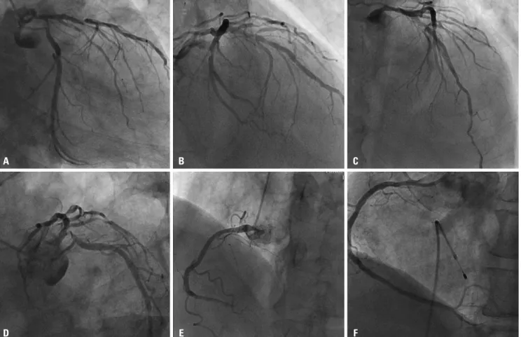
minimize the risk of CIN, intravenous normal saline infusion and usage of iso-osmolar iodine contrast was done. Initial coronary angiography revealed total occlusion of distal right coronary artery (RCA) and LM disease involving bifurcation (Fig. 1A-D). RCA lesion was successfully revascularized with 2.5×13 mm sized Orsiro (Biotronik, Berlin, Germany) stent im- plantation with total 100cc of contrast volume (Fig. 1E and F).
Staged PCI for treatment of LM bifurcation lesion was done via femoral access one week later. Initial left coronary angiog- raphy was done with 6 cc injection of contrast dye (Fig. 2A). Pri- or to PCI, initial angiogram which had already been performed one week ago with same angle of projection was uploaded to the monitor for PCI guidance. After advancing a guide wire into both left anterior descending (LAD) and left circumflex artery (LCX) without use of contrast dye, pre-stent IVUS was per- formed to determine optimal proximal and distal stent landing zones, stent diameter and length (Fig. 2B-F). Recording of po- sition of IVUS transducer on fluoroscopic image was useful to determine the distal landing zone of stent (Fig. 3A and B, ar- row). Based on the angiographic and IVUS findings, two stent technique in a culotte fashion was performed with fluoroscopic guidance, but without the use of contrast dye. Both branches were pre-dilated with 3.5×15 mm non-complaint balloon. Then,
a 3.5×23 mm XIENCE Alpine (Abbott Vascular, Santa Clara, CA, USA) stent was deployed at 11 atm in LM to LCX (Fig. 3C). The stent was re-crossed and the un-stented branch was dilated with 2.0×12 mm and 3.5×23 mm non-complaint balloons. The 3.5×38 mm XIENCE Alpine stent was positioned towards the LM to LAD at 14 atm (Fig. 3D). The first stent was re-crossed and final kissing balloon inflation was performed with two 3.5×15 mm non-compliant balloons (Fig. 3E). Post-stent IVUS confirmed optimal stent deployment without acute complica- tions; minimal lumen area was 8.5 mm2 in proximal LAD and 7.4 mm2 in ostial LCX (Fig. 2G, H, and I). After confirmation of stent optimization with post-stent IVUS examination, final second angiography with 6 cc contrast injection revealed suc- cessful results (Fig. 3F). During the two stent technique in a cu- lotte fashion for LM bifurcation lesions, successful results were achieved with minimal amount of contrast dye and only two angiographic imaging under the IVUS guidance. This patient was clinically stable for 2 days without CIN (Serum creatinine 0.74 mg/dL). This patient was discharged 2 days after PCI and has been uneventful.
Fig. 1. Initial angiography is shown. There were significant stenosis of left main coronary artery, proximal left anterior descending artery with heavy calci- fication and ostium to proximal left circumflex (A-D). Right coronary angiography showed total occlusion of distal right coronary artery (E) which was successfully treated with stent implantation (F).
A
D
B
E
C
F
DISCUSSION
Using small volumes of contrast dye (12 cc), a case of unpro- tected LM bifurcation lesion was successfully treated under the IVUS-guided PCI in an elderly patient without CIN, de- spite this patient had a higher chance to develop CIN during PCI because of old age, long and heavily calcified, LM bifurca-
tion coronary lesion of high syntax score of 33, which usually requires larger amount of contrast medium compared to rou- tine procedure.
Compared to patients without CIN, those with CIN are asso- ciated with higher in-hospital mortality (1.4% vs. 22%, respec- tively, p<0.001) and long term mortality (1-year mortality rate:
3.7% vs. 12.1%, respectively, p<0.001 and 5-year mortality rate:
Fig. 2. Pre-intervention coronary angiography is shown during staged percutaneous intervention with low contrast volume (6 cc) (A) and pre-stent intra- vascular ultrasound images are shown (B-F). Minimal lumen area of ostial left circumflex artery was 2.5 mm2 (B) and the vessel size of distal reference segment in left circumflex artery was about 4.0 mm (C). The vessel size in the left main coronary artery was about 5.5 mm (D). Minimal lumen area of proximal left anterior descending artery was 2.4 mm2 (E) and the vessel size of distal reference segment in left anterior descending artery was about 3.8 mm (F). Optimal results were initially confirmed with post-stent intravascular ultrasound examination [minimal lumen area was 8.5 mm2 in left anterior de- scending artery (H) and 7.4 mm2 in ostial left circumflex artery (I)]. Then, final post-stent angiogram was performed after confirmation of optimal results of two stent implantation in left main bifurcation lesions with intravascular ultrasound (G).
A
D
G
B
E
H
C
F
I
14.5% vs. 44.6%, respectively, p<0.001).3 Therefore, prevention of CIN is the best strategy for improving short and long term outcome after PCI. Usage of minimum volume of contrast dye is safest and most reliable method.
There are several techniques and imaging modalities to min- imize contrast volume including pre-interventional imaging to plan interventional procedure, extensive use of reference im- ages of target vessel anatomy, extensive use of diluted contrast during PCI, use of IVUS or hybrid imaging.4-6 Especially, IVUS serves as an attractive alternative imaging tool to angiography during PCI. Previous study reported that IVUS guidance sig- nificantly reduces the dose of contrast compared with an angi- ography-only approach, that contrast volume used was 3-fold lower in IVUS-guided group compared with angiography- guided group, and that there were notably no differences in flu- oroscopy time, the number of cine runs, or radiation dose.7 IVUS provided valuable information on cross-sectional coronary vascular structure, suitable landing zone, and assistance in se- lection of treatment strategy including stent diameter and length and acute complication.
We used minimal dosage of contrast at the end to confirm any injuries. We strongly believe that ultra-low contrast volume
reduces the rate of CIN development in patients with chronic renal disease who undergo coronary angiography or PCI.6,8
In conclusion, IVUS guidance is a promising method for pa- tients with renal insufficiency who need for inevitable PCI and may be useful to all patients with a high risk of CIN.
REFERENCES
1. Mehran R, Nikolsky E. Contrast-induced nephropathy: definition, epidemiology, and patients at risk. Kidney Int Suppl 2006;(100):
S11-5.
2. Lin YS, Fang HY, Hussein H, Fang CY, Chen YL, Hsueh SK, et al.
Predictors of contrast-induced nephropathy in chronic total oc- clusion percutaneous coronary intervention. EuroIntervention 2014;9:1173-80.
3. Rihal CS, Textor SC, Grill DE, Berger PB, Ting HH, Best PJ, et al. In- cidence and prognostic importance of acute renal failure after per- cutaneous coronary intervention. Circulation 2002;105:2259-64.
4. Azzalini L, Spagnoli V, Ly HQ. Contrast-induced nephropathy: from pathophysiology to preventive strategies. Can J Cardiol 2016;32:
247-55.
5. Nayak KR, Mehta HS, Price MJ, Russo RJ, Stinis CT, Moses JW, et al.
A novel technique for ultra-low contrast administration during an- giography or intervention. Catheter Cardiovasc Interv 2010;75:
1076-83.
Fig. 3. Transducer of intravascular ultrasound is located in left circumflex artery (arrow, A) and left anterior descending artery (arrow, B) on fluoroscopic images, which was useful to determine the distal landing zone of stent. Two stent technique in a culotte fashion was performed with fluoroscopic guid- ance and without use of contrast dye based on the angiographic and IVUS findings (C and D). After final kissing balloon inflation (E), final second angiog- raphy with 6 cc contrast injection revealed successful results (F).
A
D
B
E
C
F
6. Ali ZA, Karimi Galougahi K, Nazif T, Maehara A, Hardy MA, Cohen DJ, et al. Imaging- and physiology-guided percutaneous coronary intervention without contrast administration in advanced renal failure: a feasibility, safety, and outcome study. Eur Heart J 2016;
37:3090-5.
7. Mariani J Jr, Guedes C, Soares P, Zalc S, Campos CM, Lopes AC, et al. Intravascular ultrasound guidance to minimize the use of iodine contrast in percutaneous coronary intervention: the MOZART
(minimizing contrast utilization with IVUS guidance in coronary angioplasty) randomized controlled trial. JACC Cardiovasc Interv 2014;7:1287-93.
8. Okumura N, Hayashi M, Imai E, Ishii H, Yoshikawa D, Yasuda Y, et al. Effects of carperitide on contrast-induced acute kidney injury with a minimum volume of contrast in chronic kidney disease pa- tients. Nephron Extra 2012;2:303-10.