143
ABBREVIATIONS: HCC, hepatocellular carcinoma; CAV1, caveolin-1;
MDR1, multidrug resistance-1; P-gp, P-glycoprotein; si-RNA, small interfering RNA.
Corresponding to: Sung-Vin Yim, Department of Pharmacology, College of Medicine, Kyung Hee University, 1, Hoegi-dong, Dongdae- mun-gu, Seoul 130-701, Korea. (Tel) 82-2-958-9569, (Fax) 82-2-968- 0560, (E-mail) [email protected], Chang-Shin Park, Department of Pharmacology, Center for Advanced Medical Education, Inha Uni- versity College of Medicine by BK21 Project, MTRC, IRIMS, Inha University, 7-241, Shinheung-dong, Jung-gu, Incheon 400-712, Korea.
(Tel) 82-32-890-0962, (Fax) 82-32-887-7488, (E-mail) [email protected]
Role of Caveolin-1 in Indomethacin-induced Death of Human Hepato- adenocarcinoma SK-Hep1 Cells
Kyung-Nam Kim1,2, Ju-Hee Kang2,3, Sung-Vin Yim1, and Chang-Shin Park2,3
1Department of Clinical Pharmacology, College of Medicine, Kyung Hee University, Seoul 130-701, 2Department of Pharmacology, Center for Advanced Medical Education, Inha University College of Medicine by BK21 Project, 3MTRC, Inha Research Institute for Medical Science, Inha University, Incheon 400-712, Korea
Caveolin-1 (CAV1) is an integral membrane protein that may function as a scaffold for plasma membrane proteins and acts as a tumor suppressor protein. One causative factor of chemotherapy- resistant cancers is P-plycoprotein (P-gp), the product of the multidrug resistance-1 gene (M DR1), which is localized in the caveolar structure. Currently, the interactive roles of CAV1 and MDR1 expression in the death of cancer cells remain controversial. In this study, we investigated the effects of indomethacin on the cell viability and the expression levels of MDR1 mRNA and protein in a CAV1- siRNA-mediated gene knockdown hepatoma cell line (SK-Hep1). Cell viability was significantly decreased in CAV1-siRNA-transfected cells compared with that of control-siRNA-transfected cells. Furthermore, the viability of cells pretreated with CAV1 siRNA was markedly decreased by treatment with indome- thacin (400μ M for 24 h). However, the protein and mRNA levels of MDR1 were unchanged in CAV1-siRNA-transfected cells. These results suggest that CAV1 plays an important role as a major survival enzyme in cancer cells, and indomethacin can sensitively induce cell death under conditions of reduced CAV1 expression, independent of MDR1 expression.
Key Words: Caveolin-1, Multidrug resistance-1, siRNA, Indomethacin, SK-Hep1
INTRODUCTION
The caveolins (CAV1, 2, and 3) are integral mem- brane proteins of about 22∼24 kDa that directly bind cho- lesterol and are the primary structural proteins of the cav- eolar membranes (Lavie et al, 1998; Okamoto et al, 1998;
Liu et al, 2002). Of these isoforms, caveolin-1 (CAV1) is known to be a regulatory protein that interacts with the G-protein α subunit, H-Ras, the Src-family tyrosine kin- ases, protein kinase C isoforms, the EGF receptor, and eNOS, thus playing a negative regulatory role in signaling mechanisms (Li et al, 1995, 1996).
Recently, the overexpression of CAV1 has been reported in multidrug-resistant human cancer cells, including MCF-7 breast adenocarcinoma cells, HT-29 colon carcinoma cells, SKVLB1 ovarian carcinoma cells, and A549 lung carcinoma cells (Lavie et al, 1998; Yang et al, 1998; Bender et al, 2000;
Belanger et al, 2004; Zhu et al, 2004). One of the causative factors of chemotherapy-resistant cancers, P-plycoprotein (P-gp), is the product of the multidrug resistance-1 gene (MDR1), and is expressed in the caveolar structure. It is an efflux pump protein for cytotoxic medicines, playing a
survival role in cancer cells (Higgins, 1992; Scotto and Johnson, 2001).
However, the physiological effects of CAV1 on the activity or pattern of expression of MDR are controversial. In a number of human cancer cells that strongly express MDR1, CAV1 expression is increased or undetectable, suggesting that CAV1 expression levels are dependent or independent of those of MDR1 (Haber et al, 1995; Davidson et al, 2002).
Furthermore, P-gp/MDR1 is localized in the caveolae and coimmunoprecipitates with CAV1, indicating that these proteins interact with one another (Demeule et al, 2000).
Pang et al (2004) demonstrated the coordinated expression of CAV1 and MDR1 in normal and leukemic bone marrow, with a positive correlation in the expression levels of the two enzymes.
Hepatocellular carcinoma (HCC) is one of the most life- threatening cancers, and affects many of the world's pop- ulations (Farazi and DePinho, 2006). The control of MDR1 expression is a successful chemotherapeutic strategy for a variety of cancers, especially human HCC (Chen et al, 2006). CAV1 is highly expressed in the SK-Hep1 human hepatoadenocarcinoma cell line, whereas only low levels of MDR1 are expressed.
Indomethacin is a substrate used as an anticancer drug.
In this study, we hypothesized that if CAV1 interacts functionally or coordinately with MDR1 in indomethacin-
Table 1. Nucleotide sequences of primers used in RT-PCR analysis
Target Primers Sequences Product size (bp)
Cav-1 Sense 5' ACG TAG ACT CGG AGG GAC ATC TCT 3' 508
Antisense 5' CTG CAA GTT GAT GCG GAC ATT GC 3'
MDR-1 Sense 5' ATT CAA CTA TCC CAC CCG ACC G 3' 324
Antisense 5' GCT GCC CTC ACG ATC TCT TCC T 3'
18S-rRNA Sense 5' TGC ATG TCT AAG TAC GCA CG 3' 313
Antisense 5' TTG ATA GGG CAG ACG TTC GA 3' induced cell death, MDR1 expression might be altered by
modulation of CAV1 expression, especially under CAV1- siRNA-induced gene knockdown.
METHODS Chemicals and reagents
Dulbecco's modified Eagle medium (DMEM), reduced- se- rum medium (Opti-MEM I), fetal bovine serum (FBS), strep- tomycin/penicillin, LipofectamineTM 2000, and Trypan Blue stain were obtained from Invitrogen (Carlsbad, CA, USA).
Indomethacin, dimethyl sulfoxide (DMSO), protease in- hibitor mixture (P8340), and monoclonal antibody directed against β-actin were purchased from Sigma-Aldrich (St.
Louis, MO, USA). Caveolin-1 siRNA(h) (sc-29241), control siRNA-A (sc-37007), rabbit polyclonal anticaveolin-1 anti- body, and mouse monoclonal antiMDR1 antibody were pur- chased from Santa Cruz Biotechnology (Santa Cruz, CA, USA). Oligonucleotides were obtained from Genotech Co.
Ltd. (Daejeon, Korea).
Cell culture and treatment
SK-Hep1 cells, a human hepatoadenocarcinoma cell line, were purchased from the Korean Cell Line Bank (Seoul, Korea). Cells were grown in DMEM containing 10% FBS, supplemented with 100 units/ml penicillin and 100μg/ml streptomycin (Invitrogen), in a humidified atmosphere of 5% CO2 at 37oC. The cells were maintained in 100 mm cul- ture dishes (TPP, Trasadingen, Switzerland), and seeded into six-well plates with 2×10⁵ cells per well for the meas- urement of cell viability and gene expression. Indomethacin solubilized with dimethyl sulfoxide (DMSO) was stored at 20oC, and used at three different concentrations (100, 500, and 1,000μM). The same volume of DMSO was used as the vehicle control. CAV1 expression levels were also partially knocked down using a CAV1-specific siRNA to control efflux activity, instead of adjusting MDR1 ex- pression, because of the positive correlation between MDR1 and CAV1 expression described in other experiments (Lavie et al, 2001; Pang et al, 2004). Thus, SK-Hep1 cells were treated with 400μM indomethacin in the presence or ab- sence of CAV1 siRNA.
Transient transfection of CAV1 siRNA
As mentioned above, the cells were transfected with com- mercially available CAV1 siRNA using LipofectamineTM 2000 (Invitrogen), following the manufacturer's protocol.
One day before transfection, 2×10⁵ cells per well in six-well plates were seeded into DMEM (2 ml) without antibiotics,
and then incubated until 60∼70% confluence for optimum transfection. After the cells had been rinsed with Opti- MEM I reduced-serum medium (Invitrogen), they were transfected with CAV1 siRNA-LipofectamineTM 2000 or con- trol siRNA-LipofectamineTM 2000 complexes diluted to 30 nmol in the same medium for 4 h, after which growth me- dium containing 20% FBS was added. The transfected cells were maintained for 48 h.
Cell viability
To measure cell viability, the attached cells were washed with phosphate-buffered saline (PBS) and trypsinized. After the dissociated cells had been centrifuged at 2,000 rpm for 3 min, the supernatant was aspirated off, and the cells were gently resuspended with growth medium. The cells were stained with 0.4% Trypan Blue stain (Invitrogen) and counted using a Levy hemacytometer.
Reverse transcriptase-PCR (RT-PCR)
Total RNA was isolated using TRI reagent (Molcular Research Center, Cincinnati, OH) according to the manu- facturer's instructions. RT-PCR was performed to semi- quantitatively measure the expression levels of CAV1 and MDR1 mRNAs relative to that of 18s rRNA as the internal control. Total RNA (1μg) was reverse transcribed using an RNA PCR Kit (AMV version 2.1; TaKaRa, Shinga, Japan), according to the manufacture's protocol. The reaction mix- ture was incubated for 5 min at 30oC to anneal the primer to the RNA template, at 42oC for 30 min, and finally for 5 min at 99oC to inactivate the AMV reverse transcriptase.
The cDNA generated was used for PCR amplification. Table 1 shows the sequences of the primer pairs used to amplify CAV1 and MDR1 mRNAs. The PCR mixture contained 200 mM dNTPs, 0.16 mM each of the sense and antisense pri- mers, 1 unit of Taq DNA polymerase (TaKaRa), 3μl of each cDNA, and sterile distilled water to a volume of 50μl. PCR was carried out as follows: one cycle of 5 min at 94oC, 30 cycles of 1 min at 94oC, 1 min at 55oC, and 2 min at 72oC, and one final cycle of 5 min at 72oC. The amplified products were separated on 1.5% agarose gels in 1×TBE buffer (89 mM Tris, 89 mM borate, and 2 mM EDTA, pH 8.0) and stained with 1 mg/ml ethidium bromide.
Isolation of total cellular proteins and Western-blot analysis
Treated cells were washed with cold PBS and lysed in RIPA buffer [50 mM Tris-HCl (pH 7.4), 1% NP-40, 150 mM NaCl, 1 mM EDTA, 1 mM Na3VO4, 0.5% SDS] containing 10% protease inhibitor cocktail (1 mM DTT, 2μg/ml trypsin inhibitor, 2μg/ml aprotinin, 10μg/ml leupeptin, 1μg/ml
Fig. 1. Indomethacin-induced cell death. Cell viability was deter- mined by cell counting after treatment with different concen- trations of indomethacin (0, 100, 500, and 1,000μM) for 24 h.
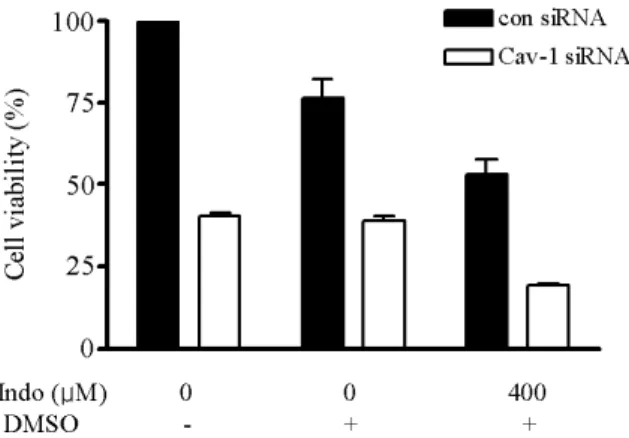
Fig. 2. Viability of CAV1-siRNA-transfected cells. Cell viability was determined by cell counting after treatment with indomethacin (400μM) for 24 h. Cell viability was decreased significantly in CAV1-siRNA-transfected SK-Hep1 cells, and was markedly decreased by indomethacin treatment compared with that in each control.
pepstatin A, and 1 mM PMSF). Total protein concentrations were determined using a Pierce BCA Protein Assay kit (Pierce, Rockford, IL, USA). Equivalent amounts of total protein from each sample were resolved by electrophoresis on SDS-10% polyacrylamide gel. The gels were loaded in running buffer containing 24.9 mM Tris, 194 mM glycine, and 1.156 mM SDS at room temperature and run at 75 V. After the proteins were resolved, they were transferred to a PVDF membrane (Millipore, Miliford, MA, USA) by incubation overnight at 4oC and 30 V in transfer buffer con- taining 25 mM Tris, 192 mM glycine, and 10% methanol.
The membranes were blocked with 5% nonfat dried milk in TBS-T buffer [10 mM Tris-HCl (pH 7.4), 100 mM NaCl, and 0.1% [v/v] Tween 20] at room temperature for 1 h. The membranes were then incubated at room temperature in fresh blocking buffer containing primary antibody directed against CAV1 (1:2,000 dilution), MDR1 (1:100 dilution), or β-actin (1:10,000 dilution). The duration of incubation at 4oC was 1 h to determine the levels of CAV1 and β-actin and overnight for MDR1. The membranes were then in- cubated for 1 h at room temperature with horse- radish-peroxidase-conjugated secondary antibodies (goat anti-rabbit or goat anti-mouse IgG). The specific bands on the membrane were visualized on X-ray film (Kodak, Roche- ster, NY, USA) activated by chemiluminescence (ECLTM Western Blotting System; Pierce, Rockford, IL, USA). The intensity of each band was determined by densitometry us- ing BIO-PROFILE software version 99.04 (Vilber Lourmat, France). Treatment of indomethacin decreased dose-de- pendently the viability of untreated cells (Fig. 1).
RESULTS
Effects of CAV1 siRNA and indomethacin on cell viability in CAV1-siRNA-treated cells
In the cells transiently transfected with CAV1-specific siRNA, cell viability was determined by direct cell counting.
The effects of indomethacin on cell viability in cells pre- treated with CAV1 siRNA were also compared. Treatment of indomethacin decreased dose-dependently the viability of untreated cells (Fig. 1). As shown in Fig. 2, CAV1 siRNA treatment for 24 h decreased cell viability by ∼60% com- pared with the viability of the control-siRNA-treated cells,
and indomethacin (400μM, a toxic concentration) de- creased the viability to ∼75% of that of cells treated with only DMSO (the vehicle for indomethacin). Furthermore, cell viability was markedly decreased by treatment with in- domethacin (400 μM for 24 h) in cells pretreated with CAV1 siRNA (Fig. 2). These results indicate that cell via- bility was significantly decreased by the synergistic effects of knockdown of CAV1 expression and the toxic activity of indomethacin.
Effects of CAV1 siRNA and indomethacin on the expression of CAV1 and MDR1 mRNAs and proteins in CAV1-siRNA-treated cells
We investigated whether MDR1 expression levels are de- pendent or independent of the levels of CAV1, because oth- er reports on normal or cancer cells have been controversial.
In SK-Hep1 cells transiently transfected with control or CAV1 siRNA, the expression of CAV1 and MDR1 mRNA and protein levels were determined by semiquantitative RT-PCR and Western-blot analysis, respectively.
In cells treated with CAV1 siRNA, CAV1 mRNA ex- pression was decreased to ∼50% of the control levels (Fig.
3). However, the levels of MDR1 mRNA expression were unchanged, as shown in Fig. 3. Furthermore, indomethacin (400μM) treatment decreased CAV1 mRNA expression by
∼50% in the control cells and by ∼70% in the CAV1-siRNA- treated cells, independently of the unchanged MDR1 levels (Fig. 3). The expression of these two enzymes at the protein level was consistent with that of the mRNA level, and CAV1 expression was independent of the expression of MDR1 (Fig. 4).
Effects of indomethacin on the expression of CAV1 and MDR1 mRNAs in untreated SK-Hep1 cells
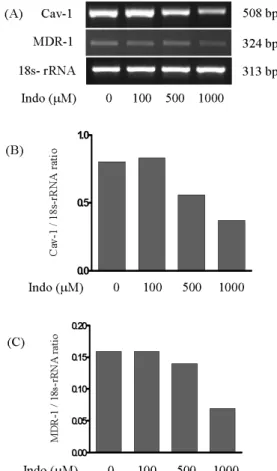
Indomethacin treatment dose-dependently decreased CAV1 mRNA expression levels, whereas those of MDR1 mRNA were unchanged except at 1 mM indomethacin, at which MDR1 mRNA was reduced (Fig. 5). Cell death was severe (>95% cell death) at 1 mM concentration of indomethacin.
However, unlike the CAV1 mRNA expression patterns,
Fig. 3. Effects of indomethacin treatment on the expression of CAV1 and MDR1 mRNAs in CAV1-siRNA-transfected cells. Cells were first transfected for 24 h with control siRNA or CAV1 siRNA.
To complete the knockdown of CAV1 gene expression, a second transfection was performed for 24 h. Forty-eight hours after the first transfection, the cells were treated with indomethacin (400μ M) for 24 h. DMSO was used as the vehicle. Expression levels of CAV1 (B) and MDR1 (C) mRNAs were determined by RT-PCR (A) and normalized against that for 18S rRNA.
Fig. 4. Effects of indomethacin treatment on the expression of CAV1 and MDR1 proteins in CAV1-siRNA-transfected cells. Crude protein was extracted from the cells. DMSO was used as the vehicle.
Expression levels of CAV1 (B) and MDR1 (C) proteins were determined by Western-blot analysis (A) and normalized against those for β-actin.
CAV1 protein expression did not change (Fig. 6). Thus, in- domethacin treatment downregulates CAV1 mRNA only, with no change in CAV1 protein levels.
DISCUSSION
In this study, we examined the effects of indomethacin on the viability of SK-Hep1 human hepatoadenocarcinoma cells transiently transfected with control siRNA or CAV1- specific siRNA. We also examined whether the control of CAV1 expression was related to MDR1 expression.
Many patients with liver cancer suffer from resistance to chemotherapeutic drugs (Nerenstone et al, 1988). A membrane enzyme encoded by the MDR1 gene acts as an efflux pump, protecting the cells from the cytotoxic effects of anticancer drugs (Higgins, 1992). Recently, several inves- tigators have reported that MDR1 is associated with CAV1 in normal and cancer cells (Lavie et al, 1998; Demeule et al, 2000; Lavie et al, 2001; Pang et al, 2004; Cai et al, 2004;
Zhu et al, 2004). However, Davidson (2002) suggested that MDR1 is not related to CAV1, particularly in ovarian cancers. Thus, the correlation between MDR1 and CAV1
enzyme activities remains controversial, because the re- lated mechanisms underlying MDR1 and CAV1 expression are not fully understood.
We used the SK-Hep1 cancer cell line, one of the human hepatoadeno-carcinoma cell lines that overexpress CAV1 but only weakly express MDR1. In these cells, the control of CAV1 expression via transient transfection with CAV1- specific siRNA did not modulate MDR1 expression, at ei- ther the mRNA or protein level, as shown in Figs. 4 and 5. Thus, our results suggest that the functional activities of CAV1 and MDR1 are independent. Importantly, in- domethacin, a known nonsteroidal anti-inflammatory drug, induces drug resistance in cancer cells via the upregulation of MDR1 expression (Rosenbaum et al, 2005). However, in our study, CAV1 was downregulated by indomethacin treatment, with no change in MDR1 expression, and in- domethacin markedly induced cell death, especially in CAV1-siRNA-treated cells, relative to that in the controls.
Furthermore, the downregulation of CAV1 only, without indomethacin treatment, also resulted in ∼50% cell death relative to that in the control siRNA-treated cells. These results suggest that CAV1 plays an important role in the survival of SK-Hep1 cells. As shown in Fig. 2, cell viability was significantly decreased by indomethacin treatment of CAV1-siRNA-transfected cells. Interestingly, Shatz and
Fig. 5. Effects of indomethacin treatment on the expression of CAV1 and MDR1 mRNAs in SK-Hep1 cells. Cells were treated with different concentrations of indomethacin (0, 100, 500, and 1,000μM) for 31 h. Expression levels of CAV1 (B) and MDR1 (C) mRNAs were determined by RT-PCR (A) and normalized against those for 18S rRNA.
Fig. 6. Effects of indomethacin treatment on the expression of CAV1 and MDR1 proteins in SK-Hep1 cells. Crude protein was extracted from cells that had been treated with different concentrations of indomethacin (0, 100, 500, and 1,000μM) for 31 h. Expression levels of CAV1 and MDR1 proteins were determined by Western- blot analysis and normalized against those for β-actin.
Liscovitch (2004) proposed that CAV1 has roles in both growth-inhibitory and survival-promoting actions, partic- ularly as a regulator of prosurvival proteins together with MDR in cancer cells.
Therefore, our results suggest that CAV1 plays a major role in a survival mechanism, at least in the SK-Hep1 can- cer cell line, independently of MDR1 expression. Further investigations are required to identify the molecular mech-
anism by which CAV1 regulates cell survival and modu- lates the expression of other genes in cancer cells.
ACKNOWLEDGEMENT
This study was supported by an intramural research fund of Inha University (2008).
REFERENCES
Belanger MM, Gaudreau M, Roussel E, Couet J. Role of caveolin-1 in etoposide resistance development in A549 lung cancer cells.
Cancer Biol Ther 3: 954−959, 2004
Bender FC, Reymond MA, Bron C, Quest AF. Caveolin-1 levels are down-regulated in human colon tumors, and ectopic expression of caveolin-1 in colon carcinoma cell lines reduces cell tumori- genicity. Cancer Res 60: 5870−5878, 2000
Bruix J, Hessheimer A J, Forner A, Boix L, Vilana R, Llovet JM.
New aspects of diagnosis and therapy of hepatocellular carci- noma. Oncogene 25: 3848−3856, 2006
Cai C, Zhu H, Chen J. Overexpression of caveolin-1 increases plasma membrane fluidity and reduces P-glycoprotein function in Hs578T/Dox. Biochem Biophys Res Commun 320: 868−874, 2004 Chen XP, Wang Q, Guan J, Huang ZY, Zhang WG, Zhang BX.
Reversing multidrug resistance by RNA interference through the suppression of MDR1 gene in human hepatoma cells. World J Gastroenterol 12: 3332−3337, 2006
Davidson B, Goldberg I, Givant-Horwitz V, Nesland JM, Berner A, Bryne M, Risberg B, Kopolovic J, Kristensen GB, Tropé CG, van de Putte G, Reich R. Caveolin-1 expression in ovarian carcinoma is MDR1 independent. Am J Clin Pathol 117: 225−
234, 2002
Dean M, Rzhetsky A, Allikmets R. The human ATP-binding cassette (ABC) t ransporter superfamily. Genome Res 11: 1156−
1166, 2001
Demeule M, Jodoin J, Gingras D, Beliveau R. P-glycoprotein is localized in caveolae in resistant cells and in brain capillaries.
FEBS Lett 466: 219−224, 2000
Farazi PA, DePinho RA. Hepatocellular carcinoma pathogenesis:
from genes to environment. Nat Rev Cancer 6: 674−687, 2006 Fielding CJ, Fielding PE. Caveolae and intracellular trafficking of
cholesterol. Adv Drug Deliv Rev 49: 251−264, 2001
Galbiati F, Volonte D, Meani D, Milligan G, Lublin DM, Lisanti MP, Parenti M. The dually acylated NH2-terminal domain of gi1alpha is sufficient to target a green fluorescent protein reporter to caveolin-enriched plasma membrane domains. Palmitoylation of caveolin-1 is required for the recognition of dually acylated g-protein alpha subunits in vivo. J Biol Chem 274: 5843−5850, Gottesman MM, Fojo T, Bates SE. Multidrug resistance in cancer: 1999 role of ATP-dependent transporters. Nat Rev Cancer 2: 48−58, 2002
Haber M, Burkhart CA, Regl DL, Madafiglio J, Norris MD, Horwitz SB. Altered expression of M beta 2, the class II beta-tubulin isotype, in a murine J774.2 cell line with a high level of taxol resistance. J Biol Chem 270: 31269−31275, 1995
Higgins CF. ABC transporters: from microorganisms to man. Annu Rev Cell Biol 8: 67−113, 1992
Kogo H, Fujimoto T. Caveolin-1 isoforms are encoded by distinct mRNAs. Identification of mouse caveolin-1 mRNA variants caused by alternative transcription initiation and splicing. FEBS Lett 465: 119−123, 2000
Krajewska WM, Maslowska I. Caveolins: structure and function in signal transduction. Cell Mol Biol Lett 9: 195−220, 2004 Lavie Y, Fiucci G, Liscovitch M. Up-regulation of caveolae and
caveolar constituents in multidrug-resistant cancer cells. J Biol Chem 273: 32380−32383, 1998
Lavie Y, Fiucci G, Liscovitch M. Upregulation of caveolin in
multidrug resistant cancer cells: functional implications. Adv Drug Deliv Rev 49: 317−323, 2001
Li L, Ren CH, Tahir SA, Ren C, Thompson TC. Caveolin-1 main- tains activated Akt in prostate cancer cells through scaffolding domain binding site interactions with and inhibition of serine/
threonine protein phosphatases PP1 and PP2A. Mol Cell Biol 23: 9389−9404, 2003
Li S, Okamoto T, Chun M, Sargiacomo M, Casanova JE, Hansen SH, Nishimoto I, Lisanti MP. Evidence for a regulated interac- tion between heterotrimeric G proteins and caveolin. J Biol Chem 270: 15693−15701, 1995
Li S, Song KS, Koh SS, Kikuchi A, Lisanti MP. Baculovirus-based expression of mammalian caveolin in Sf21 insect cells. A model system for the biochemical and morphological study of caveolae biogenesis. J Biol Chem 271: 28647−28654, 1996
Liscovitch M, Lavie Y. Multidrug resistance: a role for cholesterol efflux pathways? Trends Biochem Sci 25: 530−534, 2000 Liu J, Oh P, Horner T, Rogers RA, Schnitzer JE. Organized
endothelial cell surface signal transduction in caveolae distinct from glycosylphosphatidylinositol-anchored protein microdomains.
J Biol Chem 272: 7211−7222, 1997
Liu P, Rudick M, Anderson RG. Multiple functions of caveolin-1.
J Biol Chem 277: 41295−41298, 2002
Nerenstone SR, Ihde DC, Friedman MA. Clinical trials in primary hepatocellular carcinoma: current status and future directions.
Cancer Treat Rev 15: 1−31, 1988
Nieth C, Priebsch A, Stege A, Lage H. Modulation of the classical multidrug resistance (MDR) phenotype by RNA interference (RNAi). FEBS Lett 545: 144−150, 2003
Okamoto T, Schlegel A, Scherer PE, Lisanti MP. Caveolins, a family of scaffolding proteins for organizing "preassembled signaling complexes" at the plasma membrane. J Biol Chem 273: 5419−
5422, 1998
Pang A, Au WY, Kwong YL. Caveolin-1 gene is coordinately regulated with the multidrug resistance 1 gene in normal and leukemic bone marrow. Leuk Res 28: 973−977, 2004
Quest AF, Leyton L, Parraga M. Caveolins, caveolae, and lipid rafts in cellular transport, signaling, and disease. Biochem Cell Biol 82: 129−144, 2004
Razani B, Engelman JA, Wang XB, Schubert W, Zhang XL, Marks CB, Macaluso F, Russell R G., Li M, Pestell RG, Di Vizio D, Hou H Jr, Kneitz B, Lagaud G, Christ GJ, Edelmann W, Lisanti
MP. Caveolin-1 null mice are viable but show evidence of hyperproliferative and vascular abnormalities. J Biol Chem 276:
38121−38138, 2001a
Razani B, Zhang XL, Bitzer M, von Gersdorff G, Bottinger EP, Lisanti MP. Caveolin-1 regulates transforming growth factor (TGF)-beta/SMAD signaling through an interaction with the TGF-beta type I receptor. J Biol Chem 276: 6727−6738, 2001b Rosenbaum C, Rohrs S, Muller O, Waldmann H. Modulation of
MRP-1-mediated multidrug resistance by indomethacin analogues.
J Med Chem 48: 1179−1187, 2005
Roy S, Luetterforst R, Harding A, Apolloni A, Etheridge M, Stang E, Rolls B, Hancock JF, Parton RG. Dominant-negative caveolin inhibits H-Ras function by disrupting cholesterol-rich plasma membrane domains. Nat Cell Biol 1: 98−105, 1999
Schlegel A, Schwab RB, Scherer PE, Lisanti MP. A role for the caveolin scaffolding domain in mediating the membrane attach- ment of caveolin-1. The caveolin scaffolding domain is both necessary and sufficient for membrane binding in vitro. J Biol Chem 274: 22660−22667, 1999a
Schlegel A, Wang C, Katzenellenbogen BS, Pestell RG, Lisanti MP.
Caveolin-1 potentiates estrogen receptor alpha (ERalpha) signal- ing. caveolin-1 drives ligand-independent nuclear translocation and activation of ERalpha. J Biol Chem 274: 33551−33556, 1999b
Scotto KW, Johnson RA. Transcription of the multidrug resistance gene MDR1: a therapeutic target. Mol Interv 1: 117−125, 2001 Shatz M, Liscovitch M. Caveolin-1 and cancer multidrug resistance:
coordinate regulation of pro-survival proteins? Leuk Res 28: 907
−908, 2004
Westermann M, Leutbecher H, Meyer HW. Membrane structure of caveolae and isolated caveolin-rich vesicles. Histochem Cell Biol 111: 71−81, 1999
Williams TM, Lisanti MP. Caveolin-1 in oncogenic transformation, cancer, and metastasis. Am J Physiol Cell Physiol 288: C494−
506, 2005
Yang CP, Galbiati F, Volonte D, Horwitz SB, Lisanti MP.
Upregulation of caveolin-1 and caveolae organelles in Taxol-resistant A549 cells. FEBS Lett 439: 368−372, 1998 Zhu H, Cai C, Chen J. Suppression of P-glycoprotein gene expre-
ssion in Hs578T/Dox by the overexpression of caveolin-1. FEBS Lett 576: 369−374, 2004