1)
Introduction
Maple syrup urine disease (MSUD, OMIM#24 8600) is a rare and autosomal recessively-in- herited metabolic disorder caused by mutations in a complex enzyme system encoded by branched- chain -ketoacid dehydrogenase (BCKDH) genes. α This mitochondrial enzyme complex consists of four subunits: E1 , E1 , E2, and E3 α β
1). The defects in E1 , E1 , E2, and E3 are classified as MSUD α β
책임저자 고정민 신승한 서울시 종로구 대학로: , , 101 서울대학교 어린이병원 소아청소년과
Tel: 02)2072-3570, Fax: 02)743-3455 E-mail: [email protected]
type 1A, type 1B, type 2, and type 3, respectively.
Deficiencies in any of these subunits prevents the normal breakdown of branched-chain amino acids, such as leucine, isoleucine, and valine.
Based on the age of symptom-onset, severity of clinical symptoms or signs, and responsiveness to thiamine, MSUD can be classified as either classic, intermediate, intermittent, or thiamine- responsive
2). Classic MSUD is the most common and most severe form of MSUD. Patients with classic MSUD often present with poor feeding, vomiting, lethargy, and irritability within the first two days of life. Apnea, dystonia, and seizures
기면과 중추성 무호흡으로 나타난 단풍시럽뇨병 신생아 례
Type 1B 1
서울대학교병원 어린이병원 소아청소년과
강영태 최성환 고정민ㆍ ㆍ *ㆍ신승한 김이경 김한석ㆍ ㆍ
A Newborn Case of Maple Syrup Urine Disease Type 1B Presenting with Lethargy and Central Apnea
Youngtae Kang, Sung Hwan Choi, Jung Min Ko*
Seung Han Shin, Ee-Kyung Kim, Han-Suk Kim
Department of Pediatrics, Seoul National University Children s Hospital,’ Seoul National University College of Medicine, Seoul, Korea
Maple syrup urine disease (MSUD, OMIM#248600) is a rare and autosomal recessively-inherited meta- bolic disorder that is caused by mutations in the branched-chain -ketoacid dehydrogenase (BCKDH) α genes. It prevents the normal breakdown of branched-chain amino acids (BCAAs), such as leucine, iso- leucine, and valine, and leads to poor feeding, lethargy, abnormal movements, seizure, and death if untreated. Here, we report the case of a Korean newborn of biochemically- and genetically-confirmed MSUD manifesting lethargy and central apnea, the acute state of which was successfully treated. The molecular genetic investigation revealed two novel heterozygous mutations (p.Ala32Phefs*48 and p.Val 130Phe) in BCKDHB , and both parents were confirmed as carriers. We emphasize the importance of early diagnosis and prompt introduction of specific treatment for MSUD in life saving and prognosis.
Key words: Maple syrup urine disease, Branched-chain -ketoacid dehydrogenase, Branched-chain α
amino acid, Central apnea
may present by four to seven days of life
3). In- termittent and intermediate forms of MSUD show less severe metabolic and clinical manifestations than classic MSUD. MSUD is often initially sus- pected based on a maple syrup odor in bodily fluids, especially in urine. Diagnosis is typically confirmed through amino acid analysis showing markedly increased levels of leucine, isoleucine, alloisoleucine (a metabolite of leucine not normally found in blood), and valine. The worldwide pre- valence of MSUD is 1 in 185,000 live births. How- ever, it is much rarer in Korea, at a rate of 1 in 1,148,413 live births
4), which is related to the lack of a founder mutation cluster. In some pop- ulations such as Mennonite, the incidence is as high as 1 in 380 as a result of the representative founder variant (c.1312T>A) in BCKDHA
5).
Here, we report a case of a Korean newborn with biochemically- and genetically-confirmed classic MSUD (type 1B) manifesting with keto- acidosis, hyperammonemia, lethargy and central apnea.
Case Report
A girl was born to a 24-year-old woman with para 0-0-0-0 as a singleton at the gestational age of 39+6 weeks, with a birth weight of 2.54 kg, via normal spontaneous vaginal delivery. She was the first baby of healthy and nonconsangui- neous parents and did not have any perinatal pro- blems or family history of inherited metabolic dis- eases. At 10 days of age, she visited the emerg- ency room at the local hospital because of lethargy and poor-sucking. The initial laboratory data showed hypoglycemia (serum glucose 45 mg/dL) and ketoacidosis (VBGA pH 7.28, pCO
240 mmHg, HCO
3-20.1 mmHg, base excess -9.8 mEq/L, lactate 1.3 mmol/L), so she was admitted for in-
travenous hydration. Over the next day, the re- sults of newborn screening performed at the age of three days showed a markedly increased level of leucine (41.1 mg/dL, reference range (ref.) <
4.1 mg/dL), and MSUD was suspected. She was transferred to Seoul National University Children s ’ Hospital for further evaluation and management.
As for the findings of a physical examination upon admission to our hospital, she looked le- thargic and her Glasgow Coma Scale (GCS) score was 7 of 15 (E2V1M4). Her length, body weight, and head circumference were 47.5 cm (3-10
thpercentile), 2,610 gm (<3
rdpercentile), and 33 cm (3-10
thpercentile), respectively, and her urine smelled like maple syrup. She had central apnea and repetitive desaturation down to SpO
240%
which appeared nearly every hour. Therefore, endotracheal intubation was performed and con- ventional ventilation was begun. Intravenous glu- cose (glucose infusion rate 9.7 mg/kg/min) and lipid without protein were administered and bran- ched-chain amino acid (BCAA) low formula fee- ding was started via a gavage tube. Thiamine (10 mg/kg/d) was also tried for the evaluation of thia- mine responsiveness. The biochemical analysis upon admission showed hyperuricemia (11.3 mg/
dL, ref. 3.0-7.0 mg/dL) and hyperammonemia
(236 μ mol/L, ref. 15-51 μ mol/L). Rasburicase
and sodium benzoate were begun intravenously
and the follow up laboratory findings showed de-
creased levels of uric acid (<3.0 md/dL) and am-
monia (<150 mol/L). The initial electroenceph μ -
alography showed epileptiform discharge and phe-
nobarbital was begun for seizure prevention. The
brain sonography showed cerebral edema involving
the deep gray matter (basal ganglia/thalami), brain-
stem, and cerebellum, while the brain MRI showed
bilateral symmetrical hyperintensity with diffusion
restriction, suggesting cytotoxic edema involving
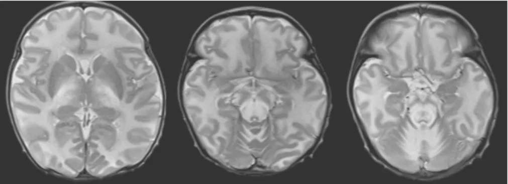
the optic chiasm, optic radiate, corticospinal tract, the anterior and posterior limb of internal capsule, thalami, posterior midbrain, pons, medulla, and the cerebellar white matter (Fig. 1). We started furo- semide 1 mg/kg q 6 hrs and mannitol 0.5 g/kg q 6 hrs in order to manage the diffuse brain edema.
The follow up brain sonography after seven days showed equivocal decreased diffuse edema. The initial serum amino acid analysis showed very high levels of isoleucine (979 mol/L, ref. 12-77 mol/ μ μ L), leucine (4,819 mol/L, ref. 46-147 mol/L), μ μ alloisoleucine (726 mol/L, ref. <2 mol/L), and μ μ valine (762 mol/L, ref. 79-217 mol/L). The ini μ μ - tial urine organic acid analysis showed increased
2-hydroxyisovaleric acid, 2-ketoisocaproic acid, 2-hydroxyisocaproic acid, and 2-hydroxy-3-me- thylvaleric acid. She could be diagnosed clinically as having the classic type of MSUD. We followed up serum amino acid analysis with specific treat- ment and a low-BCAA formula, and this was fol- lowed by decreased levels of the branched chain amino acids (Fig. 2).
After 29 days of NICU care, the GCS had in- creased to 15 and the respiration was stable without apnea in air, and a full oral feeding of low-BCAA formula was successfully achieved.
She was transferred to the general ward and dis- charged at the age of 41 days. She has since vi-
Fig. 1. The brain MRI showed bilateral symmetrical hyperintensity with diffusion restriction, suggesting cytotoxic edema involving the optic chiasm, optic radiate, corticospinal tract, the anterior and posterior limb of internal capsule, thalami, posterior midbrain, pons, medulla, and the cerebellar white matter.
Fig. 2. The follow-up serum amino acid analyses showed decreased levels of leucine with specific treatment. The level has since been kept below 800 mol/L in the patient.μ
sited the outpatient clinic on a regular basis for growth and development check-ups and for moni- toring of her BCAA levels. She is now five months old with a height of 60 cm (<3
rdpercentile), weight of 6.1 kg (3-10
thpercentile), and head circumfe- rence of 41.3 cm (25-50
thpercentile). She can babble and roll over toward both sides, although her head control is not perfect. Phenobarbital is being tapered and her levels of BCAA are kept stable below 800 mol/L of leucine. μ
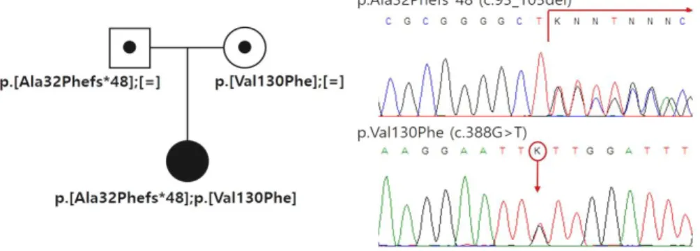
Under the clinical diagnosis of MSUD, the mo- lecular genetic investigation for BCKDHA, BCKDHB, and DBT was performed, revealing two novel he- terozygous mutations (p.Ala32Phefs*48 and p.
Val130Phe) in BCKDHB. Both parents harbored each of the two identified mutations and they were both confirmed as carriers (Fig. 3).
Discussion
Maple syrup urine disease is caused by de- creased activity of branched-chain α -ketoacid dehydrogenase (BCKAD), a multi-enzyme complex in the mitochondria. BCKAD has four subunits (E1 , E1 , E2, and E3). The gene of each subunit α β is located on different chromosomes: E1 ( α BCKDHA) on chromosome 19q13.1-q13.2, E1 ( β BCKDHB ) on chromosome 6q14.1, E2 (Dihydrolipoamide
branched chain transacylase, DBT) on chromosome 1p21.2, and E3 (Dihydrolipoamide dehydrogenase, DLD) on chromosome 7q31.1
6).
MSUD type 1A, which is a deficiency of E1 , α accounts for 45% of all cases, comprising appro- ximately 1 in 411,000 newborns. There are about 70 reported sequence variants in E1α
7,8). Type 1B, a deficiency in E1 , accounts for 35%, or ap β - proximately 1 in 528,000 newborns. This type is much more commonly seen in Ashkenazi Jews.
More than 70 sequence variants in E1 have been β reported to date
8,9). It is known that most cases of MSUD involve compound heterozygotes for rare sequence variants
10). In Korea, less than 10 MSUD patients have been reported, and only three of them have been confirmed through molecular ge- netic testing
11). In this case, the molecular genetic study revealed two novel heterozygous mutations:
c.93_103del (p.Ala32Phefs*48) from the father and c.388G>T (p.Val130Phe) from the mother, re- spectively. p.Ala32Phefs*48 leads to premature truncation of protein synthesis, and p.Val130Phe is a missense mutation. The other variant at the same location, p.Val130Gly, has been previously reported as affecting cofactor binding
12)so p.Val 130Phe is thought to be pathological. As a result, the novel variants combined together are thought to be related to very low residual BCKDHB activity.
Fig. 3. The molecular genetic study revealed two novel heterozygous mutations (p.Ala32Phefs
*48 and p.Val130Phe) in BCKDHB, and both parents were confirmed as carriers.
A deficiency of E3 produces congenital lactic acidosis because the E3 subunit shares pyruvate dehydrogenase and α -ketoglutarate dehydroge - nase. It shows increased plasma levels of pyru- vate and alanine and elevated urinary excretion of branched-chain ketoacids (BCKAs) and alpha- ketoglutarate. The clinical phenotype of E3 sub- unit deficiency differs from that of classic or inter- mediate MSUD. It ranges from early-onset neu- rologic symptoms to adult-onset liver complica- tions. Affected infants usually do not survive their first few years of life but those who survive ma- nifest intellectual ataxia, disability, spasticity and seizure. Adult-onset E3 deficiency has also been reported. If the onset is later in life, liver compli- cations are usually the only symptom
13).
Neonatal screening tests make it possible to detect MSUD early on; however, affected neonates might show clinical signs and symptoms before the results of the screening test are even re- turned, as seen in the present case. Now, if the pathogenic variants of BCKDHA, BCKDHB, or DBT have been found in a family, prenatal diag- nosis for future pregnancy is possible through the genetic testing of DNA from amniocytes or cho- rionic villi
14).
In the case of an acute MSUD crisis, sufficient nutrients and calories should be provided as soon as possible, because the patient is under a cata- bolic state. Rapid removal of BCAAs is also ne- cessary. In this case, hydration and intake restric- tion of BCAAs without dialysis improved her metabolic state, although many publications have shown that hemodialysis or peritoneal dialysis can remove BCAAs and BCKAs rapidly during the acute phase of MSUD
15). Dialysis should be con- sidered in patients presenting with severely altered mental status and cerebral edema
16).
BCAAs, especially leucine, are known to be
cytotoxic to brain cells, leading to cytotoxic ce- rebral edema affecting the myelinated white matter
17)
. As a result, brain MRI during the acute state may show marked restricted diffusion, reflecting intracellular edema, involving the corticospinal tracts (the posterior limbs of the internal capsule), thalami, globus palladi, midbrain, dorsal brain stem, and cerebellar white matter
18). In this case, the brain sonography and brain MRI showed diffusion restriction, suggesting cytotoxic edema involving the deep gray matter (basal ganglia/thalami), brain- stem, and cerebellum. Although the definite effect of mannitol and furosemide on cytotoxic edema is not known, we successfully attempted to find the decreased cerebral edema on the follow-up brain sonographies.
Even following recovery from the acute state, patients should be kept on a diet low in BCAAs for the rest of their lives
19). Dietary management should consider age-appropriate nutrition and to- lerance of BCAAs. Patients also need frequent clinical and biochemical monitoring in order to maintain stable plasma BCAA levels. Severe ke- toacidosis, cerebral edema, and even death can occur in the face of any stressful event, such as infection or injury, because infections and injuries mobilize a large endogenous muscle protein, cau- sing a metabolic crisis
3).
For patients in which diet modification and other conservative management have failed, liver trans- plantation might be considered. Only 10% of BCKDH is expressed in the liver and liver transplantation does not reverse the neurologic or psychiatric illness. However, it is known to decrease the fre- quency and the severity of life-threatening ce- rebral edema and to prevent the progression of neurocognitive impairment
20).
The prognosis of MSUD is related to the age
of the child at the time of diagnosis and to the
adequacy of metabolic control
21). Although our patient had the classic infantile form of MSUD manifested by metabolic crisis on the day of her birth, she has since caught up with development without metabolic events through the application of immediate specific treatment. Here, we empha- size the importance of early diagnosis and prompt introduction of specific treatment for MSUD in life saving and prognosis.
요 약
단풍시럽뇨병은 드문 상염색체 열성 대사 질환으로 측쇄 알파 케토산 탈수소효소 돌연변이에 의해 발생되 는 질환이다 측쇄 아미노산인 류신 이소류신 발린의 . , , 분해가 되지 않아 몸에 축적이 되고 식욕 저하 구토 , , 기면 이상행동 발작 및 심한 경우 죽음을 초래한다 , , . 세계적으로 단풍시럽뇨병의 유병율은 185,000 명당 한 명 이지만 한국에서는 극히 드물어 1,148,413 명당 한 명으로 보고된다.
이 증례의 여아는 만삭에 2.54 kg 로 주산기 특이 병 력 없이 출생한 첫째 아이로 특이 가족력 없었던 환아 이다 생후 . 10 일경 식욕저하 구토 기면으로 타병원에 , , 서 대증치료 중에 선천성 대사이상 선별 검사 결과 상 류신이 높아 전원 되었다 환아는 기면 혼수를 보였고 . , 중추 수면무호흡으로 인공호흡기를 적용하였으며 소변 에서 단풍시럽 냄새를 보였다 뇌 초음파 뇌 자기공명 . , 영상에서 뇌부종 소견을 보였으며 혈중아미노산 검사 상 류신 이소류신 발린의 수치가 정상치보다 매우 높 , , 았다 측쇄 아미노산 없는 특수분유를 시작했고 뇌부종 . 으로 만니톨과 이뇨제를 사용했으며 고암모니아혈증으 로 벤조산을 투약하였다 경련 예방을 위해 페노바비탈 . 을 투약하였으며 측쇄 알파 케토산 탈수소효소의 조효 소인 타이아민 보충을 시작하였다 치료함에 따라 혈중 . 측쇄 아미노산 수치가 감소하였고 뇌부종의 감소가 확 인되었다 . 29 일 간의 신생아중환자실 처지 후 환아는 호흡 및 구강 식이 진행이 안정적이었고 병동으로 전동 후 퇴원하였다 환아는 현재 개월로 페노바비탈을 점 . 5 차 줄여가고 있으며 측쇄 아미노산 수치도 안정적으로
유지되고 있다 고개 가누기는 완벽하지 않지만 옹알이 . 및 뒤집기 가능하며 따라잡기 성장 및 발달을 보이고 있다 유전학적 검사에서는 두 개의 . BCKDHB 이형접 합 돌연변이 p.Ala32Phefs*48 와 p.Val130Phe 를 확 인 하였으며 가족 검사를 통하여 부모 모두 보인자임 , 을 확인하였다 이 두 변이는 모두 이전에 한국에서 보 . 고되지 않은 변이이다 단풍시럽뇨병의 빠른 진단과 즉 . 각적이고 적절한 치료가 환아의 생명을 유지하고 예후 를 호전시킴을 본 증례를 통하여 다시 한 번 확인할 수 있다.
References
1) David TC. Maple syrup urine disease: It has come a long way. J Pediatr 1998;132(Suppl):17-23.
2) Lindsay CB, Sandesh CSN, Philippe MC, Brendan HL.
Branched-chain amino acid metabolism: from rare Mendelian diseases to more common disorders. Hum Mol Genet 2014;23(R1):R1-R8.
3) Morton DH, Strauss KA, Robinson DL, Puffenberger EG, Kelley RI. Diagnosis and treatment of maple syrup disease: a study of 36 patients. Pediatrics 2002;109 (6):999-1008.
4) Lee BM, Lee JY, Lee JH, Kim SY, Kim JW, Min WK, et al. 10-year Analysis of Inherited Metabolic Diseases Diagnosed with Tandem Mass Spectrometry.
J Korean Soc Inherit Metab Dis 2017;17(3):77-84.
5) Park HD, Lee DH, Hong YH, Kang DH, Lee YK, Song JH, et al. Three Korean Patients with Maple Syrup Urine Disease: Four Novel Mutations in the BCKDHA Gene. Ann Clin Lab Sci 2011;41(2):167-73.
6) Nellis MM, Kasinski A, Carlson M, Allen R, Schaefer AM, Schwartz EM, et al. Relationship of causative genetic mutations in maple syrup urine disease with their clinical expression. Mol Genet Metab 2003;80 (1-2):189-95.
7) Edelmann L, Wasserstein MP, Kornreich R, Sansaricq C, Snyderman SE, Diaz GA. Maple syrup urine dis- ease: identification and carrier-frequency determination of a novel founder mutation in the Ashkenazi Jewish population. Am J Hum Genet 2001;69(4):863-8.
8) Cooper DN, Ball EV, Stenson PD, Phillips AD, Evans K, Heywood S, el al. The Human Gene Mutation Database. Available from: http://www.hgmd.cf.ac.uk/
ac/index.php [Accessed 7th July 2018].
9) Nathan BR. Maple syrup urine disease: new insights from a zebrafish model. Dis Model Mech 2012;5(4):
417-8.
10) Nellis MM, Danner DJ. Gene preference in maple syrup urine disease. Am J Hum Genet 2001;68:232- 7.
11) Ko JM, Shin CH, Yang SW, Cheong HI, Song JH.
Identification of Two Novel BCKDHB Mutations in Korean Siblings with Maple Syrup Urine Disease Showing Mild Clinical Presentation. J Genet Med 2014;11:22-6.
12) Flaschker N, Feyen O, Fend S, Simon E, Schadewaldt P, Wendel U. Description of the mutations in 15 sub- jects with variant forms of maple syrup urine disease.
J Inherit Metab Dis 2007;30(6):903-9.
13) Shane CQ, Jess GH. Dihydrolipoamide Dehydrogenase Deficiency. Available from: https://www.ncbi.nlm.nih.
gov/books/NBK220444/ [Accessed 11th August 2018].
14) Van SC, Harding CO, Davidson SR, Barness LA, Wolff JA. Case reports of successful pregnancy in women with maple syrup urine disease and propionic acidemia. Am J Med Genet 1992;44:641-6.
15) Schaefer F, Straube E, Oh J, Mehls O, Mayatepek E.
Dialysis in neonates with inborn errors of metabolism.
Nephrology Dialysis Transplantation. Nephrol Dial Transplant 1999;14(4):910-8.
16) Atwal PS, Macmurdo C, Grimm PC. Haemodialysis
is an effective treatment in acute metabolic decompen- sation of maple syrup urine disease. Mol Genet Metab 2015;4:46-8.
17) Zinnanti WJ, Lazovic J, Griffin K, Skvorak KJ, Paul HS, Homanics GE, et al. Dual mechanism of brain injury and novel treatment strategy in maple syrup urine disease. Brain 2009;132(Pt 4):903-18.
18) Anjaneya SK, Paulo P, Mauricio C. Imaging Findings in Maple Syrup Urine Disease: A Case Report. J Pe- diatr Neurosci 2018;13(1):103-5.
19) Mazariegos GV, Morton DH, Sindhi R, Soltys K, Nayyar N, Bond G, et al. Liver transplantation for classical maple syrup urine disease: long-term follow- up in 37 patients and comparative United Network for Organ Sharing experience. J Pediatr 2012;160:
116-21.
20) George VM, Holmes M, Rakesh S, Kyle S, Navdeep N, Geoffrey B, at al. Liver Transplantation for Clas- sical Maple Syrup Urine Disease: Long-Term Follow- Up in 37 Patients and Comparative United Network for Organ Sharing Experience. J Pediatr 2012;160(1):
116-21.
21) Naughten ER, Jenkins J, Francis DE, Leonard JV.
Outcome of maple syrup urine disease. Arch Dis Child 1982;57(12):918-21.