Veterinary Science
J. Vet. Sci. (2012), 13(2), 111-118 http://dx.doi.org/10.4142/jvs.2012.13.2.111
Received: 24 Dec. 2010, Revised: 3 May 2011, Accepted: 18 Oct. 2011
Original Article
*Corresponding author: Tel: +91-1662-276151; Fax: +91-1662-276217; E-mail: [email protected]
ⓒ 2012 The Korean Society of Veterinary Science.
This is an Open Access article distributed under the terms of the Creative Commons Attribution Non-Commercial License (http://creativecommons.org/licenses/by-nc/3.0) which permits unrestricted non-commercial use, distribution, and reproduction in any medium, provided the original work is properly cited.
Isolation and genetic characterization of Japanese encephalitis virus from equines in India
Baldev R. Gulati*, Harisankar Singha, Birendra K. Singh, Nitin Virmani, Sanjay Kumar, Raj K. Singh National Research Centre on Equines, Sirsa Road, Hisar-125001, Haryana, India
Japanese encephalitis (JE) is an important vector-borne viral disease of humans and horses in Asia. JE outbreaks occur regularly amongst humans in certain parts of India and sporadic cases occur among horses. In this study, JE seroprevalence and evidence of JE virus (JEV) infection among horses in Haryana (India) is described. Antibodies against JEV were detected in 67 out of 637 (10.5%) horses screened between 2006 and 2010. Two foals exhibiting neurological signs were positive for JEV RNA by RT-PCR;
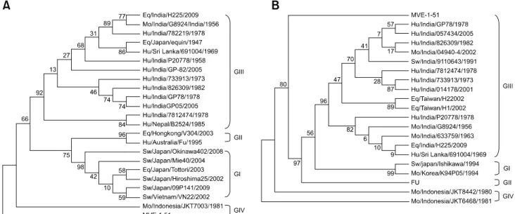
JEV was isolated from the serum of one of the foals collected on the second day of illness. This is the first report of JEV isolation from a horse in India. Furthermore, a pool of mosquitoes collected from the premises housing these foals was positive for JEV RNA by RT-PCR. Three structural genes, capsid (C), premembrane (prM), and envelope (E) of the isolated virus (JE/eq/India/H225/2009) spanning 2,500 nucleotides (from 134 to 2,633) were cloned and sequenced.
BLAST results showed that these genes had a greater than 97% nucleotide sequence identity with different human JEV isolates from India. Phylogenetic analysis based on E- and C/prM genes indicated that the equine JEV isolate belonged to genotype III and was closely related to the Vellore group of JEV isolates from India.
Keywords: envelope gene, horse, isolation, Japanese encephalitis virus, phylogenetic analysis
Introduction
Japanese encephalitis (JE) is a mosquito-transmitted viral disease caused by JE virus (JEV) belonging to genus Flavivirus of the Flaviviridae family. JE is principally a disease occurring in rural agricultural areas where vector mosquitoes proliferate in close association with pigs, wading birds, and ducks. Horses and humans are the dead-end hosts, in which JEV causes acute encephalitis. JE
occurs only sporadically in horses with low morbidity (0.045∼0.3%) and fatality rates ranging between 5 and 30% [19,41]. The virus may infect a number of other domestic animals, including cattle, sheep, goats, dogs, and cats, in which these infections are typically asymptomatic.
JE is prevalent in Southeast Asia and has also been reported in Indonesia, northern Australia, Papua New Guinea, and Pakistan [11]. Approximately, 3 billion people live in JEV-endemic areas where 50,000 cases and 15,000 human deaths due to JE are reported annually [11].
Seasonal outbreaks of JE also occur regularly in human population in several parts of India [8,10,27]. In Haryana (India), JE epidemics among humans were first reported in 1990 [34]. Subsequently, human cases have been reported regularly in this state [17,18,31].
The JEV genome is encoded by a plus-sense, single- stranded RNA of approximately 11 kb [1]. It has a single open reading frame that codes for a polyprotein of 3,432 amino acids, which is subsequently cleaved into three structural [capsid (C), premembrane (prM), and envelope (E)] and seven non-structural (NS1, NS2A, NS2B, NS3, NS4A, NS4B, and NS5) proteins [1]. The E protein is the most important structural protein and induces the production of virus-neutralizing (VN) antibodies which impart protective immunity against JEV [1]. Based on E gene phylogenetic analysis, JEV strains have been classified into five genotypes (GI-V) [25,32,35,37,40].
Genotype I (GI) includes strains isolated in Southeast Asia, Australia, northern Thailand, Cambodia, Korea, Japan, and India. Genotype II (GII) includes strains isolated in southern Thailand, Malaysia, Indonesia, and Australia.
Genotype III (GIII) includes strains isolated in Southeast
Asia, Japan, China, Korea, Taiwan, and the Central Asian
sub-continent including India [27,32,37]. GIV includes
strains isolated in Indonesia alone and GV includes a single
strain isolated in Singapore [10,14,25,37]. GIII is widely
Table 1. Japanese encephalitis seroprevalence in equines from 11 districts of the Haryana state (India) between 2006 and 2010
Year Tested Positive Percent (%)
2006 2007 2008 2009 2010
261 90 61 115 110
21 4 4 24 14
8.0 4.4 6.6 20.9 12.7
Total 637 67 10.5
distributed throughout Asian countries and most of the JEV strains isolated in India belong to GIII [27,37]. JEV GI has recently been reported in human patients from Gorakhpur (Uttar Pradesh, India) [10].
Sporadic clinical cases of JE in horses have been reported in various countries including Japan [19,41], Hong Kong [20], Taiwan [21], and India [31]. In addition, JE seropositivity among equines has been reported in Nepal, Korea, Indonesia, and India [2,9,13,23,26,28,31,33,39].
However, there is no report of JEV isolation from equines in India. Resurgence of JE cases in human populations in India during the last decade [8,27] highlights the need for JEV surveillance in horses. In this paper, we report evidence of JEV infection in horses in Haryana (India) and the first isolation of JEV from a horse.
Materials and Methods Collection of samples
Serum samples from apparently health horses in 11 districts (Ambala, Fatehabad, Gurgagon, Hisar, Jhjjar, Jind, Karnal, Panchkula, Rohtak, Sirsa and Yamunanagar) of Haryana state (India) were collected between 2006 and 2010 (Table 1). None of the horses had been vaccinated against JEV. Blood samples from jugular vein were collected and transported to the laboratory at 4
oC. Serum was separated by centrifugation at 1,000 × g for 5 min and stored at 30
oC until further use.
Blood (with EDTA) and serum samples from two horses in a farm exhibiting neurological signs (circling, pedaling followed by paralysis and recumbency) were collected aseptically for JEV isolation and RT-PCR.
Brain tissues and cerebrospinal fluid from two dead horses were collected at post-mortem and transported to the laboratory on ice. Paired serum samples were also collected from other 37 apparently healthy horses from the same equine farm.
Resting mosquitoes found around the equine farm were collected using mosquito cages measuring approximately 30 × 30 × 30 cm as previously described [7]. Briefly,
mosquitoes were collected by a series of quick forward, backward, up, and down sweeping movements of the cage through vegetation and bushes for 2 min to disturb the resting mosquitoes. This was done at least 10 times for 1 h after dusk. The mosquitoes were stored at 4
oC until engorged females of Culex spp. were identified microscopically and isolated. Female mosquitoes were pooled into six groups, each containing 100∼300 mosquitoes. Homogenate (10%
w/v) of each pool of mosquito was made in Eagle’s minimal essential medium (EMEM; Sigma-Aldrich, USA) containing 2% fetal bovine serum (FBS; Sigma-Aldrich, USA) using pestle and mortar. The homogenate was centrifuged at 12,000 × g for 5 min and the supernatant was stored at 30°C.
Viruses and cells
A JEV strain (P20778; JEV GIII strain isolated from a human patient at Vellore, India) procured from the National Institute of Virology, Pune (India) was used to prepare antigen for a hemagglutination inhibition (HI) test.
The virus was propagated in a-day old Swiss Albino suckling mice by intra-cerebral inoculation (20 μL of JEV inoculum per mouse) as described [6]. For the virus neutralization test (VNT), the virus was cultured in porcine stable kidney (PS) cells obtained from the National Centre for Cell Sciences, Pune (India). The cells were grown at 37
oC in EMEM supplemented with 10% FBS, 100 IU/mL penicillin, 100 μg/mL streptomycin and 0.25 μg/mL amphotericin-B (Sigma-Aldrich, USA).
Hemagglutination inhibition test
The HI test was carried out in duplicate in 96-well microplate (Tarson, India) with using 8 HA units/25 µL of antigen prepared from the brains of suckling mice infected with JEV P20778 strain following sucrose-acetone extraction method [6]. Serum samples were treated with cold acetone to remove non-specific inhibitors and were adsorbed with goose erythrocytes as described [6].
Hyper-immune serum against JEV (raised previously in rabbits by immunization with inactivated JEV vaccine, procured from Central Research Instititute Kasauli, India) was used as a positive control. HI titers of 1 : 20 and above were considered positive.
Virus neutralization test
Serial two-fold dilutions of JEV strain (P20778) (four wells per dilution per virus) in 100 μL volume of EMEM supplemented with 2% FBS were made in 96-well culture plates (Costar, USA). To each well, 100 μL of PS cells (2
×10
5cells/mL in EMEM supplemented with 10% FBS)
were added and incubated for 5 days at 37
oC in 5% CO
2.
The wells showing cytopathic effects (CPE) were recorded
to calculate 50% tissue culture infectivity dose (TCID
50) of
the virus. At passage 5, the virus titer was 10
7TCID
50/mL.
Table 2. Details of Japanese encephalitis virus strains used for analysis in this study Strain Geographical location
(State, Country) Year Source Genotype GenBank accession No.
prM gene E gene
G8924 782219 P20778 GP-82 733913 826309 GP78 GP05 7812474 04940-4 9110643 014178 057434 633759 691004 B2524 H225 Equine H1 H2 V304 FU
Okinawa 402 Mie40 Tottori Hiroshima 25 09P141 VN22 Ishikawa K94P05 JKT7003 JKT8442 JKT6468 MVE-1-51
†Tamil Nadu, India Tamil Nadu, India Tamil Nadu, India Gorakhpur, UP, India West Bengal, India Goa, India
Uttar Pradesh, India Uttar Pradesh, India Assam, India Maharashtra, India Karnataka, India Uttar Pradesh, India Uttar Pradesh, India Tamil Nadu, India Sri Lanka Nepal
Haryana, India Japan
Taiwan Taiwan Hong Kong Australia Japan Japan Japan Japan Japan Vietnam Japan Korea Indonesia Indonesia Indonesia Australia
1956 1978 1958 2005 1973 1982 1978 2005 1978 2002 1991 2001 2005 1963 1969 1985 2009 1947 2002 2002 2003 1995 2008 2004 2003 2002 2009 2002 1994 1994 1981 1980 1981 1951
Mosquito Human brain Human brain Human CSF Human brain Human brain Human brain Human CSF Human brain Mosquito Swine serum Human blood Human brain Mosquito Human blood Human Equine serum Equine Equine brain Equine brain Horse brain Human serum Swine serum Swine Horse brain Swine Swine Swine blood Swine Mosquito Mosquito Mosquito Mosquito Human brain
III III III III III III III III III III III III III III III III III III III III II II I I I I I I I I IV IV IV –
EF688636 NI*
EF688642 NI AB379813 D00979 AF075723 NI EF688633 EF688621 EF688635 EF688624 EF688625 D00968 D00965 NI HQ018880 NI AF373834 AF373835 NI L43565 NI NI NI NI NI NI AB051292 AF045551 NI L42159 AY184212 NC_000943
U70394 U70402 Z34096 DQ914528 Z34095 Z34094 AF075723 FJ979830 U70387 NI NI NI NI NI Z34097 U70392 GQ387646 FJ872378 NI NI AY278555 AF217620 AB471670 AB231463 AB213007 AB231465 GU108335 AY376465 NI NI U70408 NI NI
NC_000943
*NI: not included, †Murray Valley encephalitis (MVE) virus used as an outgroup for the phylogenetic analysis. prM: premembrane, E: envelope.