Molecular characterization of Japanese encephalitis virus strains prevalent in Chinese swine herds
10
0
0
전체 글
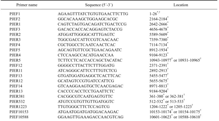
(2) 28. Hao Zheng et al.. attenuated (SA14-14-2 strain) vaccines has been widely performed in mainland China since the 1980s [33]. The number of JE cases has subsequently declined dramatically. Recently, about 5,000 JE cases in mainland China were reported each year [24]. It has been a challenge to eradicate JE because of the large populations of domestic pigs and vast rice farming areas that play key roles in JEV transmission in China. Genetic analysis of 100 JEV strains isolated from humans and mosquitoes showed that JEV strains with genotypes III (GIII) and I (GI) coexist in mainland China [26]. However, only a few JEV strains have been isolated from domestic pigs in China and genetic information for these isolates is lacking [7,29,35]. In the present study, the full-length genomes of JEV strains HEN0701 and SH0601 isolated from aborted pig fetuses in China were sequenced. Additionally, the genetic relationships between these two isolates and other previously published JEV strains were analyzed.. Materials and Methods Cells and viruses o BHK-21 cells (ATCC, CCL-10) were cultured at 37 C with 5% CO2 in Eagle’s minimal essential medium (Sigma-Aldrich, USA) supplemented with 10% fetal. bovine serum (Invitrogen, USA). The JEV strain HEN0701 was isolated from an aborted swine fetus in 2007 in Henan province (China) and the SH0601 strain was isolated from an aborted swine fetus in 2006 in Shanghai (China) [35]. Monolayers of BHK-21 cells grown in T25 Corning flasks were infected with JEV at 0.001 multiplicity of infection. Supernatants of the infected cells were harvested 3 days post-inoculation and o stored in aliquots of 0.2 mL at −75 C.. Reverse transcription-PCR and sequencing Viral RNA was extracted from 140 µL of the infected supernatant using a QIAamp Viral RNA Mini Kit (Qiagen, Germany) according to the manufacturer’s instructions. The purified viral RNA was used as the template for cDNA synthesis with the primer PJER5 (Table 1) and SuperScript reverse transcriptase (Invitrogen, USA) according to the manufacturer’s protocols. The primers (Table 1) for reverse transcription and PCR were were selected on the basis of the genomic sequences of P3 strain (GenBank accession No. U47032; National Center for Biotechnology, USA) and/or SH17M-07 strain (GenBank accession No. EU429297). Five overlapping cDNA segments of the SH0601 genome and four overlapping HEN0701 cDNA segments were amplified using PfuUltra Hotstart high. Table 1. Primers used for PCR and reverse transcription-PCR Primer name PJEF1 PJEF2 PJER1 PJEF3 PJER2 PJER3 PJEF4 PJEF5 PJER4 PJER5 PJEF12 PJER11 PJEF13 PJER12 PJEF14 PJER13 PJER381 PJER532 PJER1223 PJEF10153 PJEF10588. Sequence (5´-3´) AGAAGTTTATCTGTGTGAACTTCTTG GGCACAAAGCTGGAAGCACGC CAGTCTAGTGACAGATCTGACTCCG GACACCACCACAGGAGTCTACCG ATGGATTGGGGCATTTGAGTC TGGCGACCATTCCGTCAACAAC CGCTGGCCTCAATCAACTCAC AGCAGTGTTCGCTGAACAGAATC CTCCAAGCCACATGAACCAA TCTTCCTCACCACCAGCTACATAC GGGGCCTTACTTCTTTGGATG ATCAGGGCATTCCTTTGTCTCG GTGATGGATGAGGCTCACTTCAC GCATAGTCCGTGATCCATTCG GTCAAGGAAGTGCTCAACGAGAC CACCCCACCTCCTGAATTCTC CACGGCGTCAATGAGTGTTC ATGTCCGTGTTGTTGATGGTC TTGTGGGCTTCTCCAGTCG ATGAATGGATGATGGACAAGAC GGAAGTTGAAAGACCAACGTCAG. Location *,†. 1-26 * 2164-2184 * 2642-2666 * 4656-4678 * 5589-5609 * 7359-7380 * 7114-7134 * 8912-1934 * 9104-9123 * † 10943-10977 or 10931-10965 † 2371-2391 † 2892-2913 † 5455-5477 † 5655-5675 † 8971-8813 † 9184-9204 * † 361-380 or 362-381 * † 512-532 or 513-533 * † 1204-1222 or 1205-1223 * † 10153-10174 or 10154-10175 * † 10601-10623 or 10588-10610. *Position in the genome of Japanese encephalitis virus (JEV) P3 (GenBank accession No. U47032). †Position in the viral genome of JEV SH17M-07 (GenBank accession No. EU429297)..
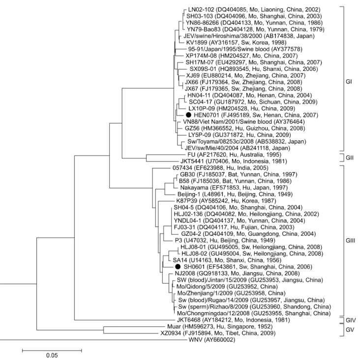
(3) Molecular characterization of swine JEV in China 29. fidelity DNA polymerase (Stratagene; Agilent Technologies, USA) and five primer pairs (PJEF1/PJER1, PJEF2/PJER2, PJEF3/PJER3, PJEF4/PJER4, and PJEF5/ PJER5) for SH0601 and four primer pairs (PJEF1/PJER11, PJEF12/PJER12, PJEF13/PJER13, and PJEF14/PJER5) for HEN0701 (Table 1). The amplifications were conducted in a total volume of 50 µL containing 10 µM each primers, 2 µL cDNA, 5 µL 10× Reaction buffer, 1 µL DNA polymerase, 4 µL dNTP (2.5 mM each) and 38 µL ddH2O. o The reactions was heated at 94 C for 5 min, followed by 27 o o o cycles of 94 C for 30 sec, 58 C for 30 sec and 72 C for 3 o min, with a final elongation step of 72 C for 10 min. Resulting cDNA segments of the expected size were cloned into a pCR-Blunt-TOPO vector (Invitrogen, USA) and then used to transform competent Escherichia coli Top10 cells (Tiangen Biotech, China). Recombinant plasmids were identified by EcoRI digestion (New England Biolabs, USA) and JEV cDNA was sequenced using a 3730 XL DNA analyzer (Applied Biosystems, USA) at Beijing Genomics Institute (China).. 5´- and 3´-rapid amplification of cDNA ends (RACE) of HEN0701 and SH0601 A 5´-Full RACE kit and the 3´-Full RACE core set (Takara, Japan) were used to sequence the 5´ and 3´ ends of HEN0701 and SH0601 according to the manufacturer’s protocols. Viral RNA was extracted with a QIAamp viral RNA mini kit (Qiagen, Germany). The primer PJER1223 (Table 1) was used for 5´-RACE, while primers PJER532 and PJER368 (Table 1) were used for nested 5´ PCR in combination with the universal primers provided in the kit. A poly(A) tail was initially added to the 3´-end of the JEV genome with poly(A) polymerase (Takara, Japan) for the 3´-RACE. The RNA was purified using a QIAamp viral RNA mini kit (Qiagen, Germany). A 3´-RACE adaptor was used as the primer for transcription. Primer PJEF10588 and the primer provided in the kit were used for 3´-PCR. The amplified PCR products were separated in 1.2% gel and purified with a gel extraction kit (Watson Biotechnology, China). The segments of the expected size were cloned into the pGEM-T vector (Promega, USA) for sequencing. Sequence analysis and multiple alignments The full-length HEN0701 and SH0601 genomes were assembled using SeqMan in the Lasergene software package (DNAStar, USA). ORFs of the JEV genomes were scanned using MapDraw (DNAStar, USA). Multiple sequence alignments and percent similarity calculations were performed using MegAlign (DNAStar, USA). Phylogenetic analysis A total of 50 JEV strains were used for phylogenetic analysis based on E gene sequences. In addition to. HEN0701 and SH0601, the genomic sequences of other 48 JEV strains were obtained from GenBank (National Center for Biotechnology Information, USA). Multiple sequence alignments and phylogenetic analysis were carried out using MEGA 4.0. A neighbor-joining tree rooted with the WNV strain Mex03 was constructed. Tree stability was established by bootstrap analysis with 1,000 replicates.. RNA structure analysis The RNA structures of HEN0701 and SH0601 were analyzed using MFOLD (Genetics Computer Group, USA) and RNADraw software. A 149-nt segment of the 5´ end and 117-nt segment of the 3´ end were subjected to secondary structure analysis to detect any potential 5´-3´ interaction using the energy minimization program on the MFOLD web server (Genetics Computer Group, USA). Codon analysis of the HEN0701 and SH0601 genomes ORFs from seven JEV GI strains and another seven JEV GIII strains were analyzed using CAI, CHIPS, CUSP (European Molecular Biology Open Software Suite), and CodonW 1.4.2 software to detect differences in codon usage between GI and GIII. Statistical analyses were carried out with SPSS software (IBM, USA).. Results HEN0701 and SH0601 genomes Four overlapping cDNA segments of HEN0701 and five overlapping SH0601 cDNA segments were sequenced and assembled into complete genomic sequences. The complete genomic sequences of HEN0701 and SH0601 were deposited into the GenBank database under accession numbers FJ495189 and EF543861, respectively. The complete SH0601 sequence was 10977 nt in length while the complete sequence of HEN0701 contained 10965 nt. CG content of the SH0601 genome was 51.5% and 51.7% in the HEN0701 genome. Both strains contained a single ORF encoding 3432 amino acid residues. The 5´ and 3´ UTRs included 95 and 586 nt in SH0601 and 96 and 573 nt in HEN0701, respectively. SH0601 and HEN0701 shared an 88.8% nucleotide sequence identity and 97.9% deduced amino acid sequence identity at the full genomic level. Phylogenetic analysis The genetic relationship of SH0601 and HEN0701 with 48 other JEV strains deposited in GenBank, including 37 strains from different areas of China and diverse hosts (human, swine, mosquito and bat) were analyzed and a phylogenetic tree based on nucleotide sequences of the E gene was constructed. The tree was rooted using the E gene nucleotide sequence of WNV as the out-group virus. As shown in Fig. 1, all 50 JEV strains were grouped into five clusters and each cluster represented one genotype. The 37.
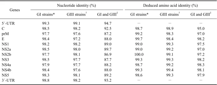
(4) 30. Hao Zheng et al.. JEV strains of Chinese origin fell into genotypes GI, GIII, and GV. HEN0701 belonged to the GI group that included LX10P-09, SC04-17, and HN04-11 while SH0601 belonged to the GIII group that contained SA14, HLJ08-01, and HLJ08-02.. Sequence analysis Sequences of all genes and UTRs of the JEV strains HEN0701 and SH0601 (Fig. 1) were analyzed and. compared with those of 12 other JEV strains including six GI (GZ56, SH17M-07, SC04-17, XJ69, JEV/sw/Mie/40/ 2004, and KV1899) and six GIII (NJ 2008, P3, SA14, GB30, 057434, and Nakayama) strains. Results of this analysis are presented in Table 2. At the full-length genomic level, HEN0701 had a high identity (≥97.7%) with the other GI strains and SH0601 shared a high identity (≥97.5%) with the other GIII strains. Similarly, high nucleotide (≥97.2%) and amino acid (≥98.3%) identities. Fig. 1. Neighbor-joining phylogenetic tree for JEV envelope genes rooted with the West Nile virus (WNV) strain Mex03. Numbers on the branches indicate bootstrap support (1,000 replicates). The GenBank accession number, origin, country/province, and year of isolation of each strain are noted. Sw: derived from swine, Mo: derived from mosquito, Hu: derived from human..
(5) Molecular characterization of swine JEV in China 31. Table 2. Nucleotide and deduced amino acid sequence identities of 14 genotype I (GI) and GIII JEV strains Genes 5´-UTR C prM E NS1 NS2a NS2b NS3 NS4a NS4b NS5 3´-UTR. Nucleotide identity (%) †. Deduced amino acid identity (%). GI strains*. GIII strains. GI and GIII‡. GI strains*. GIII strains†. GI and GIII‡. 99.3 98.5 97.7 98.4 98.2 98.5 97.7 98.5 97.9 98.4 98.3 98.8. 99.1 98.2 97.6 97.2 98.2 98.0 98.1 97.7 97.7 97.6 98.1 98.2. 94.7 92.5 87.2 88.0 89.0 89.7 86.9 87.7 88.2 88.0 89.2 93.2. − 98.7 99.2 99.7 99.0 99.0 100.0 99.3 98.7 99.3 98.6 −. − 98.9 98.3 98.4 99.3 99.2 99.1 99.3 99.2 99.4 99.3 −. − 95.0 97.0 98.2 97.5 97.0 97.2 98.2 98.3 98.1 97.9 −. *Mean identity values of seven GI strains (HEN0701, GZ56, SH17M-07, SC04-17, XJ69, JEV/sw/Mie/40/2004, and KV1899). †Mean identity values of seven GIII strains (SH0601, NJ 2008, P3, SA14, GB30, 057434, and Nakayama). ‡Mean identity values of GI and GIII strains.. Fig. 2. Alignment of the 3´ UTR variable region. Nucleotide sequences of the 3´ UTRs from 18 JEV strains were aligned with MegAlign (DNAStar, USA). A dash indicates a missing nucleotide. Nucleotides of each strain that differ from those of HEN0701 are marked with grey shading. The variable region consists of 73 nucleotides (nt) immediately downstream of the stop codon in the JEV genomic 3´ UTRs.. were found among the same genotype strains at the individual gene level. Nucleotide identity of all protein coding genes between the GI and GIII strains ranged from 86.9% to 89.7% while the amino acid identity ranged from 97.0% to 98.3% with exception of the C gene. Nucleotide. identity (92.5%) of the C gene was the highest among the ten genes analyzed while amino acid identity (95%) was the lowest. Nucleotide sequences of the 5´ and 3´ UTRs were highly conserved among strains with the same genotype. A nucleotide deletion after the stop codon was.
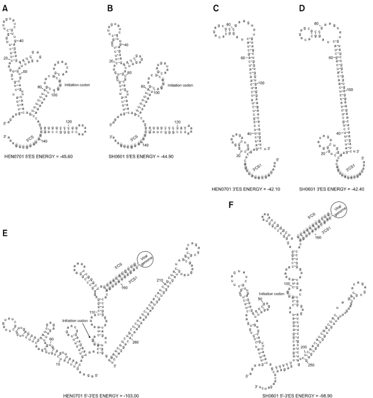
(6) 32. Hao Zheng et al.. found in the in the 3´ UTR of HEN0701 (Fig. 2). HEN0701 and SH0601 shared common conserved sequences 1 (CS1), CS2, CS3, repeated CS2 (RCS2), and RCS3 in the 3´ UTR (data not shown). A common conserved sequence. (5´ CS), which was complementary to a part of CS1, was identified in the 5´ ends of HEN0701 and SH0601. Secondary terminal structures of the HEN0701 and SH0601 genomes were similar (Fig. 3). However, their. Fig. 3. RNA structures of the 3´- and 5´-end sequences of the HEN0701 and SH0601 genomes. (A) 149 nt 5´-end sequences of the HEN0701 strain. (B) 149-nt 5´-end sequences of the SH0601 strain. (C) 117-nt 3´-end sequences of the HEN0701 strain. (D) 117-nt 3´-end sequences of the SH0601 strain. (E) Interaction between the 5´- and 3´-end sequences of HEN0701. (F) Interaction between the 5´- and 3´-end sequences of SH0601..
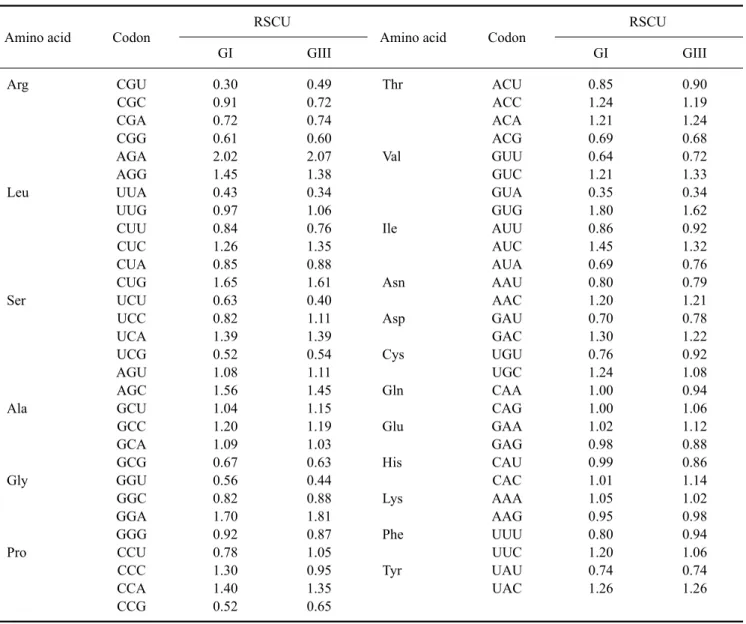
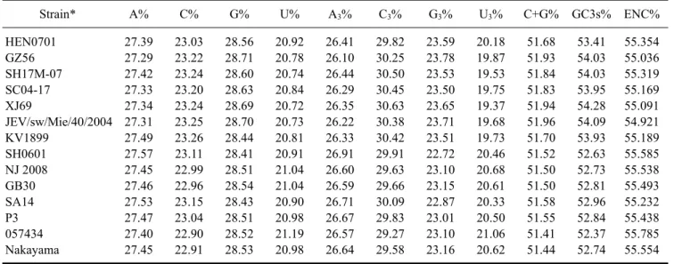
(7) Molecular characterization of swine JEV in China 33. RNA structures were different due to an interaction between the 3´- and 5´-end sequences of the genomes that caused circularization (Fig. 3).. Nucleotide composition and codon usage The nucleotide and amino acid sequence identities of 14 JEV GI and GIII strains including HEN0701 and SH0601 are presented in Table 2. In general, nucleotide sequence identities were lower than amino acid sequence identities between GI and GIII strains, indicating the presence of many synonymous mutations. We next analyzed nucleotide composition and ORF codon usage for these 14 strains. The relative synonymous codon usage (RSCU) value for every codon was determined using CodonW 1.4.2 software. Mean RSCU values of the seven GI strains and seven GIII strains are shown in Table 3. RSCU values. of strains with the same genotype were similar but varied between GI and GIII strains. Nucleotide composition and effective number of codons (ENC) are presented in Table 4. Although C% values were about 23% in the ORFs of both GI and GIII strains, the C3% values were over 29.27%, suggesting that the percentage of C bases at the third position could influence the pattern of synonymous codon usage in JEVs. Compared to GIII ORFs, the (C+G)% content and GC3% values of GI ORFs were slightly higher. The ENC for all 14 strains was about 55, indicating that both GI and GIII strains had a low codon bias. The ENC of the GIII strains had a highly significant negative correlation with GC3s% (Pearson, r = −0.956, p = 0.001 < 0.01). However, the ENC did not have a significant negative correlation with GC3s% among the GI strains (Pearson, r = −0.642, p = 0.12 > 0.05). Our results. Table 3. The relative synonymous codon usage (RSCU) values in the open reading frames (ORFs) of GI and GIII JEV strains Amino acid. Codon. Arg. CGU CGC CGA CGG AGA AGG UUA UUG CUU CUC CUA CUG UCU UCC UCA UCG AGU AGC GCU GCC GCA GCG GGU GGC GGA GGG CCU CCC CCA CCG. Leu. Ser. Ala. Gly. Pro. RSCU GI. GIII. 0.30 0.91 0.72 0.61 2.02 1.45 0.43 0.97 0.84 1.26 0.85 1.65 0.63 0.82 1.39 0.52 1.08 1.56 1.04 1.20 1.09 0.67 0.56 0.82 1.70 0.92 0.78 1.30 1.40 0.52. 0.49 0.72 0.74 0.60 2.07 1.38 0.34 1.06 0.76 1.35 0.88 1.61 0.40 1.11 1.39 0.54 1.11 1.45 1.15 1.19 1.03 0.63 0.44 0.88 1.81 0.87 1.05 0.95 1.35 0.65. Amino acid. Codon. Thr. ACU ACC ACA ACG GUU GUC GUA GUG AUU AUC AUA AAU AAC GAU GAC UGU UGC CAA CAG GAA GAG CAU CAC AAA AAG UUU UUC UAU UAC. Val. Ile. Asn Asp Cys Gln Glu His Lys Phe Tyr. RSCU GI. GIII. 0.85 1.24 1.21 0.69 0.64 1.21 0.35 1.80 0.86 1.45 0.69 0.80 1.20 0.70 1.30 0.76 1.24 1.00 1.00 1.02 0.98 0.99 1.01 1.05 0.95 0.80 1.20 0.74 1.26. 0.90 1.19 1.24 0.68 0.72 1.33 0.34 1.62 0.92 1.32 0.76 0.79 1.21 0.78 1.22 0.92 1.08 0.94 1.06 1.12 0.88 0.86 1.14 1.02 0.98 0.94 1.06 0.74 1.26.
(8) 34. Hao Zheng et al.. Table 4. Nucleotide composition and effective number of codons (ENC) for the ORFs of GI and GIII JEV strains Strain*. A%. C%. G%. U%. A3%. C3%. G3%. U3%. C+G%. GC3s%. ENC%. HEN0701 GZ56 SH17M-07 SC04-17 XJ69 JEV/sw/Mie/40/2004 KV1899 SH0601 NJ 2008 GB30 SA14 P3 057434 Nakayama. 27.39 27.29 27.42 27.33 27.34 27.31 27.49 27.57 27.45 27.46 27.53 27.47 27.40 27.45. 23.03 23.22 23.24 23.20 23.24 23.25 23.26 23.11 22.99 22.96 23.15 23.04 22.90 22.91. 28.56 28.71 28.60 28.63 28.69 28.70 28.44 28.41 28.51 28.54 28.43 28.51 28.52 28.53. 20.92 20.78 20.74 20.84 20.72 20.73 20.81 20.91 21.04 21.04 20.90 20.98 21.19 20.98. 26.41 26.10 26.44 26.29 26.35 26.22 26.33 26.91 26.60 26.59 26.71 26.67 26.57 26.64. 29.82 30.25 30.50 30.45 30.63 30.38 30.42 29.91 29.63 29.66 30.09 29.83 29.27 29.58. 23.59 23.78 23.53 23.50 23.65 23.71 23.51 22.72 23.10 23.15 22.87 23.01 23.10 23.16. 20.18 19.87 19.53 19.75 19.37 19.68 19.73 20.46 20.68 20.61 20.33 20.50 21.06 20.62. 51.68 51.93 51.84 51.83 51.94 51.96 51.70 51.52 51.50 51.50 51.58 51.55 51.41 51.44. 53.41 54.03 54.03 53.95 54.28 54.09 53.93 52.63 52.73 52.81 52.96 52.84 52.37 52.74. 55.354 55.036 55.319 55.169 55.091 54.921 55.189 55.585 55.538 55.493 55.232 55.438 55.785 55.554. *The GI and GIII strains listed are the same 14 strains shown in Table 2.. demonstrated that there were few differences in codon usage between the GI and GIII strains.. Discussion JEV has been epidemic in China since 1949. JEV GIII was the single JEV genotype reported in China before 1978 [26]. The first JEV GI strain was isolated in Yunnan province in 1979 and reported in Shanghai in 2001 [25,26]. Subsequently, GI strains were identified in Guizhou, Guangxi, Hubei, Shangdong, Shaanxi, Liaoning, Sichuan, Shanghai, Gansu, and Shanxi provinces [1,2,9,11,25-27, 29,31,32,34,35]. JEV GIII strains were also reported in Henan, Fujian, Guizhou, Yunnan, Guangdong, Shanghai, and Heilongjiang provinces [4,7,26]. More importantly, GI and GIII strains were isolated simultaneously during a JE outbreak in Shanxi province in 2006 [28], indicating that these two genotypes were coexisting. In China, most JEV strains isolated from human cases before 2005 belonged to the GIII group. However, the number of humans infected with JEV GI strains has risen recently [26-28,32,34]. In addition, the JEV GV strain FJ915894 was isolated from mosquitoes in Tibet in 2009 [10]. To date, the GV genotype has not been reported in other areas of China. The isolation and characterization of JEV from domestic pigs was not performed until recent years although clinical cases have been reported for a long time. In 2008, four strains were isolated from aborted fetuses or stillborn piglets in China and confirmed to belong to the GIII group [7]. JEV GI strains SXBJ07 and GSX09S-01 were next isolated from aborted piglet brains in China [1,29]. In the present study, the complete genomes of JEV strains HEN0701 and SH0601 isolated from aborted pig fetuses. from Henan province and Shanghai were sequenced for phylogenetic analysis. Results of our study showed that HEN0701 was grouped into the GI cluster and SH0601 was a member of the GIII cluster. Nucleotide sequence analysis indicated that HEN0701 had a high identity with GI strains and SH0601 had a high identity with GIII strains. A genotype shift from JEV GIII to GI occurred in Japan during the middle 1990s [13,15-17,19]. JEV strains isolated before 1990 belonged to the GIII group whereas those isolated after 1995 were GI [13,16,17,19]. It was believed that a JEV GI strain was transmitted from mainland China to Japan [15,16]. Additionally, JEV GIII has circulated in the eastern coastal areas of China including Shandong, Jiangsu, Shanghai, Zhejiang, Fujian, and Guangdong provinces [4,26]. In mainland China, a JEV GI strain was isolated in Yunnan province as early as 1979 and the number of isolated GI strains has risen in recent years [1,2,9,11,25-27, 29,31,32,34,35]. These findings indicate that JEV GI has perhaps become the predominant genotype in mainland China. On the other hand, it has been reported that JEV GI strains have replaced GIII in other Asian regions [5,8,18]. The factors that promoted this genotype shift are still unknown. In the present study, the complete genomic sequences of a JEV GI strain SH0601 and a JEV GIII strain HEN0701 were determined. Low nucleotide identity and high amino acid identity between GI and GIII strains were identified. The analysis of codon usage also showed a few differences in GC3%, ENC, and RSCU values between these two genotypes. However, it is unclear what roles synonymous mutations in the GI and GIII genomes might have played in JEV evolution or whether these might be related to genotype shift. An inter-genotype between GI and GIII has.
(9) Molecular characterization of swine JEV in China 35. never been detected although both genotypes have co-existed in China for 30 years. Characterization of SH0601 strain and HEN0701 strain from swine at genetic level would be helpful to understand JEV epidemic in China. Future studies may be aimed to investigate virulence, viral antigen and vector competence of SH0601 and HEN0701.. 11.. 12.. Acknowledgments This study was supported by Natural Science Foundation of Shanghai (11ZR1446200), Natural Science Foundation of China (31201917), the Science and Technology Key Project of Shanghai (09391910800), and the China Research Fund for Non-Profit Research Institutions (2012JB08), China.. References 1. Cao QS, Li XM, Zhu QY, Wang DD, Chen HC, Qian P. Isolation and molecular characterization of genotype 1 Japanese encephalitis virus, SX09S-01, from pigs in China. Virol J 2011, 8, 472. 2. Cao YX, Fu SH, Wang HY, Pan XL, Zhang JB, Liang GD. Molecular characterization of full-length genome of Japanese encephalitis virus isolated in Liaoning Province in 2008. Zhonghua Shi Yan He Lin Chuang Bing Du Xue Za Zhi 2009, 23, 248-250. 3. Chambers TJ, Hahn CS, Galler R, Rice CM. Flavivirus genome organization, expression, and replication. Annu Rev Microbiol 1990, 44, 649-688. 4. Chen D, Wang HY, Fu SH, Song H, Deng J, Yang YL, Liang GD. Molecular characteristics of three new isolates of Japanese encephalitis virus in Fujian Province. Zhonghua Shi Yan He Lin Chuang Bing Du Xue Za Zhi 2005, 19, 5-8. 5. Chen YY, Fan YC, Tu WC, Chang RY, Shih CC, Lu IH, Chien MS, Lee WC, Chen TH, Chang GJ, Chiou SS. Japanese encephalitis virus genotype replacement, Taiwan, 2009-2010. Emerg Infect Dis 2011, 17, 2354-2356. 6. Endy TP, Nisalak A. Japanese encephalitis virus: ecology and epidemiology. Curr Top Microbiol Immunol 2002, 267, 11-48. 7. Fan JM, Luo J, Chen L, Teng M, Bu D, Wang FY, Wang L, Wang CQ, Zhang GP. Genetic analysis of strains of Japanese Encephalitis Virus isolated from swine in central China. Virus Genes 2010, 40, 357-361. 8. Fulmali PV, Sapkal GN, Athawale S, Gore MM, Mishra AC, Bondre VP. Introduction of Japanese encephalitis virus genotype I, India. Emerg Infect Dis 2011, 17, 319-321. 9. Gao XY, Wang HY, Wang HY, Fu SH, Liu GF, Li Y, Li MH, Xu AQ, Liang GD. Molecular characterization of full-length genome of Japanese encephalitis virus (SD08-10) newly isolated in Shandong province. Zhonghua Shi Yan He Lin Chuang Bing Du Xue Za Zhi 2009, 23, 242-244. 10. Li MH, Fu SH, Chen WX, Wang HY, Guo YH, Liu QY, Li YX, Luo HM, Da W, Duo Ji DZ, Ye XM, Liang GD.. 13.. 14.. 15. 16.. 17.. 18.. 19.. 20.. 21.. Genotype V Japanese encephalitis virus is emerging. PLoS Negl Trop Dis 2011, 5, e1231. Li MH, Fu SH, Feng Y, Gao XY, Zhai YG, Yu DS, Li GT, Jia YX, Liang GD. Molecular characterization of Japanese encephalitis virus isolated from Gansu province in 2008. Zhonghua Shi Yan He Lin Chuang Bing Du Xue Za Zhi 2009, 23, 251-253. Li YX, Li MH, Fu SH, Chen WX, Liu QY, Zhang HL, Da W, Hu SL, Mu SDL, Bai J, Yin ZD, Jiang HY, Guo YH, Duo Ji DZ, Xu HM, Li G, Mu GGC, Luo HM, Wang JL, Wang J, Ye XM, Jin ZMY, Zhang W, Ning GJ, Wang HY, Li GC, Yong J, Liang XF, Liang GD. Japanese encephalitis, Tibet, China. Emerg Infect Dis 2011, 17, 934-936. Ma SP, Yoshida Y, Makino Y, Tadano M, Ono T, Ogawa M. Short report: a major genotype of Japanese encephalitis virus currently circulating in Japan. Am J Trop Med Hyg 2003, 69, 151-154. Mohammed MAF, Galbraith SE, Radford AD, Dove W, Takasaki T, Kurane I, Solomon T. Molecular phylogenetic and evolutionary analyses of Muar strain of Japanese encephalitis virus reveal it is the missing fifth genotype. Infect Genet Evol 2011, 11, 855-862. Morita K. Molecular epidemiology of Japanese encephalitis in East Asia. Vaccine 2009, 27, 7131-7132. Nabeshima T, Loan HTK, Inoue S, Sumiyoshi M, Haruta Y, Nga PT, Huoung VTQ, del Carmen Parquet M, Hasebe F, Morita K. Evidence of frequent introductions of Japanese encephalitis virus from south-east Asia and continental east Asia to Japan. J Gen Virol 2009, 90 (Pt 4), 827-832. Nerome R, Tajima S, Takasaki T, Yoshida T, Kotaki A, Lim CK, Ito M, Sugiyama A, Yamauchi A, Yano T, Kameyama T, Morishita I, Kuwayama M, Ogawa T, Sahara K, Ikegaya A, Kanda M, Hosoya Y, Itokazu K, Onishi H, Chiya S, Yoshida Y, Tabei Y, Katsuki K, Tabata K, Harada S, Kurane I. Molecular epidemiological analyses of Japanese encephalitis virus isolates from swine in Japan from 2002 to 2004. J Gen Virol 2007, 88 (Pt 10), 2762-2768. Nga PT, del Carmen Parquet M, Cuong VD, Ma SP, Hasebe F, Inoue S, Makino Y, Takagi M, Nam VS, Morita K. Shift in Japanese encephalitis virus (JEV) genotype circulating in northern Vietnam: implications for frequent introductions of JEV from Southeast Asia to East Asia. J Gen Virol 2004, 85 (Pt 6), 1625-1631. Tang WF, Ogawa M, Eshita Y, Aono H, Makino Y. Molecular evolution of Japanese encephalitis virus isolates from swine in Oita, Japan during 1980-2009. Infect Genet Evol 2010, 10, 329-336. Thiel HJ, Collett MS, Gould EA, Heinz FX, Houghton M, Meyers G, Purcell RH, Rice CM. Family Flaviviridae. In: Fauquet CM, Mayo MA, Maniloff J, Desselberger U, Ball LA (eds.). Virus Taxonomy: Eighth Report of the International Committee on Taxonomy of Viruses. pp. 981-998, Elsevier Academic Press, San Diego, 2005. Tsai TF. New initiatives for the control of Japanese encephalitis by vaccination: minutes of a WHO/CVI meeting, Bangkok, Thailand, 13-15 October 1998. Vaccine.
(10) 36. Hao Zheng et al.. 2000, 18 (Suppl 2), 1-25. 22. Uchil PD, Satchidanandam V. Phylogenetic analysis of Japanese encephalitis virus: envelope gene based analysis reveals a fifth genotype, geographic clustering, and multiple introductions of the virus into the Indian subcontinent. Am J Trop Med Hyg 2001, 65, 242-251. 23. Unni SK, Růžek D, Chhatbar C, Mishra R, Johri MK, Singh SK. Japanese encephalitis virus: from genome to infectome. Microbes Infect 2011, 13, 312-321. 24. Wang H, Li Y, Liang X, Liang G. Japanese encephalitis in mainland china. Jpn J Infect Dis 2009, 62, 331-336. 25. Wang HY, Fu SH, Li XY, Song H, Min JG, Deng J, Yang YL, Kurane I, Liang GD. Isolation and identification of genotype I Japanese encephalitis virus in China. Zhonghua Min Guo Wei Sheng Wu Ji Mian Yi Xue Za Zhi 2004, 24, 843-849. 26. Wang HY, Takasaki T, Fu SH, Sun XH, Zhang HL, Wang ZX, Hao ZY, Zhang JK, Tang Q, Kotaki A, Tajima S, Liang XF, Yang WZ, Kurane I, Liang GD. Molecular epidemiological analysis of Japanese encephalitis virus in China. J Gen Virol 2007, 88 (Pt 3), 885-894. 27. Wang L, Fu S, Zhang H, Ye X, Yu D, Deng Z, Yuan J, Zhai Y, Li M, Lv Z, Chen W, Jiang H, Gao X, Cao Y, Wang H, Tang Q, Liang G. Identification and isolation of Genotype-I Japanese encephalitis virus from encephalitis patients. Virol J 2010, 7, 345. 28. Wang LH, Fu SH, Wang HY, Liang XF, Cheng JX, Jing HM, Cai GL, Li XW, Ze WY, Lv XJ, Wang HQ, Zhang DL, Feng Y, Yin ZD, Sun XH, Shui TJ, Li MH, Li YX,. 29.. 30. 31.. 32.. 33. 34.. 35.. Liang GD. Japanese encephalitis outbreak, Yuncheng, China, 2006. Emerg Infect Dis 2007, 13, 1123-1125. Wang WH, Zhang X, Zhang YM, Xing FS, Xu YZ. Isolation,identification and sequence analysis of the Japanese encephalitis virus SXBJ07 strain. Xi Bei Nong Lin Ke Ji Da Xue Xue Bao Zi Ran Ke Xue Ban 2009, 37, 1-6. Weaver SC, Barrett ADT. Transmission cycles, host range, evolution and emergence of arboviral disease. Nat Rev Microbiol 2004, 2, 789-801. Xie RH, Zhu HP, Fu SH, Cheng YK, Xu F, Yao PP, Yang ZN, Zhou XL, Zhu ZY. Complete genome sequence analysis of Japanese encephalitis virus newly isolated in China. Zhonghua Shi Yan He Lin Chuang Bing Du Xue Za Zhi 2009, 23, 245-247. Xufang Y, Huanyu W, Shihong F, Xiaoyan G, Shuye Z, Chunting L, Minghua L, Yougang Z, Guodong L. Etiological spectrum of clinically diagnosed Japanese encephalitis cases reported in Guizhou Province, China, in 2006. J Clin Microbiol 2010, 48, 1343-1349. Yu Y. Phenotypic and genotypic characteristics of Japanese encephalitis attenuated live vaccine virus SA14-14-2 and their stabilities. Vaccine 2010, 28, 3635-3641. Zhang JS, Zhao QM, Guo XF, Zuo SQ, Cheng JX, Jia N, Wu C, Dai PF, Zhao JY. Isolation and genetic characteristics of human genotype 1 Japanese encephalitis virus, China, 2009. PLoS One 2011, 6, e16418. Zheng H, Zhang JW, Yuan SS. Isolation of Japanese encephalitis virus from swine and analyses of its E protein. Zhongguo Shouyi Ke-ji 2009, 39, 476-482..
(11)
수치
+3
관련 문서
Alginate appears to play a key role in the stabilization of biofilms formed by Pseudomonas aeruginosa and some other pseudomonas, with most Pseudomonas strains producing
Anti-fungal effect of isolated bactrial strains was tested in vitro by incubating in potato dextrose agar with isolates of four fungal plant pathogens
The cucumber sprouts pre-treated with all seven rhizobacterial strains at concentration of 5.0×10 6 cfu/ml were significantly increased plant height and
Phylogenetic analysis of Orientia tsutsugamushi strains based on the sequence homologies of 56-kDa type-specific antigen genes. FEMS
Tullio Scovazzi, “Open Questions on the Exploitation of Genetic Resources in Areas Beyond National Jurisdiction”, Am. Nagoya Protocol on Access to Genetic Resources and the
Full genome cloning and nucleotide sequence analysis of hepatitis C virus from sera of chronic hepatitis patients in Korea. Detection of hepatitis C virus RNA
Comparison of genotypes using REP-PCR and Integron-IS26 PCR results among 78 IRAB strains isolated from 2 university hospitals.. The result of REP-PCR genotypes and
Frequency of six efflux pump genes of adeG, adeB, adeE, adeY, abeM, and adeJ according to the year of isolation of 86 Acinetobacter baumannii clinical strains collected
