Received: March 18, 2013; Accepted: March 26, 2013 Corresponding author : Ee-Yung Chung
Tel: +82 (63) 469-1832 e-mail: [email protected] 1225-3480/24463
This is an Open Access article distributed under the terms of the Creative Commons Attribution Non-Commercial License with permits unrestricted non-commercial use, distribution, and reproducibility in any medium, provided the original work is properly cited.
Gametogenic Cycle and the Size at 50% of Group Sexual Maturity in Male Chlamys ( Azumapecten ) farreri nipponensis (Kuroda, 1932) (Bivalvia: Pectinidae) in Western Korea
Ki Yeol Park
1, Ee-Yung Chung
2, Ki-Young Lee
2and Kwan Ha Park
31Yangyang Salmon Station, Korea Fisheries Resources Agency, Yanyang 215-821, Korea
2Department of Marine Biotechnology, Kunsan National University, Gunsan 573-701, Korea;
3Department of Aquatic Life Medicine, Kunsan National University, Gunsan 573-701, Korea
ABSTRACT
We investigated the gametogenic cycle and spawning seasons of the male Chlamys (Azumapecten) farreri
nipponensis by qualitative and quantitative analyses, and also the size at 50% of group sexual maturity wascalculated by the data of first sexual maturity. In this study, the male gametogenic cycle of this species by qualitative analysis was divided into five successive stages: early active stage (January to March), late active stage (March to April), ripe stage (April to August), partially spawned stage (July to September), and spent/inactive stage (August to January). The male gametogenic cycle showed similar patterns with monthly changes in the gonadosomatic index and condition index. Particularly, spawning in male scallop occurred once a year from July to September, unlike the spawning period of this species (from June to August) reported by the previous researchers.
In quantitative statistical analysis using an image analyzer system, the patterns of monthly changes in the percent (%) of the areas occupied by spermatogenic stages to the testis areas in males showed a maximum in June, and then sharply dropped from July to September, 2006. From these data, it is apparent that the spawning season of
C. (A.) farreri nipponensis occurred once per year from July to early September, indicating a unimodalgametogenic cycle during the year. Shell heights at 50% of group sexual maturity (RM
50) fitted to an exponential equation were estimated to be 49.90 mm in males (considered to be one year old), and it was 100% for male scallops over 61.0 mm (considered to be two years old).
Keywords: Chlamys (Azumapecten) farreri nipponensis, unimodal gametogenic cycle, spawning period, size
at 50% of group sexual maturity
INTRODUCTION
The Jicon scallop, C. (A.) farreri nipponensis (Bivalvia: Pectinidae), one of the most important edible scallops in Asia is abundant along the coasts of Korea, China and Japan (Min et al., 2004). In particular, in Korea, it is mainly found in the subtidal
zones of the south and west coasts of Korea (Kwon et al., 1993; Min et al., 2004). However, the standing stock of this species has gradually been decreasing due to reckless over-harvesting. Thus, for the propagation of a living natural resource, this species has been identified as a target organism for the development of aquaculture techniques that should be carefully managed. Prior studies have considered various aspects of the biology of C. (A.) farreri nipponensis. Previously, there have been many studies on aspects of reproduction, including the reproductive cycle (Liao et al., 1983; Yacovlev and Afeichuk, 1995;
Park, 2002; Chung et al., 2005; Chung, 2008), growth and spawning (Na et al., 1995; Kang and Zhang, 2000), experimental triploids and tetraploids (Yang et
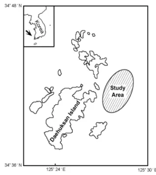
Fig. 1. Map showing the sampling area.
al., 1999a), its distribution and ecology (Whang and Kim, 1973), larval growth (Kuang et al., 1997; Yang et al., 1999b), and experimental aquaculture (Lim et al., 1995; Sun et al., 1996, 1997) of C. farreri. However, there are still significant gaps in our knowledge regarding its reproductive biology. Although some species belong to the same genus Chlamys, the spawning cycles of Chlamys species vary with the species. Recently, Jaramillo et al. (1993) reported that the gametogenic cycle of Chlamys amandi collected from Hueihue Bay, Chile showed a semiannual spawning cycle. However, in case of the Korean jicon scallop (C. (A.) farreri nipponensis), the reproductive cycle studied by qualitative histological analysis showed somewhat different spawning period (particularly, the beginning and the end months of spawning), as have been reported in other Chlamys spp.
in Korea (Park, 2002; Chung et al., 2005; Chung, 2008).
Thus, qualitative histological analysis of gonad developmental stages by individual subjectivity may lead to incorrect conclusion. To confirm the beginning and the end months during the spawning period, it is very important to investigate by quantitative statistical analysis. If the spawning periods were not confirmed by the statistical methods, it will be hard to perform an accurate age determination and assess population dynamics of this species. Thus, understanding the gametogenic cycle and spawning period of this species will provide information needed for the determination of age and recruitment period (Chung et al., 1994, 2010). Studies on the spawning season need to be accompanied by quantitative statistical analysis to determine whether a species has either unimodal or bimodal cycle of the gonads during the year.
To date, we could not find the spawning period including the beginning and the end months of spawning by quantitative statistical analysis of histological sections of C. (A.) farreri nipponensis. In addition, information on the size at 50% of group sexual maturity of this species would be very useful for resource management. Some information on the size at which individuals reach first sexual maturity could be useful in determining a prohibitory measure for adequate natural resources management (Chung et
al., 2010). Therefore, the purpose of this study is to describe the gametogenic cycle with germ cell developmental stages and compare the spawning periods by qualitative histological analysis and quantitative statistical analyses using an image analyzer system (statistical analysis).
In addition, the second aim of this study is to clarify the size at 50% of group sexual maturity of this species for aquaculture and natural living resources management.
MATERIALS AND METHODS
1. Qualitative Histological Analysis 1) Sampling Methodology
Male specimens of C. (A.) farreri nipponensis were collected monthly at the subtidal zones of Daehuksan Island coastal waters, Jeollanam-do, Korea (Fig. 1) from March 2006 to February 2007. The Jicon scallops ranging from 32.3 to 99.6 mm in shell height were used for the present study. Jicon scallops were transported alive to the laboratory, shell height, total body weight, gonad weight, and meat weight were immediately measured. Unpublished data of seawater
temperature and salinity measured daily at 10:00 a.m.
at the Fisheries Hatchery Center in Daehuksan-do were used for this study.
2) Gonadosomatic Index (GSI)
To estimate the spawning period, a total of 252 male individuals were used to calculate the gonadosomatic index (GSI). GSI was calculated by the following equation:
GSI = Gonad weight (g) x 100 / Meat weight (g)
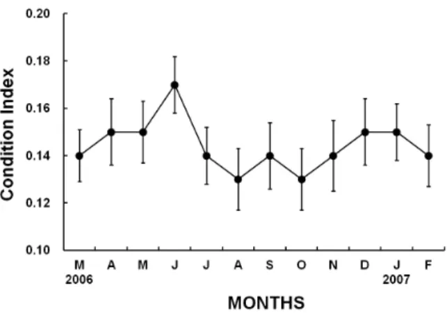
3) Condition Index (CI)
To estimate the spawning period by the indirect method, a total of 252 male individuals were used to calculate the condition index (CI). CI was calculated by the following equation:
CI = Meat weight (g) x 103 / [Shell length (cm) x Shell height (cm) x shell width (cm)]
4) Gametogenic Cycle by Histological Observation of the Gonadal Tissue Sections
For light microscopic examination of histological preparations, a total of 425 male individuals were used for histological analysis of the gonads. Gonadal tissues were removed from shells and preserved in Bouin’s fixative for 24h. They were then washed with running tap water for 24h. Tissues were then dehydrated in alcohol and embedded in paraffin molds. Embedded tissues were sectioned at 5-7 μm thickness using a rotary microtome. Sections were mounted on glass slides, stained with Hansen’s hematoxylin-0.5% eosin, and examined under a light microscope (Zeiss Axiovert 10 microscope).
2. Quantitative Statistical Analysis
Tissue slides were observed for quantitative analysis by an image analyzer system. Slides were viewed on a stereo-zoom microscope (Nikon, SMZ-U) from which the images were captured by a TOSHIBA Model IK-642K CCD camera and then viewed on a SAMSUNG color video monitor. The image analyzer (BMI plus, WINATech Co.) is capable of automatic measurement of area and diameter of polygons encircled by the operator, counting objects that are
contrasted by background color (in black and white mode), and performing statistical analysis on numerous characteristics of objects in the captured images. Measurements on female tissue were carried out for areas of total tissue, the ovary, the follicle, the oocyte, the number of the oocytes per unit area, and the diameter of each oocyte. As for males, the areas of total tissue, the testis, and the spermatogenic stages were measured. Measurements on total tissue, the testis areas were conducted at a magnification of 7.5×, at which the field area of the captured images was 60 mm2, while the other measurements were done at a magnification of 75× (field area: 0.524 mm2). Twenty individuals per month and two fields per slides were analyzed. Areas of total tissue, the testes, and the spermatogenic stages were measured by manually tracking the margins of objects with a pointer on the captured images. Counting and measuring the diameter of the oocytes was carried out by converting captured color images to black-and-white images with the appropriate threshold, and then conducting an automatic measurement procedure provided by the software.
From the measured values of image analyses, for each male slide, (1) the percent of field occupied by the testis to total tissue and (2) the percent of field occupied by the spermatogenic stages to the testis area were calculated. A one-way ANOVA (multiple comparisons by Duncan's procedure) was applied to compare the means of monthly data. One-way t-tests were used to determine significant differences in the data of two adjacent months. All statistical analyses were done using the SPSS package.
3. First Sexual Maturity by Light Microscopical Observation For determination of the size at 50% of first sexual maturity, a total of 258 individuals (135 males) of C.
(A.) farreri nipponensis, ranging from 33.3 to 99.6 mm in shell height, were examined in order to certify the size at 50% first sexual maturity (= biological minimum size) by histological observations from April to September, 2006. The percentage (%) of first sexual maturity = No. of mature individuals x 100 / No. of total individuals investigated.
Fig. 2. Monthly changes in seawater temperature and salinity in the sampling area from March 2006 to February 2007.
Fig. 3. Monthly changes in the gonadosomatic indices (GSI) in male Chlamys (Azumapecten) farreri nipponensis from March 2006 to February 2007.
4. Size at the Rate (50%) of Group Sexual Maturity (RM50) (= Biological Minimum Size)
To calculate the size at the rate (50%) of sexual maturity after fitting the rate of group sexual maturity to an exponential equation, the size equivalent to the size at 50% of sexual maturity was estimated to be the sexually mature length of the population (method used by Chung and Ryou, (2000)).
The exponential equation of the rate of sexual maturity is as follows: RM = 100 / [1+exp(a-bx)], where, RM: rate of sexual maturity; a, b: constants, x:
shell height.
RESULTS
A detailed insight into the gametogenic cycle in male C. (A.) farreri nipponensis population was ascertained from a combination of qualitative and quantitative data gathered during the study period from March 2006 to February 2007.
1. Qualitative results
Monthly qualitative assessments of the qualitative reproductive condition are illustrated in Figs. 2-6.
1) Monthly changes in the seawater temperature and salinity
Monthly changes in the surface seawater temperature and salinity are shown in Fig. 2. The range of the surface seawater temperature during the
period from March 2006 to February 2007 was 8.8-23.9°C. The surface seawater temperature began to increase from April and reached a maximum in August (24.5°C), and then gradually decreased from September to February. The range of salinity of the surface seawater during the period from March 2006 to February 2007 was from 31.86 to 32.91 psu.
Salinity of the surface seawater reached a maximum (32.91 psu) in April, and then it began to gradually decreased from June to August (31.86 psu).
Thereafter, it began to increase gradually from September, and high salinity (over 32 psu) was maintained from November to March.
2) Monthly changes in the gonadosomatic indices (GSI)
Monthly changes in the gonadosomatic indices (GSI) in male C. (A.) farreri nipponensis are shown in Fig. 3. In 2006-2007, the values of GSI corresponding to both females and males began a gradual increase in March, reaching the maximum (males mean GSI 9.22 ± 2.46) in June, when seawater temperature gradually increased. Then, the GSI gradually decreased from June to August when spawning occurred continuosly and high seawater temperature was maintained. Thereafter, the mean GSI values temporarily reached a minimum in October 2006 (males mean 3.10 ± 0.82). Monthly changes in the
Fig. 4. Monthly changes in the condition indices (CI) in male Chlamys (Azumapecten) farreri nipponensis from March 2006 to February 2007.
GSI in both sexes in 2006 showed a peak in June during the year, which indicates that the number of spawning period of Korean population of C. (A.) farreri nipponensis is once a year.
3) Monthly changes in the condition indices (CI) Monthly changes in the condition indices (CI) in male C. (A.) farreri nipponensis are shown in Fig. 4.
In 2006-2007, the values of CI in males began a gradual increase in May, reaching the maximum (mean CI 0.17 ± 0.58) in June 2006, and then the CI rapidly decreased between July and August (mean CI 0.13 ± 0.63) when spawning occurred and high seawater temperature was maintained. Monthly changes in the CI in males showed a peak in July 2006 during the year, which indicates that the number of spawning season of Korean population of C. (A.) farreri nipponensis is once a year.
4) Male Gametogenic cycle by Qualitative Histological Analysis
Spermatogenesis occurs in the acini in the testis.
Based on morphological features and sizes of the germ cells and accompanying cells, the gametogenic cycle in male individuals of this species can be classified into five successive stages: early active stage, late active stage, ripe stage, partially spawned stage, and spent/inactive stage (Fig. 5). The stages
and the criteria used in defining them are as follows:
Eearly Active Stage: In the early active stage, in particular, spermatogonia and spermatocytes proliferated along the acinus walls (germinal epithelium) of the acini in the testis. The spermatogonia and spermatocytes were 7-8 μm and 5-7 μm in diameter, respectively (Fig. 5A). In 2006-2007, male individuals in the early active stage appeared from January to March when seawater temperatures were lower than 12°C (Fig. 2).
Late Active Stage: In this stage, a few spermato- gonia and a number of spermatocytes (5-6 μm) appeared near the acinus wall. A number of spermatids, measuring about 3.7 μm in diameter appeared in the lumina of the acini. At this time a small number of spermatozoa began to transform into differentiated spermatozoa in the centre of the lumina of the acini. As spermatogenesis progressed, a number of spermatocytes, spermatids and small number of spermatozoa occupied approximately one half of the lumina of the acini (Fig. 5B). In 2006-2007, male individuals in the late active stage were found from March to April when seawater temperature gradually increased (Fig. 2).
Ripe Stage: In this ripe stage, a number of spermatids began to transform into differentiated spermatozoa in the centre of the lumen, and numerous spermatozoa appeared in the center of the lumen of the acinus (Fig. 5C). In 2006-2007, male individuals in the ripe stage were found from April to August when seawater temperature was rapidly increasing (Fig. 2).
Partially Spawned Stage: In this stage, the lumina of the acini were empty because over 60% of the spermatozoa had been discharged but undis- charged spermatozoa as well as spermatids remained in the lumina of the acini (Fig. 5D). In 2006-2007, male individuals in the partially spawned stage appeared from July to early September, with the main spawning event occurring from July to August when
Fig. 5. Photomicrographs of the histological gonadal phases in male Chlamys (Azumapecten) farreri nipponensis. A, Transverse section of the acini in the early active stage; B, Section of the acini in the late active stage; C, Section of the acini in the ripe stage; D, Section of the acini in the partially spawned stage; E, Section of the acini in the spent stage; F, Section of the acini in the inactive stage. Abbreviations: AW, acinus wall; CT, connective tissue; DSZ, degenerating spermatozoon; LU, lumen; SC, spermatocyte; SG, spermatogonium; ST, spermatid; SZ, spermatozoon; USZ, undischarged spermatozoon.
seawater temperature was higher than 20°C (Fig. 2).
Spent / Inactive Stage: During this stage, small number of remaining spermatozoa and spermatids degenerated and the products of gamete atresia were reabsorbed, thereafter, the rearrangement of a few newly formed spermatogonia and connective tissue occurred in the acini (Figs. 5 E, F).
In 2006-2007, male individuals in this stage were found from August to January when sea water
temperature was gradually decreasing and relatively low (Fig. 2).
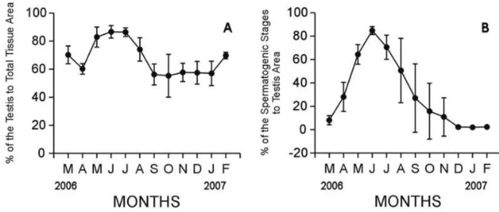
2. Quantitative results
C. (A.) farreri nipponensis in males showed a unimodal gametogenic cycle (Fig. 6). The percent of field occupied by the testis to total tissue began to increase in March. The testis area greatly increased from March to May (70.22-82.96%, p < 0.001), reached a maximum in June (86.68%), and then
Fig. 6. Monthly changes in quantitative reproductive traits in male Chlamys (Azumapecten) farreri nipponensis. A: Percent of areas occupied by the testis to total tissue area, B: Percent of areas occupied by spermatogenic stages to the testis area.
decreased between July and August (86.44-74.13%, p
= 0.005). During the winter period (December, Jan- uary and February), the percent of testis area was lower than 70%. Variations of the testis area among individuals were so high that there was no significant difference in the testis area to total tissues during May-June, June-July, September-October, October- November, November-December, December-January, and February-March (one-way ANOVA, p = 0.489, 0.941, 0.924, 0.786, 0.956, 0.947, 0.865, respectively) (Fig. 6A).
The percent of field occupied by spermatogenic stages to the testis area began to increase in April. It increased rapidly from April to May (28.06-64.37%, p
= 0.001), reached a maximum in June (84.85%), and then gradually decreased between July and August (70.62-50.63%, p = 0.122) when spawning occurred.
During the winter period (December, January and February), the percent of spermatogenic stages to testis area was lower than 3%. Variations of the testis area among individuals were so high that there was no significant difference in spermatogenic stages to the testis area during July-August, August-September, September-October, October-November, November- December, December-January, January-February, and February-March (one-way ANOVA, p=0.122, 0.306, 0.584, 0.753, 0.404, 0.674, 0.601, 0.059, respectively) (Fig. 6B). There was no significant difference in spermatogenic stages to testis area during
July-August or October-November (one-way ANOVA:
p = 0.130, 0.055, respectively) (Fig. 6B). Especially, in quantitative statistical analysis using an image analyzer system, the patterns of monthly changes in the portions (%) of the areas occupied by spermatogenic stages to the testis area in males showed a maximum in June and reached the minimum in December to February, 2007
3. Size at first sexual maturity
A total of 162 male individuals of C. (A.) farreri nipponensis were investigated histologically to determine the shell heights of the Jicon scallop that reach maturation and participate in reproduction from May (before spawning) to early September (after spawning).
As shown in Table 1, the percentage of first sexual maturity of smaller individuals ranging from 32.3-40.0 mm in shell height was 0%, and those individuals were in the early active stage, which is characterized by a small number of spermatogonia and the appearance of a number of spermatocytes in the acini of the testis. It is supposed that their sizes at sexual maturity could have not been reached until late August when spawning was completed. The percentage of first sexual maturity of female clams ranging from 40.1-50.0 mm in shell height was 41.7%, and those individuals were in the early active, late active and ripe stages during the period between June
Number of individuals by gonadal stage*
Shell height (mm) EA LA RI PS SP/IA Total Mature (%)
32.3-40.0 21 21 0.0
40.1-50.0 14 5 3 2 24 41.7
50.1-60.0 10 6 4 3 2 25 60.0
60.1-70.0 10 8 5 3 26 100.0
70.1-80.0 8 6 6 4 24 100.0
80.1-90.0 7 6 5 4 22 100.0
90.1-99.6 7 5 5 3 20 100.0
Total 162
* Gonadal stage: EA, early active stage; LA, late active stage; RI, ripe stage; PS, partially spawned stage; SP/IA;
spent/inactive stage.
Table 1. The shell heights and first sexual maturity in male Chlamys (Azumapecten) farreri nipponensis from May to September
Fig. 7. Relationship between the rates of group sexual maturities (%) and shell length (mm) in male Chlamys (Azumapecten) farreri nipponensis.
and August, when spawning was observed among older individuals. However, younger animals had a small number of spermatogonia, spermatocytes, a number of spermatids, and spermatozoa in the acini of the testis. It is supposed that their sizes at sexual maturity of most individuals could have not been reached until early October when spawning of a few mature individuals was completed. In addition, the percentage of first sexual maturity of male scallops ranging from 50.1-60.0 mm in shell height was 60.0%, but those individuals were in the early active, late active, ripe, and partially spawned stages during the breeding season. The percentage of first sexual
maturity of all individuals of shell height greater than 60.1 mm was 100%, and those individuals were in the late, ripe, partially spawned, and spent/inactive stages. Therefore, it is assumed that most individuals can reach full maturity by late August if they are larger than 60.1 mm in shell height at that time.
In this study, the percentage of first sexual maturity of male Jicon scallops ranging from 50.1 to 60.0 mm was over 50.0% (rate of group sexual maturity: 50%). The percentage was 100% for female and male scallops over 60.1 mm in shell height.
4. Size at the Rate (50%) of Group sexual Maturity (RM50)
As shown in Fig. 7, shell height of sexually mature Jicon scallop (size at 50% of group sexual maturity, RM50) that was fitted to an exponential equation wsa 49.90 mm in males.
DISCUSSION
1. Qualitative Analysis
1) Gonadosomatic index (GSI) and Condition Index (CI) To clarify the number of spawning seasons during the year by the indirect method, we calculated the gonadosomatic index in males by qualitative analysis.
As shown in Fig. 3, the male gonadosomatic index (GSI) of this species began to increase in March and reached a maximum in June when the water temperature rapidly increased, and then the GSI values then gradually decreased because of spawning.
Chung (2007) reported that the high average values of the GSI coincided with gonadal maturity, and the minimal average value following high average value were considered as an indication of spawning.
Accordingly, variations in the GSI showed a close relationship with gonadal development and gonadal activity, and showed a unimodal gametogenic cycle during the year.
In the present study, monthly variations in the condition index (CI) by qualitative analysis showed a maximum in June, and then gradually decreased from July to September, indicating the unimodal gameto- genic cycle during the year. Accordingly, as shown in monthly changes in the GSI (Fig. 3) and the CI (Fig.
4), these were clarified that the number of spawning seasons by qualitative analysis was once per year from July to September, and their variations showed the unimodal cycle.
2) Environmental Factors Associated with Gonadal Development and Maturation
Several authors (Sastry, 1966, 1968; Chung et al., 1991, 2005; Chung and Ryou, 2000) stated that exogenous factors (water temperature, food availability, salinity, day length, etc.) and endogenous factors (neuroendocrine activity) have been suggested as control factors for gonadal development and maturation in marine bivalves. Sastry (1966, 1968) stated that a wide range of exogenous factors has been suggested as controls for gonadal development and maturation in marine bivalves. Particularly, water temperature and food availability as exogenous factors seem to be particularly important. Other factors (salinity, day length, etc) probably interact with endogenous factors (neuroendocrine activity) in a complex manner to control the initiation of gametogenesis. In this study, gamete differentiation of C. (A.) farreri nipponensis began in the winter-early spring months, and reached maturity in the population from April to August when water temperature increased. After basic metabolic requirements are satisfied, gonad activity and gametogenesis of this species occur under temperature conditions that allow nutrients mobilization to the
gonads (Sastry, 1966).
In Korean coastal waters, growth and production of bivalves is relatively high from spring to early summer seasons (Chung et al., 1994, 1999; Kim, 2005) due to the abundance in phytoplankton. Thus, abundant food supply is available to C. (A.) farreri nipponensis during the period of gonadal development and maturation. Therefore, it is suggested that gonadal development and maturation of C. (A.) farreri nipponensis is closely related to water temperature and food availability. Fretter (1984) observed that in temperate zones, the seasonal temperature fluctuation associated with changing illumination is a controlling factor in gametogenesis. In consequence, gonadal development and maturation of this species may be retarded under low illumination, due to the decrease in food availability caused by diminished primary production of phytoplankton.
3) Number of the Spawning Seasons
In this study, C. (A.) farreri nipponensis belongs to the summer breeders because spawning occurs from July to September (Boolootian et al., 1962). Rand (1973) reported that breeding strategy vary with latitudinal gradients. Northern climates are characterized by a single synchronous spawning per year, temperate climates by two spawning seasons and tropical ones by year-round spawning.
In case of different local populations of R.
philippinarum, the number of spawning seasons by qualitative reproductive analysis (histological observa- tions) in most bivalves occur once a year in the northern districts of Tokyo Bay (Japan), such as Hokkaido district in Japan and all districts in Korea (Kurashige, 1943; Momoyama and Iwamoto, 1979;
Chung et al., 1994; Chung et al., 2010), while twice a year in the southern districts of Tokyo Bay. Japan (Ko, 1957; Tanaka, 1954).
Because the location of Korea belongs to northern climates, the number of spawning season of C. (A.) farreri nipponensis by qualitative histological analysis was once a year, and many results by qualitative and quantitative analyses are similar in Korea, however, in many case, the spawning season varied with
slightly different locations. Local variations and timing of spawning of this species might be related to the geographical differences in the water temperatures, time of the food production (phytoplanktons), and some other environmental factors (Ko, 1957; Momoyama and Iwamoto, 1979).
4) A Comparison of the Spawning Periods by Quantitative and Qualitative Analyses
Regarding the spawning periods of the Jicon scallop, C. (A.) farreri, there have been some studies by a few authors (Park, 2002; Chung et al., 2005).
According to the results of the spawning periods studied by Park (2002) and Chung et al. (2005), they reported the same results that the spawning period of the jicon scallop by qualitative histological analysis was from June to August. Thus, it is assumed that they could not distinguish the beginning and the end months of the spawning period because they observed the gonadal tissue sections without quantitative statistical analysis. Accordingly, we produced a number of gonadal tissue sections and confirmed the specimens of gonadal tissue sections of the Jicon scallops by quantitative statistical analysis using an image analyzer system.
According to the results of the gametogenic cycle by quantitative statistical analysis in this study, the patterns of monthly changes in the percent (%) of the areas occupied by spermatogenic stages to the testis areas in males showed a maximum in June, and then sharply dropped from July to September, 2006. From these data, it is apparent that the spawning period of C. (A.) farreri nipponensis occurred once per year from July to September, indicating the unimodal gametogenic cycle during the year.
Compared the result of the spawning period (from July to September) investigated by quantitive statistical analysis in this study with those (from June to August) studied by qualitative histological analysis in two previous studies, the beginning month and the end of month were some differences in the spawning period. Therefore, in case of C. (A.) farreri nipponensis, the spawning period should be corrected to “from July to early September” instead of “from
June to August”.
Compared to qualitative results (frequencies of gonadal phases) of other clams in previous works, the period of maturation of C. (A.) farreri nipponensis is quite similar to the May-July results of Cyclina sinensis (Chung et al., 1991), Mactra veneriformis (Chung and Ryou, 2000), Meretrix lusoria (Chung, 2007), and Saxidomus purpuratus (Chung et al., 1999). The peak of oocyte area to ovarian tissue in June implies the readiness of maturation. The spawning seemed to be initiated in June with a decrease in oocyte area. A significant decrease in oocyte area was found during June-September, indicating the major period of spawning. The spawning period of this species was also similar to other clams mentioned above (June-September for C.
sinensis, M. veneriformis, and M. lusoria, May-October for S. purpuratus).
Giese (1959) and Sastry (1979) observed latitudinal differences in timing of the reproductive cycles of marine molluscs in general. Some authors (Ropes and Stickney, 1965; Brousseau, 1978; Heffernan et al., 1989a) reported that Mya arenaria and Mercennaria mercennaria in bivalve molluscs exhibited a change from a unimodal to a bimodal cycle with decrease in latitude (Kanti et al., 1993). However, some authors (Heffernan et al., 1989b, c) reported that several other bivalves (i.e, Geukensia demisa, Crassostrea virginica and Spisular solidissima similis) showed unimodal gametogenic cycle in the southeastern U.S.
waters (Kanti et al., 1993). In this study, the gametogenic cycle of C. (A.) farreri nipponensis by quantitative statistical analysis showed unimodal gametogenic cycle in western Korea.
5) Size at First Sexual Maturity and Size at 50% of Group Sexual Maturity
Park (2002) investigated the determination of age of the Jicon scallop, C. farreri. According to the growth curves for the mean shell height fitted to the von Bertalanffy equation by Park (2002), ages (year) and mean shell lengths (mm) were estimated as follows: 1 year (44.4 mm), 2 years (67.0 mm), 3 years (80.7 mm), and 4 years (89.3 mm).
In this study, the percentages of first sexual maturity of female and male individuals of 50.1-60.0 mm in shell height were 60.0% in males. It is assumed that male scallops ranging from 50.1-60.0 mm in shell height are one year old. And the percentages of first sexual maturity of female and male individuals of over 60.1 mm in shell height were 100% in males. Accordingly, it is supposed that female and male jicon scallops ranging over 60.1 mm in shell height were approximately two years old.
According to the growth curve for shell height fitted to von Bertalanffy equation, shell heights at 50% of group sexual maturities (RM50) were 49.90 mm in males (Fig.7). In this study, these shell heights (RM50) in both sexes were considered to be one year old (Park, 2002). Therefore, we assumed that these female and male populations achieve maturity and begin reproduction at one year of age.
In the aspect of natural resource management, the present study suggests that harvesting scallops less than 49.90 mm in shell height for RM50 (one year old) can potentially lead to a drastic reduction in recruitment. Accordingly, a measure indicating a prohibitory fishing size should be taken for adequate fisheries management.
ACKNOWLEDGMENTS
Authors are grateful to Professor Emeritus, Dr.
William Heard of Florida State University and the referees for helpful comments and corrections to the manuscript. This research was supported in part by the research fund of Costal Research Center, Kunsan National University, Korea.
REFERENCES
Brousseau, D.J. (1978) Spawniong cycle. fecundity and recruitment in a pupulation of soft-shell clam. Mya arenaria from Cape Ann, Massachusetts. Fisheries Bulletin, 76:155-166.
Boolootian, R.A., Farmanfarmaina, A. and GIESE, A.C.
(1962) On the reproductive cycle and breeding habits of two western species of Haliotis. Biological Bulletin, 122: 183-192.
Chung, E.Y., Lee, T.Y. and An, C.M. (1991) Sexual maturation of the venus clam, Cyclina sinensis, on the west coast of Korea. Journal of Medical &
Applied Malacology, 3: 125-136.
Chung, E.Y., Ryou, D.K. and. Lee, J.H. (1994) Gonadal development, age and growth of the shortnecked clam, Ruditapes philippinarum (Pelecypoda:
Veneridae), on the coast of Kimje. Korean Journal of Malacology, 19: 38-54.
Chung, E.Y., Kim, Y.M. and Lee, S.G. (1999) Ultrastructure of germ cell development and reproductive cycle of the purplish Washington clam, Saxidomus purpuratus (Sowerby). Yellow Sea, 5:
51-58.
Chung, E.Y. and Ryou, D.K. (2000) Gametogenesis and sexual maturation of the surf clam, Mactra veneriformis on the west coast of Korea.
Malacologia, 42: 149-163.
Chung, E.Y., PARK, Y.J., LEE, J.Y. and Ryu, D.K.
(2005) Germ cell differentiation and sexual maturation of the hanging cultured female scallop Pationpecten yessoensis on the East coast of Korea.
Journal of Shellfish Research, 24: 913-921.
Chung, E.Y., KOO, J.G. PARK, K.Y., LEE, C.H. (2005) Seasonal changes in biochemical components of the adductor muscle, digestive diverticula and the ovary in Chlamys farreri in relation to the ovarian developmental phases. Korean Journal of Malacology, 21: 71-80 [in Korean].
Chung, E.Y. (2007) Oogenesis and sexual maturation in Meretrix lusoria (Roding, 1978) (Bivalvia: Veneridae) in western Korea. Journal of Shellfish Research, 26:
71-80.
Chung, E.Y. (2008) Ultrastructural studies of oogenesis and sexual maturation in female Chlamys (Azumapecten) Farreri farreri (Jones & Preston, 1904) (Pteriomorphia: Pectinidae) on the western coast of Korea. Malacologia, 50: 279-292.
Chung, E.Y., Lee, C.H., Choi, K.H., Choi, M.S. and Lee, K.Y. (2010) Gametogenic cycle and the number of spawning seasons by quantitative reproductive analysis in female Ruditapes philippinarum in western Korea. Korean Journal of Malacology, 26:
245-254.
Fretter, V. (1984) Prosobranchs. pp. 1-45. In: Laar, eds., The Mollusca, Vol. 7, Academic Press, New York, 352 pp.
Heffernan, P.B., Walker, R.L. and Carr, J.L. (1989a) Gametogenic cycles of three bivalves in Wassaw Sound, Georgia Ⅰ: Mercenaria mercenaria (Linnaeus, 1758). Journal of Shellfish Research, 8:
51-60.
Heffernan, P.B., WALKER, R.L. and Carr, J. L. (1989b) Gametogenic cycles of three bivalves in Wassaw Sound, Georgia II: Crassostrea virginica (Gmelin, 1971). Journal of Shellfish Research, 8: 61-70.
Heffernan, P.B., WALKER, R.L. and Carr, J.L. (1989c) Gametogenic cycles of three bivalves in Sassaw Sound, Georgia Ⅲ: Geukensia demissa (Dillwyn).
Journal of Shellfish Research, 8: 327-334.
Giese, A.C. (1959) Compartative physiology: Annual
reproductive cycles of marine invertebrates. Review of Physiology, 21: 547-576.
Jaramillo, R., Winter, J. Valencia, J. and Rivera, A.
(1993) Gametogenic cycle of the Chiloe scallop (Chlamys amandi). Journal of Shellfish Research, 12: 59-64.
Kang, T.G. and Zhang, C.I. (2000) A study on the growth and spawning of Korean scallop (Chlamys farreri) around Wando, Korea, Journal of the Korean Fisheries Society, 36: 210-221 [in Korean].
Kuang, S., Sun, H., Li, F. and Fang, J. (1997) Feeding and growth of scallop Chlamys farreri before and after spawning. Marine Fisheries Research of China, 17: 80―86.
Kanti, A., Heffernan, P.B. and Walker, R.L. (1993) Gametogenic cycle of the southern surfclam, Spisula solidissimasimilis (Say, 1822), from St. Catherine Sound, Georgia. Journal of Shellfish Research, 12:
255-261.
Kim, Y.M. (2005) A study on reproductive ecology of the hard clam, Meretrix lusoria, on the west coast of Korea. Ph. D. Thesis, Kunsan National University, 123 pp.
Ko, Y. (1957) Some histological note on the gonads of Tapes japonica Deshayes. Bulletin of Japanese Society Fisheries, 23(7, 8): 394-399.
Kurashige, H. (1943) Seasonal variation in the weight and volume as well as the chemical composition of the soft body of Tapes philippinarum with special reference to its spawning. Bulletin of Korean Fisheries Experimental Station, 8: 115-140.
Kwon, O.K., Park, G.M. and Lee, J.S. (1993) Coloured shells of Korea. Academy Publish Co. Seoul. 288 pp.
Liao, C., Xu, Y. and Wang, Y. (1983) Reproductive cycle of the scallop Clamys farreri (Jones & Prestone) at Qingdao. Journal of Fisheries of China, 7: 1-13.
Lim, H.K., Go, C.S. and Lee, Y.H. (1995) Studies on the technology development for seed production of Chlamys farreri. Pp. 355-360. In: Technical Report of South Sea Regional Fisheries Research Institute, National Fisheries Research & Development Institute, Yosu, Korea.
Min, D.K., Lee, J.S., Ko, D.B. and Je, J.G. (2004) Mollusks in Korea. Hanguel Graphics, Busan, Korea 566 pp.
Na, G.H., Jeong, W.G. and Cho, C.H. (1995) A study on seedling production of Jicon scallop, Chlamys farreri. 1. Spawning, developing and rearing of larvae. Journal of Aquaculture, 8: 307-316.
Momoyama, G. and Iwamoto, T. (1979) On the spawning season of the short necked clam in Yamaguch and Okai Bay. Bulletin of Yamaguchi Prefecture Fisheries Experimental Station, 7:19-28.
Park, K.Y. (2002) Reproductive ecology and seed
production of Jicon scallop, Chlamys farreri. Ph.D.
Thesis. Sunchunhyang University, 116 pp.
Rand, W.M. (1973) A stochastic model of the temporal aspect of breeding strategies. Journal of Theoretical Biology, 40: 337-351.
Ropes, J.W. and Stickney, P. (1965) Reproductive cycle of Mya arenaria in New England. Biological Bulletin, 128: 315-327.
Sastry, A.N. (1966) Temperature effects in reproduction of the bay scallop, Aquipecten irradians Lamarck.
Biolological Bulletin, 130: 118-134.
Sastry, A.N. (1968) Relationship among food, temperature and gonad development of the bay scallop, Aquipecten irradians Lamarck. Physiological Zoology, 41: 44-53.
Sastry, A.N. (1979) Pelecypoda (excluding Ostriedae). In:
Reproduction of marine invertebrates, Vol. V. pp.
113-292. Ed. By A. C. Giese & J. S. Pearse. New York: Academic Press.
Sun, H., Kuang, S. and. LI, F. (1996) Studies on suitable culture depths and method for scallop in Sanggou Bay. Journal of Fisheries Science of China, 3: 60-65.
Sun, J., Lin, C. LI, P. JIN, Y. and ZHOU, L. (1997) The culture experiment of scallop Chlamys farreri in Nanji Islands. Zhejiang College of Fisheries, 16:
247-255.
Tanaka, Y. (1954) Spawning season of important bivalves in Ariake Bay Ⅲ. Tapes philippinarum.
Bulletin of Japanese Society Science Fisheries, 19:
1165-1167.
Whang, H.J. and Kim, M.N. (1973) Study on the distribution and ecology of Chlamys farreri nipponensis Kuroda around the Taehuksand Island.
Bulletin of National Fisheries Research and Development Agency, 11: 25-35 [in Korean].
Yakovlev, Y.M. and Afeichuk, L.S. (1995) The reproductive cycle of the scallop Chlamys farreri in the Sea of Japan. Pp. 193-198. in : P. LUBET. J.
BARRET & J. C. DAO. Eds., Fisheries biology and aquaculture of pectinids, 8th International Pectinid Workshop, 273 pp.
Yang, A., Wang, Q., KONG, J., Liu, P., Liu, Z., SUN, H., LI, F., Wang, R. and JIANG, M. (1999a), Triploid induction in Chlamys farreri by application of 6-dimethylaminopurine. Journal of Fisheries of China, 23: 241-247.
Yang, H., ZHANG, T., WANG, J., WANG, P., HE, Y. and ZHANG, F. (1999b) Growth characteristics of Chlamys farreri and its relation with environmental factors in the intensive raft-culture areas of Sishiliwan Bay, Yantai. Journal of Shellfish Research, 18: 71-76.