• 검색 결과가 없습니다.

Gastric cancer and adenomatous colorectal polyp concomitant with pyogenic liver abscess and bacteremia

N/A
N/A
Protected

Academic year: 2021

Share "Gastric cancer and adenomatous colorectal polyp concomitant with pyogenic liver abscess and bacteremia"

Copied!
4
0
0

로드 중.... (전체 텍스트 보기)

전체 글

(1)

Introduction

Pyogenic liver abscess (PLA) is defined as a mass of cystic lesions in the liver with clinical manifestations, including fever, chills, and abdominal pain, which can be caused by either bacterial, fungal, or parasitic organisms [1-3]. The most common causative patho- gen of PLA is Klebsiella pneumoniae (KP). KP can invade the liver via many routes, including the biliary tract, hepatic arteries, and portal vein and through direct invasion from an adjacent infected organ [4-6]. Hematogenous PLA is closely associated with colon-

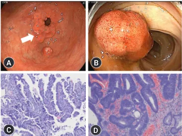
Synchronous gastric cancer and adenomatous colorectal polyp in patients with Klebsiella pneu- moniae-induced pyogenic liver abscess (KP-PLA) and bacteremia is a rare presentation. A 58-year-old man with a 6-month history of diabetes mellitus (DM) presented with febrile sensa- tion and dull abdominal pain in the right upper quadrant of the abdomen. Subsequent to labora- tory test results and abdominal computed tomography findings, KP-PLA with bacteremia was di- agnosed. After intravenous antibiotic administration, his symptoms improved, and upper endos- copy and colonoscopy were performed to evaluate the cause of KP-PLA. Biopsy specimens of the prepyloric anterior wall revealed a moderately differentiated adenocarcinoma. Endoscopic muco- sal resection of the colon revealed high-grade dysplasia. Early gastric cancer (EGC) and adeno- matous colorectal polyps with high-grade dysplasia concomitant with KP-PLA and bacteremia were diagnosed in our patient who had DM. Intravenous antibiotic treatment for KP-PLA, subto- tal gastrectomy for EGC, and colonoscopic mucosal resection for the colon polyp were performed.

After 25 days of hospitalization, subtotal gastrectomy with adjacent lymph node dissection was performed. Follow-up ultrasound imaging showed resolution of the abscess 5 weeks post-antibi- otic treatment, as well as no tumor metastasis. Upper gastrointestinal endoscopy and colonosco- py should be performed to evaluate gastric cancer in patients with PLA or bacteremia, accompa- nied with DM or an immunocompromised condition.

Keywords: Adenomatous polyps; Klebsiella pneumoniae; Pyogenic liver abscess; Stomach cancer

Gastric cancer and adenomatous colorectal polyp

concomitant with pyogenic liver abscess and bacteremia

Min Kyu Kang

1

, Hee Jung Kwon

2

, Min Cheol Kim

3

1

Department of Internal Medicine, Yeungnam University College of Medicine, Daegu, Korea

2

Department of Pathology, Yeungnam University Hospital, Daegu, Korea

3

Department of Internal Medicine, Yeungnam University Hospital, Daegu, Korea

Case report

eISSN 2384-0293 Yeungnam Univ J Med 2020;37(3):246-249 https://doi.org/10.12701/yujm.2020.00094

Received: February 27, 2020 Revised: March 24, 2020 Accepted: March 25, 2020 Corresponding author:

Min Kyu Kang

Department of Internal Medicine, Yeungnam University College of Medicine, 170 Hyeonchung-ro, Nam-gu, Daegu 42415, Korea Tel: +82-53-620-3316 Fax: +82-53-654-8386

E-mail: [email protected]

ic mucosal injuries such as colon cancer, intestinal tuberculosis, colonic diverticulitis, and inflammatory bowel disease (IBD) [3,7]. However, there are few reported cases of PLA with gastric cancer [3,7].

Herein, we present a rare case of early gastric cancer (EGC) and adenomatous colonic polyp with high-grade dysplasia concomi- tant with KP-induced PLA (KP-PLA) and bacteremia in a patient with diabetes mellitus (DM).

https://doi.org/10.12701/yujm.2020.00094 246

Copyright © 2020 Yeungnam University College of Medicine

This is an Open Access article distributed under the terms of the Creative Commons Attribution Non-Commercial License (http://creativecommons.org/licenses/by-nc/4.0/)

which permits unrestricted non-commercial use, distribution, and reproduction in any medium, provided the original work is properly cited.

(2)

Case

This study was approved by the Institutional Review Board of the Yeungnam University Hospital (IRB No: 2019-10-031). All au- thors declare that written informed consent was obtained from the patient for publication of this case report and accompanying images.

A 59-year-old man with a history of continuous dull pain in the right upper quadrant of the abdomen and chilling sensations for the previous 2 days visited our hospital. He had a 6-month history of DM and 2-year history of hypertension, for which he had not been prescribed any medication. His vital signs were as follows:

blood pressure, 142/86 mmHg; heart rate, 98 beats/min; respira- tory rate, 22 breaths/min; and body temperature, 39.6°C. Physical examination revealed tenderness in the right upper quadrant of the abdomen without a positive Murphy’s sign. Blood chemistry results were as follows: white blood cell count, 15,520/μL (neu- trophil count 82.8%, lymphocyte count 6.8%); hemoglobin, 11.8 g/dL; platelets, 188× 10

3

/μL; erythrocyte sedimentation rate, 120 mm/hr (range, < 25 mm/hr); C-reactive protein, 32.3 mg/

dL (range, < 0.5 mg/dL); total bilirubin, 1.16 mg/dL; aspartate aminotransferase, 102 IU/L; alanine aminotransferase, 87 IU/L;

alkaline phosphatase, 130 IU/L; gamma-glutamyl transferase, 56 IU/L; albumin 3.66 g/dL; and creatinine, 0.7 mg/dL. Abdominal computed tomography revealed a low attenuated lesion measur- ing approximately 2.1× 1.8 cm in size with an irregular margin in the eight segments of the liver with no intrahepatic biliary abnor- malities (Fig. 1). The patient was diagnosed with PLA, and anti- biotics, including intravenous ceftriaxone and metronidazole,

were preemptively administered. His clinical symptoms im- proved, and after KP was identified in blood cultures, ceftriaxone monotherapy was administered.

After 15 days of hospitalization, upper endoscopy and colonos- copy were performed to determine the cause of KP-PLA. Upper endoscopy and colonoscopy revealed ill-defined nodular lesions measuring approximately 2 cm in size from the anterior wall to the lesser curvature of the prepyloric antrum and a pedunculated polyp in the sigmoid colon measuring approximately 1.5 cm in size (Fig. 2A, 2B). Biopsy specimens of the prepyloric anterior wall revealed a moderately differentiated adenocarcinoma. Endo- scopic mucosal resection of the colon revealed high-grade dyspla- sia (Fig. 2C, 2D). After 25 days of hospitalization, the patient un- derwent subtotal gastrectomy with adjacent lymph node dissec- tion. His final diagnosis was a moderately differentiated adenocar- cinoma in type 1 EGC (pT1aN0) that was classified by the American Joint Committee on Cancer. Follow-up ultrasound im- aging showed resolution of the abscess 5 weeks post-antibiotic treatment and no tumor metastasis.

Discussion

Liver abscesses, characterized by the spreading of various infec-

Fig. 1. Initial abdominal computed tomography shows a low attenuated lesion (arrow) measuring approximately 2.1×1.8 cm in size with an irregular margin in the right liver lobe in the portal phase.

Fig. 2. (A) Upper gastroscopy shows several nodular lesions (arrow) measuring 2 cm in size with an irregular margin with erosion from the anterior wall to the lesser curvature of the prepyloric antrum. (B) Colonoscopy shows a pedunculated polyp measuring approximately 1.5 cm in size at the sigmoid colon. (C) The prepyloric nodular lesions show moderately differentiated adenocarcinoma (hematoxylin and eosin [H &E] stain, ×100). (D) The resected colon polyp shows tubular adenoma with high- grade dysplasia. Tumor cells have hyperchromatic nuclei, with cribriform pattern (H&E stain, ×100).

A B

C D

247 https://doi.org/10.12701/yujm.2020.00094

Yeungnam Univ J Med 2020;37(3):246-249

(3)

tious pathogens via either the biliary tract, portal vein, or hepatic artery; infection from an adjacent organ; or the accumulation of exudate, are grouped into either PLA or amebic liver abscess [1,2]. The recent improvement in public health in Korea has re- sulted in reducing the prevalence of amebic liver abscess, whereas PLA accounts for 73%–96% of liver abscesses, with KP being the primary causative pathogen [8]. In particular, immunocompro- mised patients with KP-PLA and conditions such as DM or can- cer are at a risk of severe complications, including bacteremia, sep- tic shock, and multiple organ failure [8].

Classically, PLA pathogenesis develops because of an ascending bacterial infection acquired via the biliary tract and typically origi- nates from pylephlebitis accompanied by appendicitis and chol- angitis owing to biliary tract surgery or gallbladder stones [5,6,8].

Hematogenous PLA is acquired from the hepatic portal vein or artery, which occurs because of a mucosal injury owing to colon cancer, colonic diverticulitis, or IBD [9,10]. Through this same mechanism, gastric cancer can lead to PLA via the hematogenous spread of infection from the hepatic portal vein [3,7].

However, because gastric acid eliminates the main causative pathogens, including KP, PLA rarely occurs despite the disruption of mucosal barriers caused by conditions such as gastric ulcers in immunocompetent hosts. Therefore, immunosuppressive condi- tions, including DM or malignant tumors, may accompany PLA and allow for the migration of pathogens via the compromised gastric mucosa, leading to distant-site infections such as KP-PLA [11].

In our study, mucosal injury owing to EGC enabled the hema- togenous spread of KP via the portal vein, leading to KP-PLA with bacteremia. The putative mechanisms underlying KP-PLA with bacteremia are concomitant immunosuppressive conditions such as DM and gastric cancer. As previously mentioned, immu- nosuppressive conditions may be related to reduced eradication rates of stomach bacteria and reduced opsonized functions of the reticular endothelial systems such as Kupffer cells in the liver [7].

Finally, the decreased phagocytic activity associated with a weak- ened immune system may have led to the development of PLA with bacteremia in our case.

This study had some limitations. First, adenomatous colorectal polyps with high-grade dysplasia as precancerous lesions are asso- ciated with the ascending hematogenous spread of bacterial infec- tion via the portal vein. However, definite evidence of mucosal in- jury of the colon polyp unlike EGC was not documented in the pathology report. Second, it is unclear whether the major cause of PLA is DM or mucosal injury, and it is difficult to explain the cause-and-effect relationship. It is clear that DM alone is a trigger for PLA. Despite the short duration of DM, mucosal injury in pa-

tient with DM is thought to accelerate the formation of PLA.

To the best of our knowledge, this is the first English-written case of EGC and adenomatous colorectal polyp with high-grade dysplasia concomitant with KP-PLA and bacteremia. Based on our case findings, upper gastrointestinal endoscopy can be per- formed for immunocompromised patients or patients with DM and PLA or bacteremia to screen for gastric cancer. However, fur- ther studies are needed to establish the association between PLA and gastric cancer in patients with PLA.

Acknowledgments

Conflicts of interest

No potential conflict of interest relevant to this article was report- ed.

Funding

This work was supported by the 2019 Yeungnam University Re- search Grant.

Author contributions

Conceptualization: MKK, MCK; Data curation: all authors; For- mal analysis: MCK; Methodology: MKK, MCK; Investigation:

MCK; Project administration, HJK; Visualization: HJK; Writ- ing-original draft, Writing-review & editing: MKK, MCK.

ORCID

Min Kyu Kang, https://orcid.org/0000-0002-1435-3312 Hee Jung Kwon, https://orcid.org/0000-0002-8800-7690 Min Cheol Kim, https://orcid.org/0000-0002-2234-8070

References

1. Li J, Zhao D, Lei L, Zhang L, Yu Y, Chen Q. Liver abscess caused by ingestion of fishbone: a case report. Medicine (Baltimore) 2019;98:e16835.

2. Morioka H, Iguchi M, Kuzuya T, Mikamo H, Yagi T. Recurrent bacteremia and liver abscess caused by Clostridium difficile: a case report. Medicine (Baltimore) 2017;96:e7969.

3. Youn GJ, Choi Y, Kim MJ, Lee JS, Ko UW, Joo YH. Liver ab- scess and septic complications associated with advanced gastric cancer. Yeungnam Univ J Med 2015;32:38–41.

4. Jung HG, Kim DH, Lee CH. A case of subcapsular liver abscess secondary to perforating ulcer of gastric cancer. Korean J Gas- troenterol 2010;56:109–13.

5. Kim SJ, Chu ST, Lee KS, Nam SW, Choi JK, Chung JW, et al.

Metastatic endophthalmitis and thyroid abscess complicating https://doi.org/10.12701/yujm.2020.00094 248

Kang MK et al. Synchronous gastric cancer and adenomatous colon polyp in liver abscess patient

(4)

Klebsiella pneumoniae liver abscess. Clin Mol Hepatol 2018;

24:88–91.

6. Lee KW, Kim HY, Kim CW, Kim YK, Kwon O, Kim MA, et al.

Hepatogastric fistula as a rare complication of pyogenic liver ab- scess. Clin Mol Hepatol 2017;23:87–90.

7. Park DH, Heo NY, Sa-Kong H, Jeong NR, Jeong SJ, Oh SJ, et al.

A case of advanced gastric cancer concomitant with pyogenic liver abscess in the patient with subtotal gastrectomy. Korean J Gastroenterol 2017;69:143–6.

8. Jun JB. Klebsiella pneumoniae liver abscess. Infect Chemother

2018;50:210–8.

9. Jeong SW, Jang JY, Lee TH, Kim HG, Hong SW, Park SH, et al.

Cryptogenic pyogenic liver abscess as the herald of colon can- cer. J Gastroenterol Hepatol 2012;27:248–55.

10. Margalit M, Elinav H, Ilan Y, Shalit M. Liver abscess in inflam- matory bowel disease: report of two cases and review of the lit- erature. J Gastroenterol Hepatol 2004;19:1338–42.

11. Lecube A, Pachon G, Petriz J, Hernandez C, Simo R. Phagocyt- ic activity is impaired in type 2 diabetes mellitus and increases after metabolic improvement. PLoS One 2011;6:e23366.

249 https://doi.org/10.12701/yujm.2020.00094

Yeungnam Univ J Med 2020;37(3):246-249

수치

Fig. 1.  Initial abdominal computed tomography shows a low  attenuated lesion (arrow) measuring approximately 2.1×1.8 cm in  size with an irregular margin in the right liver lobe in the portal  phase.

참조

관련 문서

Results: 44 of 1048 patients with gastric cancer(4.1%) had synchronous and metachronous cancers. The average time interval between gastric cancer and secondary primary cancer

For my study, this being diagnosed with prostate cancer receiving treatment in patients with complementary and alternative therapies for their experience

(Method) Among 102 patients with metastatic liver carcinoma due to colorectal cancer who were treated at Chosun University Hospital from January 2003 to

GDP impact of COVID-19 spread, public health response, and economic policies. Virus spread and public

Randomised comparison of fluorouracil, epidoxorubicin and methotrexate (FEMTX) plus supportive care with supportive care alone in patients with non-resectable

현재 간암(Hepatocellular carcinoma) 1차치료제, 대장암(Colorectal cancer) 3차 치료제, 위암(Gastric cancer) 2차 치료제, 선양낭성암(Adenoid cystic

Patients with breast cancer; ovarian cancer; renal cell carcinoma; pancreatic neuroendocrine cancer; colorectal cancer; head and neck cancer; non-small cell lung

Micro- and nano-sized pores were formed on the surface of the alloy using PEO and anodization methods, and the pore shape change according to the Zr