Ⅰ. 서 론
종양이나 낭종 등의 외과적 적출이나 외상으로 인한 골 결 손부의 치유를 위해서는 골 이식 등의 조직 이식을 필요로 하며, 조직 재생은 기능 회복 및 심미적인 관점에서 매우 중
요하다. 질환으로 인한 결손부의 창상 치유기전에 의한 자 연적 회복을 기대할 수도 있으나 크기가 광범위한 경우 부 가적인 조직 이식이 필요하다. 최근에 와서 줄기세포를 이 용한 조직 재생에 대한 연구가 활발히 진행되고 있으며, 골 결손부의 재생이나 골 이식술 및 임프란트의 치료에 있어서 김병렬∙장현석∙임재석∙이의석∙김동현
고려대학교 의과대학 치과, 구강악안면외과학교실
PLLA/HA Composite Scaffold와 골수 줄기세포를 이용한 조직공학적 골재생에 대한 연구
BONE TISSUE ENGINEERING USING PLLA/HA COMPOSITE SCAFFOLD AND BONE MARROW MESENCHYMAL STEM CELL
Byeongyol Kim, Hyonseok Jang, Jaesuk Rim, Euiseok Lee, Donghyun Kim
Department of Oral & Maxillofacial Surgery, School of Medicine, Korea University
Aim of the study: Scaffolds are crucial to tissue engineering/regeneration. Biodegradable polymer/ceramic composite scaffolds can overcome the limitations of conventional ceramic bone substitutes such as brittleness and difficulty in shaping. In this study, poly(L-lactide)/hydroxyapatite(PLLA/HA) composite scaffolds were fabricated for in vivo bone tissue engineering.
Material & methods: In this study, PLLA/HA composite microspheres were prepared by double emul- sion-solvent evaporation method, and were evaluated in vivo bone tissue engineering. Bone marrow mes- enchymal stem cell from rat iliac crest was differentiated to osteoblast by adding osteogenic medium, and was mixed with PLLA/HA composite scaffold in fibrin gel and was injected immediately into rat cranial bone critical size defect(CSD:8mm in diameter). At 1. 2, 4, 8 weeks after implantation, histological analy- sis by H-E staining, histomorphometric analysis and radiolographic analysis were done.
Results: BMP-2 loaded PLLA/HA composite scaffolds in fibrin gel delivered with osteoblasts differenti- ated from bone marrow mesenchymal stem cells showed rapid and much more bone regeneration in rat cra- nial bone defects than control group.
Conclusion: This results suggest the feasibility and usefulness of this type of scaffold in bone tissue engineering.
Key words: Bone marrow mesenchymal stem cell, Bone regeneration, Bone tissue engineering, Poly(L- lactide)/hydroxyapatite(PLLA/HA) composite scaffolds
Abstract
골 융합(osseointegration)을 증진시키기 위한 조직 공학적 기법의 적용에 널리 관심을 가지게 되었다. 조직공학 기법 을 이용한 골 재생에는 다양한 종류의 scaffold 들이 이용되 고 있다. 골조직의 재생에 널리 사용되고 있는 인공적인 polymer scaffold에는 hydroxyapatite(HA)
1,2), polygly- colide(PGA)
3,4), polylactides(PLA)
5,6)와 polycaprolac- tone(PCL)
7,8)등이 있는데 이들은 생체적합성이 우수하고 생분해성을 지니고 있어 우수한 재료로 알려지고 있다. 또 한 천연 polymer로 collagen
9,10)이나 alginate
11,12)등이 사 용되기도 한다. 골재생에 있어서 여러 가지의 polymer들이 사용되는데 흔히 사용되는 polymer는 sponge나 fiber형태 로 이용되는 PLA, PGA, PLGA 등이 있다. 이런 생분해성 고분자들은 생체적합성이 좋고, 봉합사 등의 사용에서 보듯 이 생체재료로 많이 이용되고 있어 안전성도 입증되었으며, 쉽게 원하는 형태로 제조도 용이하고, 용해되는 시간조절도 가능하며, 성장인자들의 방출정도를 조절할 수 있는 이점이 있다.
일반적으로 생체재료를 이식하면 주변골과의 사이에 apatite layer를 형성함으로 골과 결합하는 것으로 알려져 있고
13), 생체재료에 의한 골 형성능을 증진시키기 위해 상온 에서 simulated body fluid (SBF)에 이식재를 담가두어 apatite coating을 유도하는 방법이 Kokubo 등에 의해 개 발되고 그 유용성에 대해 보고되고 있어
14,15)생체재료 표면 에서의 apatite의 효과에 대한 많은 연구가 이루어지고 있 다. HA 등의 생활성 세라믹은 우수한 골 재생 능력에 비하 여 기계적으로 취약하여 그 임상적 응용에 한계가 있으며 polymer 등의 고분자는 골 형성 능력이 세라믹에 비하여 떨어지는 등 생체 특성적인 면에서 개선해야 될 부분이 많 은 실정이다. 이에 본 연구에서는 생활성 세라믹과 생체 고 분자와의 복합화를 통하여 역학적 생물학적 최적화를 통한 골 조직공학에의 유용성에 대하여 연구하고자 하였다.
Ⅱ. 연구재료 및 방법
1. PLLA/HA Composite Scaffold Fabrication
PLLA/HA composite scaffold는 Poly(L-lactic acid)(PLLA)(Boehringer-Ingelheim, Ingelheim, Germany, viscosity 1.4-1.8 dl/g)를 이용하여 double emulsion-solvent evaporation 법을 사용하여 제조하였 다. HA와 1:1 volume으로 혼합하여 PLLA/HA compos-
에 위치시키고 gold coating을 시행하였다. JSM 6330F(JEOL, Tokyo, Japan)을 이용하여 주사전자 현미 경 소견을 촬영하고 microsphere는 ethylene oxide gas 소독을 시행하였다.
2. 줄기세포의 배양 및 골모세포로의 분화
백서의 장골에서 간엽 골수 줄기세포를 채취하여 직경 150mm의 배양접시에 plating 하고 10% FCS, 100U penicillin/ml과 100mg streptomycin/ml(Gibco, Grand Island, NY, USA)을 함유하고 있는 DMEM(Gibco, Grand Island, NY, USA)에서 5-7일 동안 배양하였다.
Confluent 해지면 0.05% Trypsin/EDTA (Invitrogen, Carlsbad, CA, USA)를 이용하여 직경 100mm의 배양접 시에 plating하여 계대배양을 시행하였다. 세포들은 10%
FCS, 100U penicillin/ml, 100mg streptomycin/ml, 0.2mM ascorbic acid, 10mM β-glycerophosphate, 0.1μ M dexamethasone 등의 osteogenic supplement를 함유 한 DMEM (Gibco, hg, minus calcium, without L-glu- tamine and pyruvate, Grand Island, NY, USA)으로 3 주간 배양하여 골모세포로의 분화를 유도하였다.
3. 동물실험
체중 250-300g 내외의 백서를 ketamine hydrochlo- ride(8mg/kg body weight, 유한양행, 서울, 대한민국)와 xylazine hydrochloride(1.15 mg/kg body weight, 한국 바이엘주식회사, 서울, 대한민국)를 근주하여 마취를 시행 하고 두개부의 정중선에 절개를 가하고 8mm trephine drill을 이용하여 8mm 크기의 critical size defect(CSD) 를 형성한 다음 장골에서 채취한 줄기세포에서 분화시킨 골 모세포와 PLLA/HA scaffold를 혼합하여 이식하고 3-0 black silk를 이용하여 피부를 봉합하였다. 아무런 이식을 시행하지 않은 경우를 대조군으로 하고 실험군은 fibrin glue(Greenplast
�, 녹십자, 서울, 대한민국), PLLA, HA, PLLA/HA만을 각각 이식한 경우와 여기에 골모세포를 혼 합하여 이식한 군, 여기에 추가적으로 1ug의 rhBMP-2를 혼합하여 이식한 군으로 분류하여 실험을 시행하였다. 각 군당 1, 2, 4, 8주마다 5마리씩의 백서를 연구에 이용하여 총 220마리의 백서를 연구에 이용하였고 골 결손부 당 5×
10
5개의 골모세포를 혼합하여 이식하였으며 100 mg/ml의
4. 조직학적 소견의 관찰
실험 1,2,4,8 주 경과 후 실험동물을 희생하여 주위 정상 골을 포함한 이식부를 치과용 드릴을 이용하여 채취하고 생 리 식염수로 세척하고 10% neutral buffered formalin 에 고정한 뒤 탈회 및 탈수 과정을 거쳐서 paraffin에 매입한 후 5-6㎛의 두께로 박절 표본을 만들어 Hematoxylin- Eosin에 중염색하여 조직 표본을 제작하여 신생골 형성을 평가하였다.
5. Soft X-ray & 3D CT Analysis
신생골 형성을 soft x-ray 분석을 위하여 Movix-1000를 이용하여 50 Kvp, 5mA로 촬영하고 디지털 영상을 Photoshop 7.0 프로그램을 이용하여 주위 정상골에 대한 신생골의 상대적인 density 분석을 시행하고 채취된 표본 에 대한 3D CT를 촬영하여 신생골 형성을 비교 분석하였 으며 그 결과를 Microsoft Excel의 paired t-test를 이용하 여 통계처리를 시행하였다. 대조군과 실험군의 1, 2, 4, 8 주 표본을 각군당, 채취 시기별로 3개씩을 분석에 사용하 였다.
Ⅲ. 연구 결과 1. 전자 현미경 소견
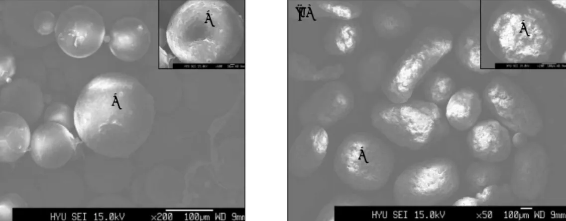
Double emulsion-solvent evaporation 법을 사용하여 제조한 PLLA/HA composite scaffold의 전자 현미경 소견 에서 PLLA 표면에 hydroxyapatite 결정의 균일한 침착을 보이고 있었다(Fig. 1).
2. 조직학적 소견
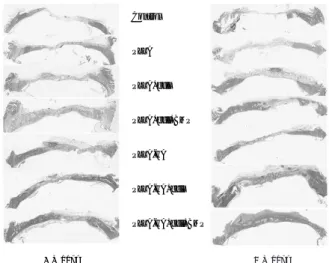
조직학적 소견에서 초기 1,2 주에서는 아무런 이식을 시 행하지 않거나 fibrin glue 만을 이식한 경우 염증세포의 침 윤이 다량 보였으며 4주 경과 후부터 신생골 형성이 관찰되 나 8주 경과 후에도 그 양이 미미하였다. 그러나 HA, PLLA 이식군에서는 신생골 형성 속도 및 양이 대조군에 비하여 증가된 양상이었지만 8주 경과 후에도 결손부의 완 전한 재생은 관찰되지 않고 있었다. PLLA/HA 이식군에서 는 4주 경과 후부터 신생골 형성이 왕성하였고 골모세포를 동시에 투여한 경우와 커다란 차이를 관찰할 수 없었지만 특히 1ug의 rhBMP를 동시에 투여한 경우 8주 소견에서 골 결손부가 거의 재생이 완료되고 bone remodeling이 상 당히 진행되어 있었다(Fig. 2).
Fig. 1. SEM findings of PLLA/HA composite scaffold.
(a: Only PLLA(×200), b: PLLA/HA(×50)) (a)
*
*
* (b)
*
Fig. 2. H&E staining findings.
Control
PLLA
PLLA+cell
PLLA+cell+BMP
PLLA-HA
PLLA-HA+cell
PLLA-HA+cell+BMP
4 Weeks
1
1 WWeeeekk 22 WWeeeekkss 44 WWeeeekkss 88 WWeeeekkss 11 WWeeeekk 22 WWeeeekkss 44 WWeeeekkss 88 WWeeeekkss 8 Weeks
Control (no graft)
Fibrin glue
Fibrin glue+cell
HA
HA+cell
PLLA
PLLA+cell
PLLA+cell+BMP
PLLA-HA
PLLA-HA+cell
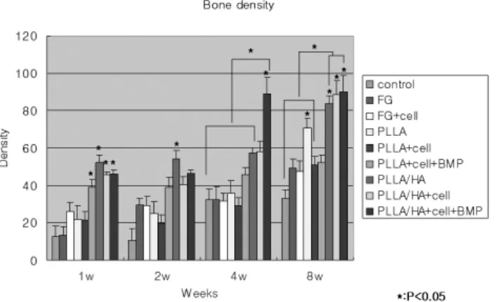
Fig. 5. Result of bone density analysis.
Fig. 6. 3-D CT findings(4 weeks). Fig. 7. 3-D CT findings(8 weeks).
Control
FG
FG+cell
HA
HA+cell
HA+cell+BMP
PLLA
PLLA+cell
PLLA+cell+BMP
PLLA/HA
PLLA/HA+cell
PLLA/HA+cell+BMP
Control
Fibrin Glue (FG)
FG+Cell
HA
HA+Cell
HA+Cell+BMP
PLLA
PLLA+Cell
PLLA+Cell+BMP
PLLA-HA
PLLA-HA+Cell
PLLA-HA+Cell+BMP