Yeungnam Univ J Med 2018;35(1):45-53
Ultrasound-guided superficial cervical plexus block under dexmedetomidine sedation versus general anesthesia for carotid
endarterectomy: a retrospective pilot study
Wangseok Do
1, Ah-Reum Cho
1, Eun-Jung Kim
2, Hyae-Jin Kim
1, Eunsoo Kim
1, Heon-Jeong Lee
11
Department of Anesthesia and Pain Medicine, Pusan National University, School of Medicine, Busan;
2
Department of Dental Anesthesia and Pain Medicine, Pusan National University Dental Hospital, Dental Research Institute, Yangsan, Korea
Background: Carotid endarterectomy (CEA) has been performed under regional and general anesthesia (GA).
The general anesthesia versus local anesthesia for carotid surgery study compared the two techniques and concluded that there was no difference in perioperative outcomes. However, since this trial, new sedative agents have been introduced and devices that improve the delivery of regional anesthesia (RA) have been developed. The primary purpose of this pilot study was to compare intraoperative hemodynamic stability and postoperative outcomes between GA and ultrasound-guided superficial cervical plexus block (UGSCPB) under dexmedetomidine sedation for CEA.
Methods: Medical records from 43 adult patients who underwent CEA were retrospectively reviewed, inclu- ding 16 in the GA group and 27 in the RA group. GA was induced with propofol and maintained with sevo- flurane. The UGSCPB was performed with ropivacaine under dexmedetomidine sedation. We compared the intraoperative requirement for vasoactive drugs, postoperative complications, pain scores using the numerical rating scale, and the duration of hospital stay.
Results: There was no difference between groups in the use of intraoperative antihypertensive drugs. How- ever, intraoperative inotropic and vasopressor agents were more frequently required in the GA group (p<0.0001). In the GA group, pain scores were significantly higher during the first 24 h after surgery (p<
0.0001 between 0-6 h, p<0.004 between 6-12 h, and p<0.001 between 12-24 h). The duration of hospital stay was significantly more in the GA group (13.3±4.6 days in the GA group vs. 8.5±2.4 days in the RA group, p<0.001).
Conclusion: In this pilot study, intraoperative hemodynamic stability and postoperative outcomes were better in the RA compared to the GA group.
Keywords: Carotid endarterectomy; Cervical plexus block; Dexmedetomidine; General anesthesia;
Ultrasonography
Copyright ©2018 Yeungnam University College of Medicine
This is an Open Access article distributed under the terms of the Creative Commons Attribution Non-Commercial License (http://creative- commons.org/licenses/by-nc/4.0/) which permits unrestricted non-commercial use, distribution, and reproduction in any medium, provided the original work is properly cited.
Received: January 29, 2018, Revised: March 28, 2018 Accepted: April 9, 2018
Corresponding Author: Ah-Reum Cho, Department of Anesthesia and Pain Medicine, School of Medicine, Pusan National University, 179, Gudeok-ro, Seo-gu, Busan 49241, Korea
Tel: +82-51-240-7399, Fax: +82-51-242-7466 E-mail: [email protected]
INTRODUCTION
Carotid endarterectomy (CEA) has been successfully per-
formed under regional anesthesia (RA) and general anesthesia
(GA). A large randomized multicenter study of general anes-
thesia versus local anesthesia for carotid surgery (GALA) was
conducted to determine whether GA or RA resulted in the
better outcomes, including the proportion of patients with stroke, myocardial infarction (MI), or death within 30 days after anesthesia; survival free of stroke, MI or death up to 1 year after anesthesia; length of stay in hospital and blood pressure manipulation [1]. The GALA study concluded that the anesthetic technique does not affect perioperative out- comes. However, the trial has been criticized due to the wide variability in methodology; general anesthetic techniques were compared with a variety of regional anesthetic techniques.
In addition, clinical practice had changed over the 8-year trial period. Since this study, there have been improvements in regional anesthetic techniques, intraoperative sedation, and the use of ultrasound guidance for RA [2,3]. In the GALA trial, RA was performed using landmark-guided superficial or deep cervical plexus block, which has been replaced by ultrasound-guided block, a method that allows direct visual- ization of structures and injection of local anesthetic [3].
The α
2-adrenoreceptor agonist sedative, dexmedetomidine, is widely used in anesthetic practice. The α
2-adrenoreceptors mediate sedation, sympatholysis, inhibition of vasoconstric- tion, and antinociception. The advantage of dexmedetomidine is that it has minimal respiratory depression [4]. Moreover, compared to sedation with a combination of midazolam and propofol, dexmedetomidine is less likely to cause periopera- tive hypertension and tachycardia. It also decreases intraope- rative and postoperative anxiety and the need for opioids for postoperative pain control [5]. However, perioperative hypo- tension and bradycardia can occur [6].
Although the GALA trial did suggest a tendency towards a better hemodynamic profile in the RA group, the trial did not focus on the use of sedatives or perioperative hemo- dynamic status. The purpose of this pilot study was to test the hypothesis that for CEA, intraoperative hemodynamic stabil- ity is improved using an ultrasound-guided superficial cervical plexus block (UGSCPB) with dexmedetomidine sedation com- pared to GA. The primary outcome of this study was the re- quirement for vasoactive drugs between the two groups. Post- operative hemodynamic stability, pain severity, and complica- tions within 1 year were also compared.
MATERIALS AND METHODS
1. Study design
This study was approved by the Institutional Review Board
and exempted from the need to obtain informed consent.
We retrospectively reviewed the electronic medical records of adult patients (aged between 46 and 91 years) who under- went CEA from June 2012 to July 2015. We chose this period because a single surgeon performed all CEA procedures during this time. We excluded patients who underwent a combined surgical procedure with CEA or underwent emergency sur- gery. The anesthetic technique was routinely determined by the anesthesiologist and the surgeon depending on the clinical condition of the patient. Patients were divided into the GA and the RA groups.
2. Anesthetic technique
Anesthesia was performed according to hospital protocols.
In both groups, intraoperative monitoring included electro- cardiography, invasive blood pressure (BP) measured from the radial artery, pulse oximetry, bispectral index monitoring (BIS;
Bispectral Index
TM, Aspect Medical System Inc., Norwood, MA, USA), and near-infrared spectroscopy (NIRS; INVOS 5100; Somanetics Corp., Troy, MI, USA). GA was induced with intravenous propofol (1-2 mg/kg), remifentanil (0.1 μg/
kg/min), and rocuronium (1.0 mg/kg) and maintained with sevoflurane and remifentanil. Sevoflurane was titrated to a BIS score of 40-60. Remifentanil was titrated to maintain the mean arterial pressure at 80% of the baseline. Mechanical ventilation was carried out with an oxygen/air mixture (frac- tion of inspired oxygen, 0.5) to maintain the end-tidal carbon dioxide at 30-40 mmHg. RA was performed by UGSCPB.
After negative aspiration, 8-12 mL of 0.375-0.5% ropivacaine was injected beneath the sternocleidomastoid muscle. The sternocleidomastoid muscle was directly visualized sonogra- phically (Vivid i, GE Healthcare, Wauwatosa, WI, USA) using a 12L-RS, 5-13 MHz linear probe. Prior to incision, a loading dose of dexmedetomidine (1.0 μg/kg) was administrated over 10 minutes, followed by a maintenance infusion at 0.5 μg/kg/h, and adjusted to maintain a BIS score of 60-80 (infusion rate:
0.1-1.0 μg/kg/h). Adequacy of the RA was not documented;
however, lidocaine (0.5%) administered by the surgeon to treat
intraoperative pain is common practice in our institution. Hy-
pertension and tachycardia were managed with diltiazem
(5-10 mg), esmolol (10-20 mg), labetalol (10 mg), or nicardi-
pine (1-2 mg). To treat hypotension and bradycardia, intrave-
nous ephedrine (10 mg bolus) or atropine (0.5 mg bolus) was
Fig. 1. Flow diagram of the patients.
administered. Persisting hypotension and bradycardia were treated with a continuous infusion of dopamine, dobutamine, norepinephrine, or phenylephrine. Postoperative pain was ma- naged by the surgeon; morphine, 3 mg, and ketorolac, 30 mg were administered according to the numerical rating scale (NRS) until the pain was controlled.
3. Outcome variables
Demographic data, including the American Society of Anes- thesiologists physical status, age, sex, weight, height, and co- existing diseases were obtained from the electronic medical records. Details on surgery-related outcomes, including the side of surgery, duration of surgery, type of anesthesia, carotid artery cross-clamping, and the use of a shunt were also ob- tained. Hemodynamic instability was categorized into three groups: none, no intervention; mild, 1 or 2 doses of vasoac- tive medication; severe, ≥3 doses or a continuous infusion of vasoactive medication. We identified the frequency and ti- ming of vasoactive drugs administered during surgery: T1, during the administration of dexmedetomidine or after intu- bation; T2, at skin incision; T3, before carotid artery cross- clamping; T4, after carotid artery cross-clamping; T5, at the completion of surgery. Hemodynamic profiles during the 24 h postoperative period and the duration of hospital stay were recorded. Postoperative pain was rated using the NRS and recorded at 0-6 h, 6-12 h, and 12-24 h after surgery. Major complications including cardiovascular, respiratory, and neu- rological complications within 1 year were also compared.
4. Statistical analysis
Data are presented as number (%), mean±standard devia- tion, or median (interquartile range). All continuous variables were checked for normal distribution with a Q-Q plot and the Kolmogorov-Smirnov test. Continuous variables that were normally distributed were compared with the Student’s t-test.
The frequency of interventions for hemodynamic instability and complications were compared with the chi-square or the Fisher’s exact test. Postoperative NRS was compared using two-way repeated measures ANOVA with Bonferroni correc- tion. For Bonferroni correction, a p -value of less than 0.0083 (0.05/6) was considered significant. For other statistical tests, a p -value of less than 0.05 was considered significant. All
analyses were performed using SPSS version 18.0 (SPSS Inc., Chicago, IL, USA).
RESULTS
Forty-eight patients underwent CEA during the study pe- riod. Five patients were excluded as they had combined or emergency surgery. Of the remaining 43 patients, 16 received GA and 27 received RA (Fig. 1).
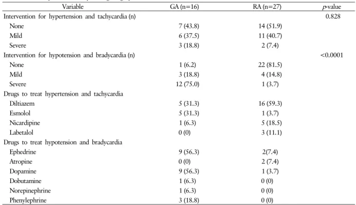
Demographic data were not different between the two groups (Table 1). In the RA group, no patient required con- version to GA and no local anesthetic-related adverse effects were noted. Duration of surgery, anesthesia, and carotid artery cross-clamping were longer, and a shunt was used more fre- quently in GA group (Table 1). The frequency of intervention for hypertension and tachycardia was similar between groups;
however, inotropic and vasopressor agents were more fre- quently used in the GA group ( p <0.0001, Table 2). In the RA group, an antihypertensive drug was required in 51.9%
of patients at the time of administration of the loading dose of dexmedetomidine. In the GA group, 50% of patients re- quired an antihypertensive drug after intubation and at the time of the skin incision. An infusion of inotropic or vasopre- ssor agents was required in 75% of patients in the GA group.
In contrast, 18.5% of patients in the RA group required in-
otropic or vasopressor agents (Fig. 2). Intervention for hyper-
tension was more frequently required in the GA group at
Table 1. Demographic data and intraoperative outcomes
Variable GA (n=16) RA (n=27) p -value
ASA physical status (II/III) 12 (75)/4 (25) 21 (77.8)/6 (22.2) 1.000
Sex (male) 14 (87.5) 23 (85.2) 1.000
Age (year) 72.7±8.2 68.1±9.8 0.124
Weight (kg) 62.9±9.0 65.7±8.6 0.318
Height (cm) 164.9±8.0 164.7±5.4 0.919
Coexisting diseases
Diabetes 7 (43.8) 8 (29.6) 0.348
Hypertension 11 (68.8) 21 (77.8) 0.768
Ischemic heart disease 4 (25.0) 12 (44.4) 0.202
Hyperlipidemia 3 (18.8) 4 (14.8) 0.929
TIA/stroke 3 (18.8) 6 (22.2) 0.906
COPD 3 (18.8) 1 (3.7) 0.291
Renal failure 1 (6.3) 2 (7.4) 0.635
Indication for surgery 0.878
Asymptomatic carotid stenosis 1 (6.3) 4 (14.8)
TIA 9 (56.3) 16 (59.3)
Stoke 6 (37.5) 7 (25.9)
Location of surgery (rt) 8 (50.0) 17 (63.0) 0.303
Duration of surgery (min) 147.8±29.6 125.6±32.0 0.029
Duration of anesthesia (min) 191.6±30.0 164.1±29.4 0.005
Duration of clamping (min) 49.1±10.4 31.5±9.8 <0.0001
Shunt 15 (93.8) 5 (18.5) <0.0001
Data are presented as mean±SD or number (%).
ASA, American Society of Anesthesiologists; GA, general anesthesia; RA, regional anesthesia; TIA, transient ischemic attack; COPD, chronic obstructive pulmonary disease; Rt, right; SD, standard deviation.
Fig. 2. Frequency and timing of antihypertensive drugs (A) and inotropic or vasopressors (B) administrating during surgery. T1, during administration of dexmedetomidine or after intubation; T2, at skin incision; T3, before carotid artery cross-clamping; T4, after carotid artery cross-clamping; T5, at the end of the operation.
a)p <0.005 compared with regional anesthesia group.
the time of skin incision (50% in the GA group vs. 3.7% in the RA group, p =0.001) and at the completion of surgery
(31.3% in the GA group vs. 3.7% in the RA group, p =0.039).
Use of inotropic and vasopressor agents was significantly more
Table 2. Hemodynamic stability during surgery
Variable GA (n=16) RA (n=27) p -value
Intervention for hypertension and tachycardia (n) 0.828
None 7 (43.8) 14 (51.9)
Mild 6 (37.5) 11 (40.7)
Severe 3 (18.8) 2 (7.4)
Intervention for hypotension and bradycardia (n) <0.0001
None 1 (6.2) 22 (81.5)
Mild 3 (18.8) 4 (14.8)
Severe 12 (75.0) 1 (3.7)
Drugs to treat hypertension and tachycardia
Diltiazem 5 (31.3) 16 (59.3)
Esmolol 5 (31.3) 1 (3.7)
Nicardipine 1 (6.3) 5 (18.5)
Labetalol 0 (0) 3 (11.1)
Drugs to treat hypotension and bradycardia
Ephedrine 9 (56.3) 2(7.4)
Atropine 0 (0) 2 (7.4)
Dopamine 9 (56.3) 1 (3.7)
Dobutamine 1 (6.3) 0 (0)
Norepinephrine 1 (6.3) 0 (0)
Phenylephrine 3 (18.8) 0 (0)
Data are presented as number (%).
GA, general anesthesia; RA, regional anesthesia; n, number; None, no intervention; Mild, 1 or 2 injections of drugs; Severe, 3 injections of drugs or infusion.
frequent in the GA group throughout the surgery (All p <0.01, Fig. 2).
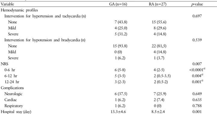
Postoperative hemodynamic stability was not different be- tween groups. Only 6.2% of patients in the GA group and 18.5% of patients in the RA group required intervention for hypotension, while 56.2% of patients in the GA group and 44.4% in the RA group received antihypertensive agents.
NRS during the first 24 hours postoperatively was significan- tly higher ( p <0.0001 between 0-6 h, p <0.004 between 6-12 h, and p <0.001 between 12-24 h), and the duration of hospi- tal stay was significantly longer (13.3±4.6 vs. 8.5±2.4 days, p <0.001) in the GA group. The incidence of major complica- tions was similar in both the groups (Table 3).
DISCUSSION
In this study, we found that intraoperative hemodynamic stability was better in the RA group compared to the GA group.
The NRS during the first 24 h postoperatively and the dura- tion of hospital stay were higher in the GA group. However,
major postoperative complications within 1 year were not significantly different between groups.
RA has the advantage of ease of assessment of neurological status during carotid cross-clamping. RA has been in use for CEA for over 15 years. The GALA [1] and the other studies [7,8] have not identified a statistically significant difference in the incidence of major perioperative outcomes, such as stroke, MI, or death between RA and GA. However, if major peri- operative outcomes were similar between GA and RA, hemo- dynamic stability, postoperative pain, minor complications, and duration of hospital stay may be appropriate outcomes to determine the better anesthetic technique for CEA. Thus, well-standardized RA with advanced techniques needs to be compared with GA to evaluate perioperative outcomes.
Despite the ease of perioperative neurological assessment
with RA, this technique has several risks. Systemic toxicity
from local anesthetics can be a life-threatening complication
resulting from intravascular injection or overdose [9]. Punc-
ture-related complications include vertebral artery injection,
subarachnoid or epidural injection, Horner’s syndrome, phre-
Table 3. Postoperative outcomes
Variable GA (n=16) RA (n=27) p -value
Hemodynamic profiles
Intervention for hypertension and tachycardia (n) 0.697
None 7 (43.8) 15 (55.6)
Mild 4 (25.0) 8 (29.6)
Severe 5 (31.2) 4 (14.8)
Intervention for hypotension and bradycardia (n) 0.539
None 15 (93.8) 22 (81,5)
Mild 0 (0) 4 (14.8)
Severe 1 (6.2) 1 (3.7)
NRS 0.007
0-6 hr 6 (5-8) 4 (2-5) <0.0001
a)6-12 hr 5 (3-5) 2 (0.5-3.5) 0.004
a)12-24 hr 3 (2-3) 2 (0.5-2) 0.001
a)Complications
Neurologic 6 (37.5) 7 (25.9) 0.649
Cardiac 1 (6.2) 2 (7.4) 0.635
Respiratory 1 (6.2) 0 (0) 0.788
Hospital stay (day) 13.3±4.6 8.5±2.4 0.001
Data are presented as mean±SD, median (interquartile range) or number (%).
GA, general anesthesia; RA, regional anesthesia; NRS, n, number; numeric rating scale; SD, standard deviation; None, no intervention;
Mild, 1 or 2 injections of drugs; Severe, 3 injections of drugs or infusion.
a)