CASE REPORT eISSN 2384-0293 https://doi.org/10.12701/yujm.2017.34.1.111 Yeungnam Univ J Med 2017;34(1):111-114
YUJM VOLUME 34, NUMBER 1, JUNE 2017 111
베체트병 환자에서 저용량 스테로이드 사용과 관련하여 발생한 중심성장액맥락망막병증
차성욱, 김경진, 권성민, 이신애, 민병철, 김은성, 이정욱 부산성모병원 내과
Central serous chorioretinopathy associated with low dose systemic corticosteroid treatment of Behcet’s disease
Sungwook Cha, Kyung Jin Kim, Seongmin Kweon, Sinae Lee, Byungchul Min, Eunsung Kim, Jungwook Lee
Department of Internal Medicine, Busan St.Mary’s Hospial, Busan, Korea
Central serous chorioretinopathy may induce poor eyesight and serous retinal detachment. However, its exact cause has not been well established thus far. It can be associated with systemic high-dose corticosteroid treatment mainly for young and middle-aged men and may spontaneously regress or recur after withdrawal from corticosteroid. After corticosteroid administration for Behcet’s disease, it is necessary to identify any ocular symptoms. Behcet’s disease can lead to the development of ocular complications, such as uveitis, hypopyon, retinal vasculitis, optic neuritis, angiogenesis, secondary cataract, and glaucoma. It is possible to diagnose any of these complications via optical coherence tomography and digital indocyanine green angio- graphy. It is easy to neglect an ocular symptom that may appear after a low-dose corticosteroid treatment as an ocular complication in patients with Behcet’s disease. Thus, we report on a case concerning high-dose corticosteroid treatment with a literature review.
Keywords: Steroid; Behcet’s syndrome; Central serous chorioretinopathy
Copyright ©2017 Yeungnam University College of Medicine
This is an Open Access article distributed under the terms of the Creative Commons Attribution Non-Commercial License (http://creative- commons.org/licenses/by-nc/4.0/) which permits unrestricted non-commercial use, distribution, and reproduction in any medium, provided the original work is properly cited.
Received: February 24, 2016, Revised: June 20, 2016 Accepted: June 23, 2016
Corresponding Author: Jung wook Lee, Department of Internal Medicine, Busan St.Mary’s Hospial, 25-14 Yongho-ro 232beon-gil, Nam-gu, Busan 48575, Korea Tel: +82-51-933-7235, Fax: +82-51-932-8600
E-mail: [email protected]
서 론
중심성장액맥락망막병증(central serous chorioretinopathy) 은 원인은 아직 정확하게 알려지지 않았다. 하지만 다른 안과 적 또는 전신적 질환이 없으면서 맥락막 모세혈관으로부터
삼출물이 망막 하로 누출되고, 장액성 감각 신경 박리를 보이 는 질환으로 외층 혈액망막장벽의 특발성 이상으로 발생한 다고 알려졌다[1,2]. 이 질병의 원인과 병태생리학적 기전은 정확히 알려진 바가 없으나 여러 요인이 이 질병의 발생과 관계가 있을 것으로 추측되며, 고용량의 전신적 스테로이드 투여가 시력 저하와 장액성 망막박리를 유발했다는 증례들 이 보고되었다. 스테로이드 치료를 받는 동안 발생한 중심성 장액맥락망막병증의 증례들에서 보고된 스테로이드의 용량 은 프레드니솔론 10 mg 이상의 경우였다.
본 증례에서는 고령이 아닌 41세의 남자 환자에서 저용량 의 스테로이드(7.5 mg/day) 사용 후 32일째 발생한 중심성장 액맥락망막병증을 진단하게 되었다.
Sungwook Cha et al.
112 YUJM VOLUME 34, NUMBER 1, JUNE 2017
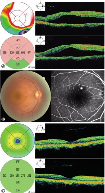
Fig. 1. The patient’s optical coherence tomography (OCT). (A) OCT of the right eye showed a serous sensory retinal detachment and edema. (B) Fundus photograph and fluorescein angiogram of right eye shows serous retinal detachment and focal areas of hyperfluorescene. (C) OCT of the left eye showed no retinal
detachment and edema. Fig. 2. Post treatment of optical coherence tomography showed resolution of retinal detachment and edema.
증 례
환 자: 41세 남자
주 소: 최근 발생한 시력저하, 어지럼증
과거력 및 현 병력: 내원 3년 전부터 피로감 및 이주하는 근육통 및 관절통이 있었으나 정밀검사 없이 지냈다고 한다.
2주 이상 지속하는 다발성 관절통, 구강궤양, 피로감을 주소 로 내원하였고, 3회 이상 재발하는 구강궤양, 피부상 결절홍반
(erythema nodosum), 이상초과민검사(pathergy test)상 양성 으로 베체트병을 진단하였다. 이후, 콜키신(colchicine 0.6 mg/
day), 아자씨오프린(azathioprine 100 mg/day), 저용량의 경구 용 스테로이드(프레드니솔론 7.5 mg/day) 복용 후 32일째 발생 한 어지럼증과 시력저하를 주소로 외래 내원하였다.
사회력 및 가족력: 특이소견 없었다.
계통적 검사 및 이학적 소견: 내원 시 활력징후는 특이소 견이 없었다. 문진상 시력저하, 어지럼증이 있었고, 신체검사 상 구강궤양이 남아있는 것 외에 이상소견은 보이지 않았다.
안과 검사소견: 내원 당시 시력은 우안 0.75, 좌안 1.0이었 고, 안압은 우안 15 mmHg, 좌안 16 mmHg였다. 안저검사에 서 우안 황반부에 장액망막박리가 관찰되었고, 빛간섭단층 촬영(optical coherence tomography)에서도 우측에 장액망막 박리(serous retinal detachment)가 관찰되었다. 형광안저촬 영술에서 형광물질의 누출 소견이 보였다(Fig. 1).
검사실 소견: 말초혈액 검사에서 백혈구 7,100/μL, 혈색소 11.7 g/dL, 혈소판 354,000/μL였다. 생화학 검사에서 나트륨 140 mmol/L, 칼륨 4.8 mmol/L, 염소 104 mmol/L, 크레아티닌 1.1 mg/dL, 알부민 4.3 g/dL, 아스파르테이트아미노전달효소 19 IU/L, 알라닌아미노전달효소 13 IU/L, 총빌리루빈 0.6 mg/
dL, 혈당 97 mg/dL였다. 면역학적 검사를 포함한 기타 검사에 서 갑상샘자극호르몬 4.248 μU/mL, 류마티스인자 5 IU/mL, 면역글로불린E (immunoglobulinE, IgE) 139 U/mL, 형광항핵 항체 음성, 항중성구세포질항체 정상, 사람백혈구항원 음성 이었다.
치료 및 임상경과: 프레드니솔론 중단 후 2주 후부터 환자 의 증상이 호전되었고, 2개월 후 빛간섭단층촬영에서 크게 호전되는 양상이 관찰되었다(Fig. 2).
Systemic Corticosteroid Treatment and CSCR
YUJM VOLUME 34, NUMBER 1, JUNE 2017 113
고 찰
중심성장액맥락망막병증은 주로 20-50세의 젊은 남자에 서 많이 발생하고, 중심 시력 저하, 변시증 그리고 중심암점 등의 시력저하를 초래한다[3]. 그리고 특정한 삼출 현상의 원인 없이 망막색소상피의 국소적인 기능 장애와 맥락막으 로부터 망막하 공간에 장액성 액체의 유입으로 후극부에 감 각 신경 망막박리가 일어나는 질환이다[4,5].
예후는 상당히 양호하여 특별한 치료 없이도 증상 호전이 가능하지만 35-45% 정도에서 재발할 수 있으며, 만성적인 경과를 보일 경우 망막색소상피와 감각 신경 망막층의 위축 으로 비가역적인 시력 감소가 가능하다[6]. 이 질병의 원인과 병태생리학적 기전은 정확히 알려진 바가 없으나 여러 요인 이 이 질병의 발생과 관계가 있을 것으로 추측된다. 그 중 쿠싱증후군 등 전신적인 질환이나 A형 성격, 임신, 스트레스 등 혈류 내 코티솔 수치가 증가하는 경우와 관련이 있다고 알려졌고 이에 대한 것은 보고되어 있다[7]. 증가된 코티솔이 혈액 내 아드레날린성 물질과 교감신경계에 작용하여 맥락 막과 망막색소 상피층의 투과성을 직접적으로 증가시키거 나, 맥락막의 혈관수축을 일으킴으로써 상대적 허혈을 유발 하고, 삼투압의 변화와 망막색소상피의 손상을 일으켜 투과 성을 증가시킬 것이라는 주장도 있다[1,8].
최근에는 인도사이아닌그린혈관조영술(digital indocya- nine green angiography)에서 맥락막 모세혈관의 투과성이 증가하여있는 것이 중심성장액맥락망막병증의 병태생리로 알려졌다[9,10]. 이는 아마도 스트레스와 관련하여 아드레날 린 분비가 증가하고, 이에 의해 동맥의 관류압이 하강하거나 맥락막이 허혈 상태가 되어 이차적으로 맥락막 모세혈관이 나 정맥의 울혈이 발생하는 것으로 생각되고 있다[11].
그리고 고용량의 전신적 스테로이드 투여가 시력 저하와 장 액성 망막박리를 유발했다는 증례들이 보고되었다. Wakakura 등[12]이 분석한 스테로이드와 관련되어 발생한 중심성장액 맥락망막병증 33예의 경우 잠복 기간이 70일 이내였던 군에 서는 증상이 발현된 시점에서 하루 스테로이드 용량이 20 mg 이상이었다. 잠복 기간이 6개월 이상이었던 군에서는 증상 이 발현된 시점에서 하루 스테로이드 용량이 20 mg 미만이 었다. Chaine 등[13]이 발표한 예들에서도 Wakakura 등과 마찬가지로 증상의 발현 시기가 빠를수록 하루 스테로이드 용량이 많았다. Koyama 등[14]이 분석한 중심성장액맥락망 막병증 17예의 경우 잠복 기간이 3일에서 23년이었고, 하루 스테로이드 용량은 5-1,000 mg이었다. Loo 등[15]은 뇌하수체
선종 수술 후 10여 년간 코티솔과 티록신 치료를 하였던 50 세 환자, 신장이식 후 10여 년간 하루 10 mg의 프레드니솔론 과 사이클로스포린을 복용하였던 55세 환자와 보통천포창 (pemphigus vulgaris)으로 7년간 하루 120 mg의 프레드니솔 론을 복용하였던 36세 환자에서 발생한 중심성장액맥락망 막병증을 보고하였다. Levy 등[16]은 심한 갑상샘 눈확병증 (thyroid orbitopathy)으로 수술 후 6개월 동안 하루 20 mg의 프레드니솔론을 복용한 52세 환자와 천식 악화로 10일간 하 루 10 mg의 프레드니솔론을 복용한 38세 환자에서 발생한 중심성장액맥락망막병증 2예를 보고하였다. Fawzi 등[17]은 신장이식을 받은 후 스테로이드와 면역억제제 병합요법으로 치료를 하면서 중심성장액맥락망막병증이 발생한 27-55세 의 환자들 15예를 보고하였다.
한국에서의 증례를 살펴보면, 32세의 우측 벨 마비 환자에 서 경구용 스테로이드(프레드니솔론, 60 mg/day)를 사용 4일 째 240 mg (60 mg/day, 4일간) 투여 후 우측 시야 한가운데가 꺾여 보이는 증상으로 안과 협진 후 중심성장액맥락망막병 증으로 진단하였다[18]. 다른 증례의 경우는 만성 사구체 질 환의 환자들이다. 초점성 분절성 사구체경화증 진단을 받은 29세 남자와 IgA 신병증 진단을 받은 33세 남자 환자에서 경구용 스테로이드(프레드니솔론 60 mg/day) 복용 후 각각 9일과 10일째 발생한 시력 저하로 안과 검사 후 중심성장액 맥락망막병증으로 진단하였다[19].
본 증례의 경우는 베체트병으로 32일째 치료를 받은 30대 의 젊은 남성이다. 베체트병에서는 포도막염, 앞방축농(hy- popyon), 혈관염 등의 안구 합병증이 동반될 수 있어, 이에 대한 감별이 필요하다. 포도막염의 경우 보통 양측성으로 나타나고, 포도막 전반에 영향을 미치게 된다. 앞방축농의 경우 전방(anterior chamber)의 화농성 물질 및 망막 혈관염 (retinal vasculitis)의 양상이 관찰된다. 후방 포도막염, 망막 혈관염, 시신경염에서는 전신적 면역억제 치료가 필요하다.
다른 변화로 혈관신생, 이차성 백내장, 녹내장 등이 나타날 수 있다. 이 환자는 안과 협진에서 빛간섭단층촬영, 안저촬영 및 형광안저촬영술을 시행하였다. 보통 양측성으로 경계가 명확하지 않은 부종을 동반하는 포도막염과는 달리 경계가 명확한 편측의 망막 부종이 있었고, 화농성 물질이나 망막 혈관염의 양상은 관찰되지 않았다. 망막 부종등의 특이소견 이 없는 좌측에 비해 우측에서 장액망막박리가 관찰되었고, 형광안저촬영술에서 잉크 반점 형태의 형광 누출이 관찰되 어 중심성장액맥락망막병증을 진단받았다. 이전의 증례들 과는 다르게 30대의 젊은 남성에서 하루 스테로이드 용량
Sungwook Cha et al.
114 YUJM VOLUME 34, NUMBER 1, JUNE 2017
7.5 mg이라는 저용량에서 장액성망막박리가 발생하였다. 이 환자는 베체트병 관련 포도막염과는 달리 스테로이드 사용 후 안구 증상이 발생하였다가 스테로이드 중단 이후 호전되는 양 상을 보였다.
중심성장액맥락망막병증은 스테로이드를 투여하지 않더 라도 A형 성격, 임신, 스트레스 등의 다양한 원인에 의해 발 병할 수 있고, 예후는 상당히 양호하여 특별한 치료 없이도 증상 호전이 가능하다. 하지만 베체트병과 같은 자가면역질 환의 경우 면역계 억제를 위해 스테로이드 치료가 필요하고, 기저질환이 없는 환자에 비해 스테로이드에 더욱 민감할 것 이다[20]. 저용량의 스테로이드 사용에 따른 합병증을 기저 질환에 의한 합병증으로 오인하여 스테로이드 사용을 지속 할 수 있다는 점에서 본 증례는 가치가 있다.
스테로이드를 사용하는 환자의 수는 많지만, 중심성장액 맥락망막병증이 발생하는 경우는 매우 드물고, 이로 인해 간과할 우려가 높다. 내과 의사로서 다양한 질병 및 임상 증상에서 흔하게 스테로이드를 사용하게 된다. 본 증례에서 는 저용량의 경구용 스테로이드를 사용한 젊은 환자에게서 장액성망막박리가 발생했다는 점에서 의미가 있으며, 이 증례 를 통해 저용량 스테로이드의 사용에서도 환자의 임상 증상 에 대해 주의를 기울이는 계기가 되기를 바란다.
CONFLICT OF INTEREST
No potential conflict of interest relevant to this article was reported.
REFERENCES
1. Kleinberger AJ, Patel C, Lieberman RM, Malkin BD. Bilateral central serous chorioretinopathy caused by intranasal cortico- steroids: a case report and review of the literature. Laryngo- scope 2011;121:2034-7.
2. Schatz H, Madeira D, Johnson RN, McDonald HR. Central serous chorioretinopathy occurring in patients 60 years of age and older. Ophthalmology 1992;99:63-7.
3. Ryan SJ. Retina. 2nd ed. St. Louis: SV Mosby; 1994. p. 1158- 68.
4. Gass JD. Pathogenesis of disciform detachment of the neuro- epithelium. Am J Ophthalmol 1967;63(3 Suppl):1-139.
5. Yannuzzi LA, Gitter KA, Schatz H. The macula: a compre-
hensive text and atlas, 2nd ed. Baltimore: William & Wilkins;
1982. p. 145-65.
6. Ueoka T. A pathologic study of central serous chorioretino- pathy. Acta Soc Ophthalmol Jpn 1949;53:222-6.
7. Bouzas EA, Karadimas P, Pournaras CJ. Central serous cho- rioretinopathy and glucocorticoids. Surv Ophthalmol 2002;
47:431-48.
8. Cho WB, Chung H, Kim HC. Spectral domain OCT findings of asymptomatic fellow eyes in central serous chorioretino- pathy. J Korean Ophthalmol Soc 2010;51:1345-53. Korean.
9. Lafaut BA, Salati C, Priem H, De Laey JJ. Indocyanine green angiography is of value for the diagnosis of chronic central serous chorioretinopathy in elderly patients. Graefes Arch Clin Exp Ophthalmol 1998;236:513-21.
10. Piccolino FC, Borgia L. Central serous chorioretinopathy and indocyanine green angiography. Retina 1994;14:231-42.
11. Taban M, Boyer DS, Thomas EL, Taban M. Chronic central serous chorioretinopathy: photodynamic therapy. Am J Ophthalmol 2004;137:1073-80.
12. Wakakura M, Song E, Ishikawa S. Corticosteroid-induced central serous chorioretinopathy. Jpn J Ophthalmol 1997;41:
180-5.
13. Chaine G, Haouat M, Menard-Molcard C, Favard C, Vignal- Clermont C, Campinchi-Tardy F, et al. [Central serous cho- rioretinopathy and systemic steroid therapy]. J Fr Ophtalmol 2001;24:139-46.
14. Koyama M, Mizota A, Igarashi Y, Adachi-Usami E. Seventeen cases of central serous chorioretinopathy associated with sys- temic corticosteroid therapy. Ophthalmologica 2004;218:
107-10.
15. Loo JL, Lee SY, Ang CL. Can long-term corticosteriods lead to blindness? A case series of central serous chorioretinopathy induced by corticosteroids. Ann Acad Med Singapore 2006;
35:496-9.
16. Levy J, Marcus M, Belfair N, Klemperer I, Lifshitz T. Central serous chorioretinopathy in patients receiving systemic corti- costeroid therapy. Can J Ophthalmol 2005;40:217-21.
17. Fawzi AA, Holland GN, Kreiger AE, Heckenlively JR, Arroyo JG, Cunningham ET Jr. Central serous chorioretinopathy af- ter solid organ transplantation. Ophthalmology 2006;113:
805-13.e5.
18. Park WI, Jung HW, Cho JE, You JR. A case of central serous chorioretinopathy associated with systemic corticosteroid treatment of Bell’s palsy. Korean J Otorhinolaryngol-Head Neck Surg 2012;55:458-60. Korean.
19. Moon YR, Kim YK, Kim YS, Lee YS. Two cases of central serous chorioretinopathy (CSCR) following corticosteroid therapy for chronic glomerulonephritis. Korean J Med 2008;
75:221-4. Korean.
20. Quax RA, van Laar JA, van Heerebeek R, Greiner K, Ben- Chetrit E, Stanford M, et al. Glucocorticoid sensitivity in Behçet’s disease. Endocr Connect 2012;1:103-11.