2021 The Korean Society of Veterinary Science.
This is an open-access article distributed under the terms of the Creative Commons Attribution Non-Commercial license (http://creativecommons.
org/licenses/by-nc/4.0/), which permits unrestrict- ed non-commercial use, distribution, and repro- duction in any medium, provided the original work is properly cited.
Introduction
Porcine epidemic diarrhea virus (PEDV) causing porcine epidemic diarrhea (PED), is a large-enveloped RNA virus, which belongs to the genus Alphacorona- virus, family Coronaviridae, order Nidovirales. The PEDV genome is approximate- ly 28 kb long containing at least 7 open reading frames (ORF1a, ORF1b, and ORF2–6), and a 3' untranslated region [1,2]. PEDV infection in pigs causes watery diarrhea, anorexia, and depression leading to high mortality rates in suckling pig- lets [1,3].
It is critical to propagate PEDV isolates efficiently in cell culture to perform re- search on pathogenesis and vaccine development, as growing PEDV in cell culture has proven difficult. PEDV can be isolated from clinical samples; however, the vi- rus may gradually lose infectivity upon further passages in cell culture [4].
The spike (S) protein of PEDV interacts with the cellular receptor in the entry process of virus and involves in production of neutralizing antibodies in pigs [1].
The S protein is considered a primary target antigen for the induction of neutral- izing antibodies and development of an effective vaccine against PEDV [5,6]. Re- cent reports have demonstrated the diversity of S gene in many isolates. The pathogenicity and antigenicity of PEDV can be changed according to mutations,
activity of porcine epidemic diarrhea viruses from Vietnam
Van Tan Do
1,†, Quang Lam Truong
2,†, Hoai Thu Dao
1, Thi Lan Nguyen
2, Jini Kim
1, Tae-Wook Hahn
1,*1 College of Veterinary Medicine and Institute of Veterinary Science, Kangwon National University, Chuncheon 24341, Korea
2 Key Laboratory of Veterinary Biotechnology, Faculty of Veterinary Medicine, Vietnam National University of Agriculture, Ha Noi 12406, Vietnam
pISSN 2466-1384 · eISSN 2466-1392 Korean J Vet Res 2021;61(3):e23 https://doi.org/10.14405/kjvr.2021.61.e23
*Corresponding author:
Tae-Wook Hahn
College of Veterinary Medicine and Institute of Veterinary Science, Kangwon National University, 1 Kangwondaehak-gil, Chuncheon 24341, Korea Tel: +82-33-250-8671 Fax: +82-33-244-2367 E-mail: [email protected]
Current affiliation (Van Tan Do and Hoai Thu Dao): Institute of Veterinary Research and Development of Central Vietnam, Nha Trang, Vietnam
ORCID:
https://orcid.org/0000-0002-3919-1153
† These authors contributed equally to this work.
Conflict of interest:
The authors declare no conflict of interest.
Received: May 14, 2021 Revised: July 14, 2021 Accepted: July 27, 2021
Original Article
Porcine epidemic diarrhea (PED) is characterized by acute enteritis, watery diarrhea, weight loss, dehydration, and death with high mortality in neonatal piglets. In this study, 3 virus isolates collected in Vietnam between 2016 and 2017 were successfully propagated in Vero cells at high virus titers. Sequence analysis of the full-length spike (S) gene revealed that all 3 isolates belong to genogroup 2a, which is closely related to other prevalent Asian strains. Amino acid sequence comparisons revealed 98.19% to 99.13% homology with the Vietnam isolates circulating during 2013–2015, suggest- ing that field PED viruses (PEDVs) evolve continuously. Experiments in animals demon- strated that antisera from guinea pigs immunized with the vaccine strain resulted in higher levels (5 log2) of neutralizing antibody against the homologous strain, and showed a relatively lower level of neutralizing antibody against the field isolates. This finding would be helpful in choosing a PEDV strain for vaccine development.
Keywords: porcine epidemic diarrhea virus; neutralizing activity; spike gene; Vietnam isolates
deletions and/or insertions of amino acids in the S protein. As a result, viral antigenicity and viral neutralizing activity may be changed [7,8]. Zhang et al. [8] in 2015 stated that a field CHN/
FL2013 strain reduced virulence in cell culture with a 7-amino acid deletion at N-termination of S protein.
In this study, PEDV isolates from Vietnam were propagated in Vero cells, and full-length S genes were sequenced to investi- gate the diversity of PEDVs. In addition, the cross-neutralizing activity of the serum of guinea pigs immunized with a PEDV vaccine strain against the isolates was evaluated.
Materials and Methods
Ethics statement
Animal experiments were approved by the Institutional Ani- mal Care and Use Committee (approval number: KW-181031- 1) and performed at the Center for Animal Experiment, Kang- won National University.
Isolation of porcine epidemic diarrhea virus in Vero cells Stool specimens and small intestine were taken from suckling piglets and post-weaning pigs exhibiting acute watery diarrhea.
The PEDV HID9047 and HID9048 isolates used in this study were collected from infected samples in Ha Nam and Thai Binh provinces, Vietnam in 2016, and the PEDV HID9049 isolate was collected from infected samples in Hung Yen province, Vietnam in 2017.
Isolation and propagation of PEDV in Vero cells was con- ducted as adapted from methods in Park et al. [9] in 2018 with modifications. Briefly, 80% confluent Vero cells were inoculated with virus at 37°C for 1 hour. Infection medium (Dulbecco’s modified Eagle’s medium-DMEM supplemented with 0.02%
yeast extract, 0.3% tryptose phosphate broth, 5 μg/mL trypsin and 1% antibiotic–antimycotic) was added to the infected cell flask without removing the inoculum. The infected cells were incubated at 37°C for 7 days until extensive cytopathic effects (CPEs) were observed; viruses were harvested by 3 freeze-thaw cycles.
The presence of PEDV was confirmed by a reverse transcrip- tion polymerase chain reaction (RT-PCR) amplifying approxi- mately 900 bp of the N gene with primers PEDV-N–F (5'-CGG TTC TCA CAG ATA GTG A-3') and PED-N-R (5'-CTC CTC CAC TCT GGG ATG T-3').
Virus titration
Vero cells (1 × 10⁵) were seeded in each well of a 96-well plate (Corning, USA) and grown at 37°C 1 day before the assay.
Virus was diluted in infection medium and 0.2 mL of 10-fold serial virus dilutions was added to each well after removing the medium. Each virus dilution was used to infect 10 wells of monolayer Vero cells. CPE was examined under a microscope every day for 7 days postinfection and the virus titer was calcu- lated as the 50% tissue culture infective dose (TCID50) using the Reed-Muench method [7].
Immunofluorescent assay
Vero cells were infected with PEDV 24 hours before the assay.
The control and infected cells were fixed with 4% paraformal- dehyde and permeabilized with 0.1% Triton X-100. Cells were then blocked with 5% bovine serum albumin, followed by incu- bation with 1/1,000 monoclonal mouse anti-PEDV (Median Diagnostics, Korea) and 1/1,000 fluorescein isothiocyanate (FITC)-conjugated goat anti-mouse immunoglobulin G (IgG) (MP Biomedicals, USA). The signals were observed using fluo- rescence microscopy [9].
Nucleotide sequence analysis
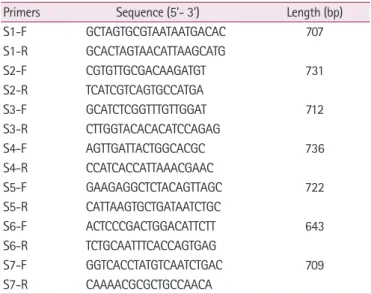
The full-length S genes of PEDVs were amplified by RT-PCR using the primers described in Table 1. The individual fragment was cloned into T&A cloning vector (RBC; Real Biotech Corp., Taiwan), and sequenced in both directions. The sequences of full-length S genes of PEDV isolates in this study and reference PEDVs were used in sequence alignments and phylogenetic analyses with the Serial Cloner 2.6, the ClustalX 2.0 program, and the Mega 6.0 program [10].
Table 1. List of primers used for the full-length spike gene sequenc- ing in this study
Primers Sequence (5'- 3') Length (bp)
S1-F GCTAGTGCGTAATAATGACAC 707
S1-R GCACTAGTAACATTAAGCATG
S2-F CGTGTTGCGACAAGATGT 731
S2-R TCATCGTCAGTGCCATGA
S3-F GCATCTCGGTTTGTTGGAT 712
S3-R CTTGGTACACACATCCAGAG
S4-F AGTTGATTACTGGCACGC 736
S4-R CCATCACCATTAAACGAAC
S5-F GAAGAGGCTCTACAGTTAGC 722
S5-R CATTAAGTGCTGATAATCTGC
S6-F ACTCCCGACTGGACATTCTT 643
S6-R TCTGCAATTTCACCAGTGAG
S7-F GGTCACCTATGTCAATCTGAC 709
S7-R CAAAACGCGCTGCCAACA
Serum neutralization
The neutralizing activity of antisera collected from guinea pigs immunized with a PEDV vaccine strain SM98 against PEDV isolates HID9047, HID9048, and HID9049 was evaluat- ed. Briefly, 3 to 4 month-old guinea pigs (n = 3) were immu- nized twice intramuscularly at a 2-week interval with 105 TCID50 inactivated vaccine strain SM98. Two other guinea pigs were given phosphate-buffered saline (PBS) as control. Serum samples were collected 2 weeks after the last immunization for the neutralizing assay.
Vero cells were grown at 2 × 10⁵ cells per well in 96-well plates. Viruses were diluted to 200 TCID50 in 50 µL DMEM, mixed with 50 µL of twofold diluted serum and incubated at 37°C for 1 hour. The cells were rinsed 3 times with PBS, fol- lowed by inoculation of 100 µL of the mixture at 37°C for 1 hour. After removing the mixture, the cells were rinsed 3 times with PBS, and the infection medium was added to the cells and incubated at 37°C, 5% CO2. The neutralization titer was calcu- lated as the highest dilution of serum that inhibited virus-spe- cific CPE [10].
Statistical analysis
One-way analysis of variance in GraphPad Prism 6 (Graph- Pad Software Inc., USA) was used, and p-values of less than 0.05 were considered statistically significant.
Results
Virus isolation and characterization
PEDV-PCR positive samples were processed and incubated in Vero cells. Three PEDV isolates, designated HID9047, HID9048, and HID9049, were propagated successfully in Vero cells with clear CPEs such as cell fusion, large syncytia and cell detachment (Fig. 1A). The viral titers of HID9047, HID9048, and HID9049 were 107.5, 108.2, and 108.5 TCID50/mL at passage 10, respectively. To confirm virus growth in Vero cells, an im- munofluorescence assay was performed using the monoclonal mouse anti-PEDV and FITC-conjugated goat anti-mouse IgG antibodies. A specific fluorescence signal was detected in the cytoplasm of Vero cells infected with viruses, but not in the non-infected cells (Fig. 1B).
HID9047
HID9047
1 kb 500 bp
HID9048
HID9048
HID9049
HID9049
Vero cells
Vero cells
C B A
Fig. 1. (A) Isolation of Vietnam porcine epidemic diarrhea virus (PEDV) isolates in Vero cells at 5 days postinfection with obvious cytopathic effects such as cell fusion and large syncytia.
(B) Immunofluorescence assay in Vero cells using monoclonal mouse anti-PEDV and fluores- cein isothiocyanate-conjugated goat anti-mouse immunoglobulin G antibodies. (C) Reverse transcription polymerase chain reaction amplifying a 900 bp fragment of the N gene. M, DNA standard 3 kb plus; –, Vero cell control; 1, SM98; 2, HID9047; 3, HID9048; 4, HID9049. Scale bar: (A, B) 100 μm.
Phylogenetic analysis
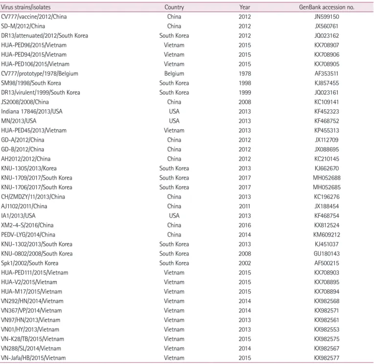
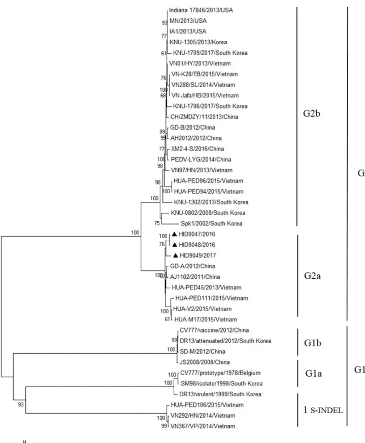
A total of 40 PEDVs were used for the construction of a phy- logenetic tree (Table 2). In this study, phylogenetic analysis based on the nucleotide sequences of the S gene revealed that PEDV strains could be divided into 2 genogroups, the classical genogroup 1 represented by prototype CV777 and the variant genogroup 2 of pandemic strains. Each group can be divided into subgroups 1a, 1b, 1 S-INDEL, 2a and 2b. Three isolates
(HID9047, HID9048, and HID9049) were found to belong to the subgroup 2a, which is most closely related to the Asian strains designated HUA-PED45/2013/Vietnam, HUA- PED111/2014/Vietnam; GD-A/2012/China and AJ1102/2011/
China (Fig. 2).
Spike gene sequence comparisons
The entire DNA sequences of the S genes and the deduced
Table 2. PEDV reference strains used in this study
Virus strains/isolates Country Year GenBank accession no.
CV777/vaccine/2012/China China 2012 JN599150
SD-M/2012/China China 2012 JX560761
DR13/attenuated/2012/South Korea South Korea 2012 JQ023162
HUA-PED96/2015/Vietnam Vietnam 2015 KX708907
HUA-PED94/2015/Vietnam Vietnam 2015 KX708906
HUA-PED106/2015/Vietnam Vietnam 2015 KX708905
CV777/prototype/1978/Belgium Belgium 1978 AF353511
SM98/1998/South Korea South Korea 1998 KJ857455
DR13/virulent/1999/South Korea South Korea 1999 JQ023161
JS2008/2008/China China 2008 KC109141
Indiana 17846/2013/USA USA 2013 KF452323
MN/2013/USA USA 2013 KF468752
HUA-PED45/2013/Vietnam Vietnam 2013 KP455313
GD-A/2012/China China 2012 JX112709
GD-B/2012/China China 2012 JX088695
AH2012/2012/China China 2012 KC210145
KNU-1305/2013/Korea South Korea 2013 KJ662670
KNU-1709/2017/South Korea South Korea 2017 MH052688
KNU-1706/2017/South Korea South Korea 2017 MH052685
CH/ZMDZY/11/2013/China China 2013 KC196276
AJ1102/2011/China China 2011 JX188454
IA1/2013/USA USA 2013 KF468754
XM2-4-S/2016/China China 2016 KX812524
PEDV-LYG/2014/China China 2014 KM609212
KNU-1302/2013/South Korea South Korea 2013 KJ451037
KNU-0802/2008/South Korea South Korea 2008 GU180143
Spk1/2002/South Korea South Korea 2002 AF500215
HUA-PED111/2015/Vietnam Vietnam 2015 KX708903
HUA-V2/2015/Vietnam Vietnam 2015 KX708895
HUA-M17/2015/Vietnam Vietnam 2015 KX708894
VN292/HN/2014/Vietnam Vietnam 2014 KX982568
VN367/VP/2014/Vietnam Vietnam 2014 KX982571
VN97/HN/2013/Vietnam Vietnam 2013 KX982561
VN01/HY/2013/Vietnam Vietnam 2013 KX982553
VN-K28/TB/2015/Vietnam Vietnam 2015 KX982575
VN288/SL/2014/Vietnam Vietnam 2014 KX982567
VN-Jafa/HB/2015/Vietnam Vietnam 2015 KX982577
PEDV, porcine epidemic diarrhea virus.
Fig. 2. Phylogenetic analysis of porcine epidemic diarrhea virus (PEDV) isolates and the reference strains. The phylogenetic tree was gen- erated by the neighbor-joining method using Mega 6.0 program with nucleotide sequences of the full-length S gene and 1,000 replicates bootstrapping.
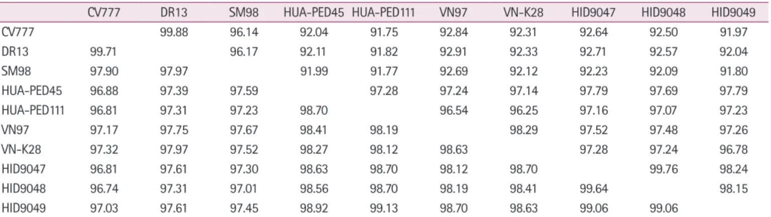
amino acid sequences of 3 isolates from Vietnam were com- pared with the sequences of the reference strains (CV777, SM98, DR13) and other Vietnam isolates. As shown in Table 3, the nucleotide identities of the S gene between 3 Vietnam iso- lates and reference strains ranged from 91.80% to 92.71%, and the deduced amino acid sequences were 96.74% to 97.61% ho- mologous. Furthermore, compared with the Vietnam isolates (HUA-PED45, HUA-PED111, VN97, and VN-K28), the 3 iso- lates shared 96.78% to 97.52% nucleotide homology and 98.19%
to 99.13% amino acid homology. The homology of nucleotide and amino acid of 3 isolates showed 98.15% to 99.76% and 99.06% to 99.64%, respectively (Table 3).
Sequence alignment of amino acids in the S protein illustrat- ed that mutations in amino acid sequence including substitu- tions, insertions and deletions were observed in 3 Vietnam iso- lates. Among them, 4 insertions (58NQGV61) and 2 deletions (163DI164) were found in the N-terminal of S protein (Fig. 3).
Alignment analysis of 4 neutralizing epitope regions COE, SS2, SS6 and 2C10 revealed that all 3 Vietnam isolates in this study have 4 substitutions in COE (502I → 502T, 509L → 509S, 529S → 529G,
607A → 607E), 1 substitution in SS6 (766Y → 766S), and no mutations in SS2 and 2C10 compared with CV777, DR13, and SM98 strains (Fig. 3). Comparison with other Vietnam strains (VN288, VN-Jafa, VN01, VN97 isolated during 2012-2015) in- dicated the substitution in COE (502I → 502T), and the conserva- tion in SS2, SS6, and 2C10 region. PEDV VN288, VN-Jafa, VN01, VN97 showed addition substitutions in COE, while HUA-PED45 showed more homology with the 3 isolates, sug- gesting that 3 isolates HID9047, HID9048, and HID9049 are closely related to PEDV HUA-PED45.
Neutralizing activity
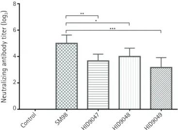
To determine whether vaccine strain SM98 can neutralize the 3 PEDV isolates (HID9047, HID9048, and HID9049), guinea pig antisera collected 2 weeks after the final immunization were used for neutralizing tests. The antisera effectively inhibited in- fection with the vaccine strain SM98 with mean neutralizing antibody (NA) titers of 5 log2. The antisera showed lower dilu- tions with mean of NA titers of 3.67 log2 (p < 0.01), 4 log2 (p <
0.05), and 3.27 log2 (p < 0.001) for inhibiting infection with isolates HID9047, HID9048, and HID9049, respectively (Fig. 4).
There was a significant difference in the mean NA titers against the SM98 strain and 3 isolates, suggesting that the antisera strongly reacted with the homologous virus, but moderately with the heterologous field isolates regarding to antigenic varia- tions between PEDVs.
Discussion
The S protein is an essential factor mediating PEDV entry into host cells and is considered a primary target antigen for in- duction of neutralizing antibodies and the development of an effective vaccine against PEDV [5-7]. Several previous studies indicated that the S gene is prone to mutations including re- placement, insertion and/or deletion of nucleotide sequences [7,8,11,12].
PEDV has been reported in Vietnam since 2010 and has spread rapidly [13]. The disease has been circulating and be- coming a significant issue in the pork industry. This study was conducted to isolate PEDVs from infected pigs in Vietnam, in- vestigate the genetic variation of PEDV isolates based on the full-length S gene and assess the neutralizing activity of the tra- ditional vaccine strain with the circulating isolates.
Table 3. Pairwise comparisons of the nucleotide and deduced amino acid sequences of the full-length S gene of porcine epidemic diarrhea virus isolates compared with the reference strains
CV777 DR13 SM98 HUA-PED45 HUA-PED111 VN97 VN-K28 HID9047 HID9048 HID9049
CV777 99.88 96.14 92.04 91.75 92.84 92.31 92.64 92.50 91.97
DR13 99.71 96.17 92.11 91.82 92.91 92.33 92.71 92.57 92.04
SM98 97.90 97.97 91.99 91.77 92.69 92.12 92.23 92.09 91.80
HUA-PED45 96.88 97.39 97.59 97.28 97.24 97.14 97.79 97.69 97.79
HUA-PED111 96.81 97.31 97.23 98.70 96.54 96.25 97.16 97.07 97.23
VN97 97.17 97.75 97.67 98.41 98.19 98.29 97.52 97.48 97.26
VN-K28 97.32 97.97 97.52 98.27 98.12 98.63 97.28 97.24 96.78
HID9047 96.81 97.61 97.30 98.63 98.70 98.12 98.70 99.76 98.24
HID9048 96.74 97.31 97.01 98.56 98.70 98.19 98.41 99.64 98.15
HID9049 97.03 97.61 97.45 98.92 99.13 98.70 98.63 99.06 99.06
Nucleotide and amino acid homology (%) are presented in the upper right and the lower left, respectively.
In this study, 3 PEDV isolates were successfully isolated from infected samples and propagated in Vero cells with high virus titers. Nucleotide sequencing and a phylogenetic tree based on the S gene revealed that the 3 isolates from Vietnam (HID9047, HID9048, and HID9049) belong to subgroup 2a, which in- cludes PEDV isolates that circulated in China from 2011 to 2012 and in Vietnam from 2013 to 2015. In a previous study conducted by Diep et al. [13] in 2018, highlighting the genetic heterogeneity of PEDVs in Northern Vietnam from 2012–2015, 6 out of 25 isolates were found to belong to subgroup 2a, which is closely related to Asian strains. Our 3 PEDVs were isolated in 2016 and 2017, suggesting that they are novel PEDVs that share unique genetic features with the subgroup 2a PEDV strains cir- culating in Vietnam from 2013 to 2015. In addition, the 3 iso- lates showed less mutations than PEDV HUA-PED111, HUA-V2, and HUA-M17 which were isolated from in the northern provinces of Vietnam in 2015, while showed higher homology in amino acid sequences with strain HUA-PED45, suggesting that the 3 isolates and PEDV HUA-PED45 are ge- netically closely related. Amino acid sequences alignment indi- N terminal
58NQGV61 163DI164
COE
SS2 SS6 2C10
Fig. 3. Analysis of amino acid mutations in the S protein of isolates from Vietnam with reference strains. Insertion and deletions are highlighted in blue and red, respectively. The core neutralizing epitope (COE) and 3 other neutralizing epitopes SS2 (yellow), SS6 (green) and 2C10 (purple) were aligned.
Neutralizing antibody titer (log2)
Control SM98
HID9047 HID9048 HID9049 8
6
4
2
0
Fig. 4. Neutralizing antibodies in serum samples of guinea pigs immunized against porcine epidemic diarrhea virus (PEDV) iso- lates. Serum samples were collected at 2 weeks after the second immunization and subjected to the virus neutralization assay using isolates PEDV HID9047, HID9048, and HID9049. Data rep- resent the mean ± standard deviation. Statistical significance was assessed by one-way analysis of variance. *p < 0.05, **p < 0.01,
***p < 0.001.
cated mutations such as substitutions, mutations, insertions and deletions in S protein, especially in N-terminal region. N-termi- nal domain interacts with 5-N-acetylneuraminic acid, muta- tions in N-terminal may have impact on PEDV infection in the host [14].
Neutralizing activity of antisera against the 3 Vietnam isolates was determined. Immunized sera showed the higher neutraliz- ing activity against the homogenous strain SM98, but moderate against the isolates HID9047, HID9048, and HID9049. This finding is consistent with a previous study that revealed that the antisera strongly recognized the homologous strain regarding to antigenic variations [10]. In a study of Lee et al. [10] in 2015, antisera of guinea pigs given 2 immunization with strain KNU- 141112 effectively inhibited the homogenous strain with mean NA titers of 1:112, but relatively low NA titers of 1:37 against strain SM98-1. In this study, the 3 isolates have 1 and 4 amino acid substitutions in SS6 and COE regions, respectively, which may support the hypothesis of variations in neutralizing epitope and speculate that the changes may influence the antigenicity of PEDV. Further studies on pathogenicity and neutralizing activi- ty of these 3 isolates need to be carried out in pigs.
Acknowledgments
We would like to acknowledge the veterinarians who collect- ed the infected samples and the contribution of all authors for this study.
ORCID
Van Tan Do, https://orcid.org/0000-0003-4512-7622 Quang Lam Truong, https://orcid.org/0000-0002-9403-2614 Hoai Thu Dao, https://orcid.org/0000-0002-2797-9955 Thi Lan Nguyen, https://orcid.org/0000-0002-1409-9967 Jini Kim, https://orcid.org/0000-0001-7802-6110 Tae-Wook Hahn, https://orcid.org/0000-0002-3919-1153
References
1. Lee C. Porcine epidemic diarrhea virus: an emerging and re-emerging epizootic swine virus. Virol J 2015;12:193.
2. Song D, Park B. Porcine epidemic diarrhoea virus: a compre- hensive review of molecular epidemiology, diagnosis, and vaccines. Virus Genes 2012;44:167–175.
3. Diel DG, Lawson S, Okda F, Singrey A, Clement T, Fernandes MHV, Christopher-Hennings J, Nelson EA. Porcine epidemic
diarrhea virus: an overview of current virological and serolog- ical diagnostic methods. Virus Res 2016;226:60–70.
4. Chen Q, Li G, Stasko J, Thomas JT, Stensland WR, Pillatzki AE, Gauger PC, Schwartz KJ, Madson D, Yoon KJ, Stevenson GW, Burrough ER, Harmon KM, Main RG, Zhang J. Isolation and characterization of porcine epidemic diarrhea viruses as- sociated with the 2013 disease outbreak among swine in the United States. J Clin Microbiol 2014;52:234–243.
5. Chang SH, Bae JL, Kang TJ, Kim J, Chung GH, Lim CW, Laude H, Yang MS, Jang YS. Identification of the epitope re- gion capable of inducing neutralizing antibodies against the porcine epidemic diarrhea virus. Mol Cells 2002;14:295–299.
6. Sun DB, Feng L, Shi HY, Chen JF, Liu SW, Chen HY, Wang YF. Spike protein region (aa 636789) of porcine epidemic di- arrhea virus is essential for induction of neutralizing antibod- ies. Acta Virol 2007;51:149–156.
7. Sun J, Li Q, Shao C, Ma Y, He H, Jiang S, Zhou Y, Wu Y, Ba S, Shi L, Fang W, Wang X, Song H. Isolation and characteriza- tion of Chinese porcine epidemic diarrhea virus with novel mutations and deletions in the S gene. Vet Microbiol 2018;
221:81–89.
8. Zhang X, Pan Y, Wang D, Tian X, Song Y, Cao Y. Identifica- tion and pathogenicity of a variant porcine epidemic diarrhea virus field strain with reduced virulence. Virol J 2015;12:88.
9. Park JE, Kang KJ, Ryu JH, Park JY, Jang H, Sung DJ, Kang JG, Shin HJ. Porcine epidemic diarrhea vaccine evaluation using a newly isolated strain from Korea. Vet Microbiol 2018;221:19–
26.
10. Lee S, Kim Y, Lee C. Isolation and characterization of a Kore- an porcine epidemic diarrhea virus strain KNU-141112. Virus Res 2015;208:215–224.
11. Sun RQ, Cai RJ, Chen YQ, Liang PS, Chen DK, Song CX. Out- break of porcine epidemic diarrhea in suckling piglets, China.
Emerg Infect Dis 2012;18:161–163.
12. Wang L, Byrum B, Zhang Y. New variant of porcine epidemic diarrhea virus, United States, 2014. Emerg Infect Dis 2014;
20:917–919.
13. Diep NV, Sueyoshi M, Izzati U, Fuke N, Teh APP, Lan NT, Ya- maguchi R. Appearance of US-like porcine epidemic diar- rhoea virus (PEDV) strains before US outbreaks and genetic heterogeneity of PEDVs collected in Northern Vietnam during 2012-2015. Transbound Emerg Dis 2018;65:e83–e93.
14. Kim SH, Cho BH, Lee KY, Jang YS. N-terminal domain of the spike protein of porcine epidemic diarrhea virus as a new can- didate molecule for a mucosal vaccine. Immune Netw 2018;
18:e21.