- 39 - 대한두경부종양학회지, 제33권 제2호, 2017. pp.39-42
Korean Journal of Head & Neck Oncology, Vol.33, No.2
http://doi.org/10.21593/kjhno/2017.33.2.39 ISSN 1229-5183(Print) / ISSN 2586-2553(Online)
혀에서 발생한 일차성 결핵
한준1⋅김지훈1⋅정순희2⋅봉정표1+
연세대학교 원주의과대학 이비인후과학교실1, 연세대학교 원주의과대학 병리학교실2
Primary Tuberculosis of the Oral Tongue: A Case Report and Literature Review
Chun Han, MD1, Ji Hoon Kim, MD1, Soon Hee Jung, MD, PhD2, Jeong Pyo Bong, MD1+
Department of Otorhinolaryngology - Head and Neck Surgery1,
Department of Pathology Yonsei University Wonju College of Medicine, Wonju, Republic of Korea2
= Abstract =
Tuberculosis is primarily a pulmonary disease and extrapulmonary involvement of the oral cavity is an infrequent occurrence accounting for 0.2 - 1.5% of all extrapulmonary sites. The authors report a case of primary tuberculosis of the oral tongue in a 60-years-old male patient who visited the clinic for ongoing tongue pain that he experienced for several months. The lesion was initially indistinguishable from the malignancy, but differential diagnosis was performed on the biopsy with Ziehl-Neelseen stain. After treatment with anti-tuberculosis, the oral lesion healed completely without surgery.
Key W ords:Primary tuberculosis, Tongue, Oral cavity, Extrapulmonary tuberculosis
R eceived R e v i s e d A ccepted
: June 15, 2017 : July 10, 2017 : July 11, 2017
+Corresponding author: Jeong Pyo Bong
Department of Otorhinolaryngology-Head and Neck Surgery, Yonsei Universitiy Wonju College of Medicine
20 Ilsan-ro, Wonju, Gangwon-do, 26426, South Korea Tel: +82-33-741-0642 Fax: +82-33-732-8287 E-mail: [email protected]
Introduction
Tuberculosis, is primarily a pulmonary chronic infectious granulomatous disease caused by acid-fast Mycobacterium tuberculosis, and less commonly by Mycobacterium bovis and other atypical mycobacteria.1) Extrapulmonary tuber- culosis lesions of the oral cavity are extremely rare, account- ing for 0.2-1.5% of all extrapulmonary involvement. Among the oral cavity components, the tongue is the most com- monly affected site. Others include the lips, cheek, soft pal- ate, uvula, and gingival mucosa.2) Primary tuberculosis of
the oral tongue is rare and only few case reports have been published. Here, we report a case of 60-years-old male pa- tient with oral tongue tuberculosis, together with a literature review.
Case report
A 60-years-old male patient visited the clinic for ongoing tongue pain that he experienced for several months with deep ulceration on the right lateral side of the oral tongue. The patient experienced severe pain that interfered intake of meals, causing dysphagia. He had a medical history of alco- holic pancreatitis diagnosed 5 years previously and history of smoking one pack of cigarettes a day for 40 years, as well as history of alcohol intake for 40 years. Comprehensive physical examination revealed no other specific abnormal- ities; the patient was overall in good health, weighing 55kg and was 162cm tall. Oral examination revealed poor oral hygiene with multiple dental caries and 3 right upper molar
- 40 -
Fig. 1. Initial physical examination findings of the patient. The right ventral side of the tongue had a deep ulceration meas- uring about 3 cm in length 4 mm in depth Appearance of tongue ulceration on the first clinic visit
A
B
C
Fig. 2. Axial view of the computed tomography scan. (A) Ill-defined oral tongue lesion at right lateral tongue. (Black dotted circle), (B) Enlarged lymph nodes at right neck level Ia (8.5 x 6.7 x 6.5mm, white arrow), (C) Enlarged lymph nodes at right neck level IIa (10 x 7.7 x 13 mm, white arrow)
A
B
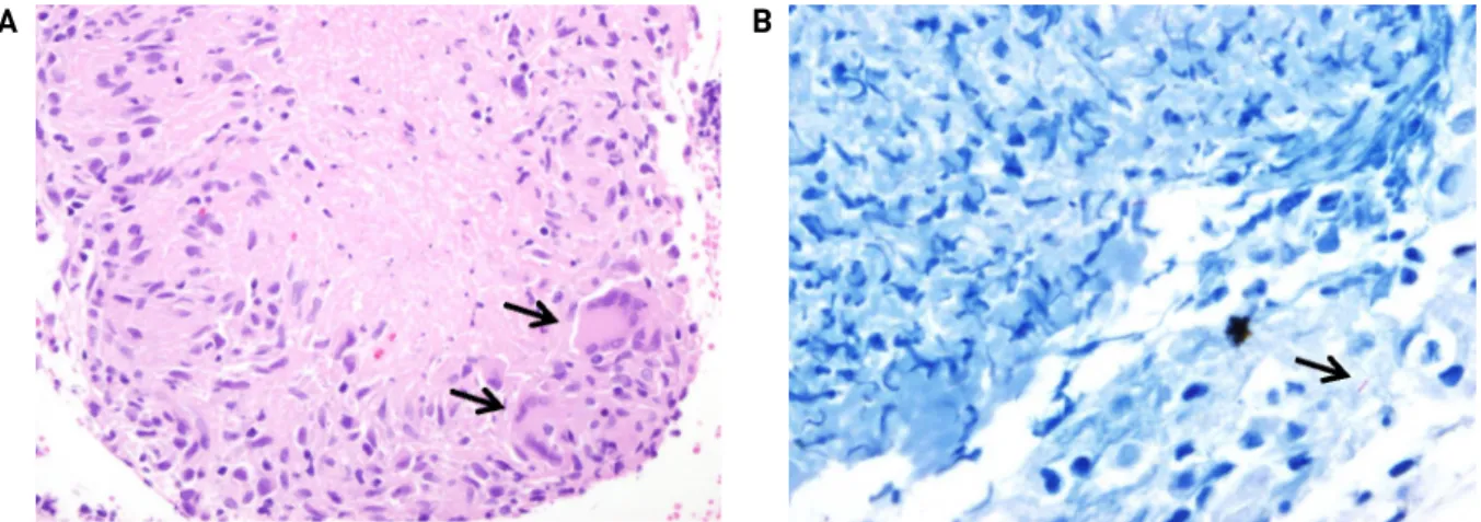
Fig. 3. (A) The microscopic examination revealed multiple areas of chronic granulomatous inflammation with caseous necrosis.
Some Langhans giant cells are also seen in Hematoxylin and eosin (H&E) stain (black arrow, x400). (B) A few acid-fast bacilli are found in Ziehl-Neelson stain (black arrow, x1000).
teeth missing. The right ventral side of the tongue had deep ulceration measuring about 3 cm in length and 4mm in depth, elliptical in shape with severe tenderness and sur- rounded by soft edges (Fig. 1). Physical examination of the
neck revealed no enlarged lymph nodes, no tenderness or fixation to surrounding tissues. Based on the patient's long smoking history combined with his poor hygiene, it was important to distinguish the lesion from the oral cavity squ- amous cell carcinoma. We further examined the patient with computed tomography(CT) scan and found relatively en- larged lymph nodes on the right neck at level I and II (Ia:
8.5 x 6.7 x 6.5 mm, IIa: 10 x 7.7 x 13 mm, Fig. 2B, 2C), and an ill-defined mass at the right lateral border of tongue without definite midline crossing or tongue base extension (Fig. 2A). Malignancy could not be ruled out based on the CT scan finding. Therfore, an incisional biopsy was per- formed and a specimen was taken from the central origin of the ulceration. Bleeding was minimal and hemostasis was quickly achieved with bosmine-soaked gauze and cold ice.
The final pathological diagnosis was chronic granulomatous inflammation with caseous necrosis, which was consistent
- 41 -
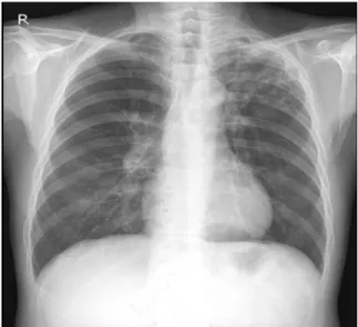
Fig. 4. Chest X-ray. Nonspecific finding, No abnormality is seen.
Fig. 5. Tongue ulceration observed on the second clinic visit, after 45 days of anti-tuberculosis treatment
with tuberculosis (Fig. 3A). Ziehl-Neelsen stain was per- formed on the biopsy specimen and was positive for acid-fast bacilli with this histochemical stain (Fig. 3B).
DNA was extracted from the paraffin sample of the tongue, and the DNA concentration was 193.3 ng/µL. The polymer- ase chain reaction showed that the sample was positive for Mycobacterium tuberculosis. Pulmonary origin of the tuber- culosis was suspected, but the patient’s chest X-ray showed no abnormality of the lung. Furthermore, the patient showed no signs of fever, sputum nor dyspnea, night sweats, and weight loss(Fig. 4). Without any signs of pulmonary tuber- culosis further chest investigations were unnecessary. Thus, our final diagnosis was tuberculosis of the tongue without any primary focus. Anti-tuberculosis treatment was started with isoniazid (400 mg/day), ethambutol (1000 mg/day), ri- fampin (600 mg/day), and pyrazinamide (1500 mg/day).
The patient revisited the outpatient clinic 1 month later with greatly reduced oral pain and the size of the ulcer visibly
decreased from its original size. The patient was given a total of 6 months of anti-tuberculosis medication and re- visited the clinic 4 months after treatment had commenced, with the ulcer site improved greatly, and almost completely healed (Fig. 5). No further follow up was required after 6 month of treatment.
Disscusion
The incidence of tuberculosis has decreased greatly in well-developed countries worldwide due to the improve- ment in public healthcare and the development of anti-tuber- culosis medication. This includes South Korea, where the prevalence of bacteriologically confirmed pulmonary tuber- culosis decreased from 940 to 219 cases per 100,000 pop- ulation during the period from1965 to 19953) following the implementation of the National Tuberculosis Program in 1962.
Most oral tuberculosis is secondary to primary pulmonary tuberculosis. However, when oral tuberculosis occurs as a primary lesion, manifestations of tongue ulceration occur- ring along the lateral margin with history of trauma are commonly seen. These tongue ulcers are usually progressive in pain and interfere severely with absorption of nutrition and ultimately, quality of life.4)
Primary tongue tuberculosis is commonly misdiagnosed as cancer due to its malignancy-like ulcerating pattern and its relatively rare incidence. Radiological examination even can be misleading, therefore biopsy is vital for final diagnosis.2) Our initial impression was also oral cavity squ- amous cell carcinoma, but the patient was later finally diag- nosed with tuberculosis after histochemical staining showed caseous necrosis. Most oral tuberculosis lesions are located in the anterior portion of the oral cavity, such as the buccal mucosa or vestibule area near the corner of the mouth or lip, while oral squamous cell carcinoma usually exists in the lateral border of the tongue and retromolar area. Our case of oral tuberculosis lesion was located in the lateral border of the tongue, which was different from its known usual preferential location.5)
It is believed that tuberculosis-infected sputum somehow infiltrates oral soft tissue during coughing episodes and causes secondary infection. However, the continuous secre- tion of saliva, presence of various normal flora, and oral
- 42 - antibodies makes it difficult for external invasion to occur;
however, any break or loss of this natural barrier, which may be the result of trauma, inflammation, or poor hygiene could be the reason for entry of the organism.6)
Treatment of tongue tuberculosis is similar to that of pul- monary tuberculosis, including the standard anti-tuberculosis therapy, which is a combination of isoniazid, rifampicin, pyr- azinamide, and ethambutol. The administration of these 4 drugs daily for the 2 months, followed 2 drugs for an addi- tional 4 month (isoniazid and rifampicin) is the accepted and effective therapy.4) We followed the same protocol with our patient who showed great improvement after 4 months of treatment, which was concluded with complete resolution after 6 months. While our patient was fortunately treated successfully, tuberculosis treatment is sometimes difficult because of drug resistance. Drug resistance is the result of genetic mutation that causes a loss of gene factors that con- trol drug susceptibility. If drug resistance is suspected, then treatment duration may be prolonged up to 9 to 12 months.
중심 단어:일차성 결핵, 혀, 구강, 폐외 결핵
References
1) Kapoor S, Gandhi S, Gandhi N, Singh I. Oral manifestations of tuberculosis. CHRISMED J Health Res. 2014;1:11-14.
2) Ammar C, Al-Rikabi, Maria ARA. Tuberculosis of the Tongue Clinically Masquerading as a Neoplasm: A Case Report and Literature Review, Oman Med J. 2011;26:267-268.
3) Hong YP, Kim SJ, Lew WJ, Lee K, Han YC. The seventh nation- wide tuberculosis prevalence survey in Korea, 1995. Int J Tuberc Lung Dis. 1998;2:27-36.
4) Garg RK, Singhal P. Primary Tuberculosis of the Tongue: A Case Report. J Contemp Dent Pract. 2007;8:74-80.
5) Wen-Chen W, Jin-Yi C, Yuk-Kwan C, Li-Min L. Tuberculosis of the head and neck: a review of 20 cases. Oral Surg Oral Med Oral Pathol Oral Radiol Endod. 2009;107:381-386.
6) Iype EM, Ramdas K, Pandey M, Jayasree K, Thomas G, Sebastian P, et al. Primary tuberculosis of the tongue: report of three cases. Br J Oral Maxillofac Surg. 2001;39:402-403.