• 검색 결과가 없습니다.

Cutaneous asthenia (Ehlers-Danlos syndrome) in a Korean short-haired cat

N/A
N/A
Protected

Academic year: 2021

Share "Cutaneous asthenia (Ehlers-Danlos syndrome) in a Korean short-haired cat"

Copied!
3
0
0

로드 중.... (전체 텍스트 보기)

전체 글

(1)

pISSN 2466-1384 eISSN 2466-1392 大韓獸醫學會誌 (2016) 第 56 卷 第 1 號 Korean J Vet Res(2016) 56(1) : 53~55 http://dx.doi.org/10.14405/kjvr.2016.56.1.53

53

<Case Report>

Cutaneous asthenia (Ehlers-Danlos syndrome) in a Korean short-haired cat

Sang-Hyuk Seo

1,2

, Miru Choi

2

, Changbaig Hyun

2,

*

1

VIP Animal Medical Center, Seoul 02830, Korea

2

Section of Small Animal Internal Medicine, College of Veterinary Medicine, Kangwon National University, Chuncheon 24341, Korea

(Received: January 29, 2016; Revised: March 4, 2016; Accepted: March 8, 2016)

Abstract : A 1-year-old Korean domestic short-haired cat presented with skin hyperextensibility and a severely macerated wound on the skin of the dorsal part of the neck. Diagnostic studies including histopathology and skin extensibility index revealed congenital cutaneous asthenia (Ehlers-Danlos syndrome). In this cat, the skin wounds and defects were successfully managed with standard wound management and cosmetic surgery. Although skin hyperextensibility is persistent, the cat has lived well without other complications to date. To the best of our knowledge, this is the first report of cutaneous asthenia in a cat in Korea.

Keywords : cat, cutaneous asthenia, Ehlers-Danlos syndrome, joint laxity, skin hyperextensibility

Ehlers-Danlos syndrome (EDS) is a type of inherited cuta- neous asthenia, which is caused by collagen production [4].

Although EDS in cats has rarely been reported, inherited EDS has been described in the Himalayan breed, which is similar EDS VIIc in man and dermatosporaxis in calves and sheep [1, 10]. Although the EDS is not fatal in cats, it often causes hanging folds of skin, extensive scarring, joint laxity, delayed wound healing with hematoma and hygroma forma- tion [11]. Although the diagnosis is challenging, clinical signs and histopathologic studies of the collagen structure are important to identify this disease. This case study described a rare case of EDS in a Korean domestic short-haired cat.

A 1-year-old castrated male Korean domestic short-haired cat weighing 4.2 kg, was presented with severe macerated wound on the skin of dorsal part of neck (Fig. 1A and B).

The cat had medical history of skin maceration by self- scratching and frequent grooming on the abdomen and hind limb. On the physical examination, the skin lesion was con- sisted of 8.5 × 5.5 cm skin defects by ulcerative wound on the dorsal part of neck (Fig. 1A and B). The margin of defected skin was necrotized and ulcerative. Skin elasticity was markedly increased (hyperextensibility) on the lesion (Fig. 1). Skin in other body part was also thin and was hyper- extensible and fragile (Fig. 2C and D). Complete blood cell count revealed no particular abnormalities except moderate polycythemia (red blood cell 10.45 M/µL; reference range, 5.5–8.5 M/uL; hematocrit 57.3%; reference range, 37–55%).

Serum biochemistry found no particular abnormalities except

increased glucose (151 mg/dL; reference range, 60–120 mg/

dL) and decreased albumin (2.7 g/dL; reference range, 3.1–

4.1 g/dL).

Emergency surgery for covering skin defects of the neck was performed (Fig. 2A and B). After removal of necrotized wound margin, the skin defected area was covered by extending skin nearby with anchored suture (Fig. 2A and B).

After installing wound drainage, bacterial culture with antibi-

*Corresponding author

Tel: +82-33-250-8681, Fax: +82-33-244-2367 E-mail: [email protected]

Fig. 1. Skin lesion of this case. (A and B) The skin lesion was

consisted of 8.5 × 5.5 cm skin defects by ulcerative wound on

the dorsal part of neck. The margin of defected skin was necro-

tized and ulcerative. Skin elasticity was markedly increased

(hyperextensibility) on the lesion. (C and D) Skin in other body

part was also thin and was hyperextensible and easily torn.

(2)

54 Sang-Hyuk Seo, Miru Choi, Changbaig Hyun

otics sensitivity test and histopathological test of wound mar- gin removed were performed. To rule out hyperadrenocor- ticism, ectoparacitism and fungal infection, low dose dexam- ethasone suppression (LDDST), skin scraping and fungal culture were also performed. The cat was managed with sugar dressing and skin nutrient supplement (1 pump/day;

Coatex; VetPlus, UK), along with prescription diet (Hill’s Prescription Diet z/d; Hill’s Pet Nutrition, USA) until the final diagnosis has been made.

Skin scraping found no particular ectoparasites. However, fungal culture with DTM culture media found strong posi- tive reaction, suggesting dermatophyte infection. Bacterial culture found gram negative cocci (Actinetobacter spp.) sen- sitive to amikacin and imipemem. The LDDST revealed neg- ative for hyperadrenocorticism (< 1 µg/dL at 0 h, < 1 µg/dL at 4 h, < 1 µg/dL at 8 h). Histopathological exam on the skin lesion found prominent looseness to the dermis due to edema, wide separation of the collagen bundles, inactive hair follicle structures and macrophage infiltration within dermal stroma, suggesting a heritable collagen disorder (e.g., EDS;

Fig. 3). Based on the diagnostic tests, the cat was diagnosed as EDS complicated with bacterial and fungal infection.

The cat was treated with itraconazole (5 mg/kg per oral [PO], q24h; Sponazole; Nelson Pharm Korea, Korea) and amikacin (10 mg/kg subcutaneously, q24h; AMIKACIN injection; Samu Median, Korea) for skin infection along with wound manage- ment. After 30 days of treatment, the skin lesion was fully restored. However, the skin was still fragile with increased elasticity (Fig. 2C and D). The skin extensibility index (SEI) was measured to confirm the collagen detect as described previously [3]. In this dog, the SEI was increased to 22%

(reference < 19%) [3].

Cutaneous asthenia, called as dermatosparaxis or EDS refers a disease complex resulting in hereditary congenital defects of dermal connective tissue and has been reported in

man [7] and in cats and dogs [1, 5, 9, 11]. Clinical features of feline cutaneous asthenia are skin hyperextensibility and fra- gility along with various skin problems. Young age, clinical features, the SEI and histopathologic results in our case were well compatible with a congenital collagen defect. One study found histopathological features of feline cutaneous asthenia included alterations of collagen fibers in arrangement and length (i.e., shortening, disarray, curling, and uneven size) and disorganization of the collagen fibrils [2]. In this case of cat, similar histological findings were all clearly noticed.

Some forms of cutaneous asthenia can be occurred by acquired diseases (i.e., Cushing’s syndrome, hepatic cholang- iocarcinoma, and hepatic lipidosis). To rule out acquired dis- eases, several tests were also performed in this case. The LDDST and ACTH stimulation tests revealed the intact adre- nal function in this cat. Normal hepatic enzyme levels ruled out the possibility of hepatic cholangiocarcinoma and hepatic lipidosis. Although the cat was positive in bacterial and fun- gal culture, authors believed the positive results might be due to secondary bacterial and fungal infection responded well to antibiotic and anti-fungal medications. The SEI value over the reference range and histopathological features with young age of this cat were well supported to the congenital cutane- ous asthenia.

Two forms of feline congenital cutaneous asthenia have been found [2]. In the autosomal dominant form of the dis- ease, type I collagen was poorly packaged and thus it caused skin hyperextensibility and joint hypermobility in affected cats. In the autosomal recessive form of the disease, it is caused by a deficiency of procollagen peptidase or a struc- tural abnormality at its cleavage site, resulting in extreme skin hyperextensibility only. In this recessive form, joint hypermobility is rarely occurred. Because our case of cat was mixed breed and adopted from the cat shelter, we could not Fig. 2. (A and B) Emergency surgery for covering skin defects

of the neck was performed. After removal of necrotized wound margin, the skin defected area was covered by extending skin nearby with anchored suture. (C and D) After 30 days of treat- ment. Although the skin lesion and defect was fully treated and resorted, the skin was still fragile with increased elasticity.

Fig. 3. Histopathology of the skin lesion. The skin lesion con- sisted of prominent looseness to the dermis due to edema, wide separation of the collagen bundles, inactive hair follicle struc- tures and macrophage infiltration within dermal stroma, sug- gesting a heritable collagen disorder (e.g., Ehlers-Danlos syndrome).

(A) The epidermis is thin with mild orthokeratotic hyperkera-

tosis. (B) The dermis has loosely arranged with thin collagenous

bundle separated by dermal edema. (C) The dermis has increase

numbers of foamy macrophages, few lymphocytes, plasma cells

and rare neutrophils. H&E stain. 2× (A), 10× (B), and 40× (C).

(3)

Cutaneous asthenia (Ehlers-Danlos syndrome) in a Korean short-haired cat 55

perform the genetic analysis for inheritance pattern. Since the cat had no joint hypermobility at the first presentation, authors suspected autosomal recessive form of feline congenital cuta- neous asthenia in this case.

Unfortunately, no specific treatment for feline cutaneous asthenia has been reported, although the oral administration of vitamin C (i.e., necessary for collagen synthesis in cats, 50 mg/head, PO, q24h) has been suggested [11]. Because the affected cat can easily have skin tear or skin wound by scratching, mating and playing with other cats, affected cats should be declawed not to injure themselves by scratching, It is also recommended neutering all affected cat not to use for breeding (i.e., because of the heritability of the disease) and to reduce the chance of injury during mating. If the affected cats have skin tear, it should be sutured if possible, along with conventional wound management with antibiotics.

According to literatures [6, 8, 11], the affected cat without signs of joint hypermobility (i.e., recessive form) can live long lives. However, if the affected cats have secondary man- ifestations of the disease (i.e., joint laxity), the prognosis is usually poor to guarded [6, 8, 11]. In our case of cat, the skin wound and defects were successfully managed with standard wound management and cosmetic surgery. Although skin hyperextensibility is persistent, the cat lives well without other complication, to date. To authors’ best knowledge, this is the first report of cutaneous asthenia in cats in Korea.

References

1. Counts DF, Byers PH, Holbrook KA, Hegreberg GA.

Dermatosparaxis in a Himalayan cat: I. Biochemical studies

of dermal collagen. J Invest Dermatol 1980, 74, 96-99.

2. Hansen N, Foster SF, Burrows AK, Mackie J, Malik R.

Cutaneous asthenia (Ehlers-Danlos-like syndrome) of Burmese cats. J Feline Med Surg 2015, 17, 954-963.

3. Hnilica KA. Small Animal Dermatology: A Color Atlas and Therapeutic Guide. 3rd ed. pp. 340-341, Elsevier/Saunders, Amsterdam, 2011.

4. Malfait F, Wenstrup RJ, De Paepe A. Clinical and genetic aspects of Ehlers-Danlos syndrome, classic type. Genet Med 2010, 12, 597-605.

5. Paciello O, Lamagna F, Lamagna B, Papparella S. Ehlers- Danlos–like syndrome in 2 dogs: clinical, histologic and ultrastructural findings. Vet Clin Pathol 2003, 32, 13-18.

6. Patterson DF, Minor RR. Hereditary fragility and hyperextensibility of the skin of cats. A defect in collagen fibrillogenesis. Lab Invest 1977, 37, 170-179.

7. Proske S, Hartschuh W, Enk A, Hausser I. Ehlers-Danlos syndrome – 20 years experience with diagnosis and classification. J Dtsch Dermatol Ges 2006, 4, 308-318.

8. Scott DW, Miller WH, Griffin CE. Muller and Kirk’s Small Animal Dermatology. 6th ed. pp. 35-36, 979-984, WB Saunders, Philadelphia, 2001.

9. Sequeira JL, Rocha NS, Bandarra EP, Figueiredo LMA, Eugenio FR. Collagen dysplasia (cutaneous asthenia) in a cat. Vet Pathol 1999, 36, 603-606.

10. Suregaonkar SN, Kotikalapudi R, Patel RK, Sunkara PSS. Screening of cattle breeds for 17bp deletion in a gene causing Ehlers-Danlos syndrome, Type VII (dermatosparaxia).

Int J Vet Sci 2013, 2, 96-98.

11. Szczepanik M, Go y ski M, Wilko ek P, Popiel J, Œmiech A, Pomorska D, Nowakowski H. Ehlers-Danlos syndrome (cutaneous asthenia) – a report of three cases in cats. Bull Vet Inst Pulawy 2006, 50, 609-612.

l n í l S í

수치

Fig. 1. Skin lesion of this case. (A and B) The skin lesion was consisted of 8.5 × 5.5 cm skin defects by ulcerative wound on the dorsal part of neck
Fig. 3). Based on the diagnostic tests, the cat was diagnosed as EDS complicated with bacterial and fungal infection

참조

관련 문서

In addition, measurements at 0 and 12 weeks (body composition, α-amylase, metabolic syndrome related factors) were performed twice. For 12 weeks, the

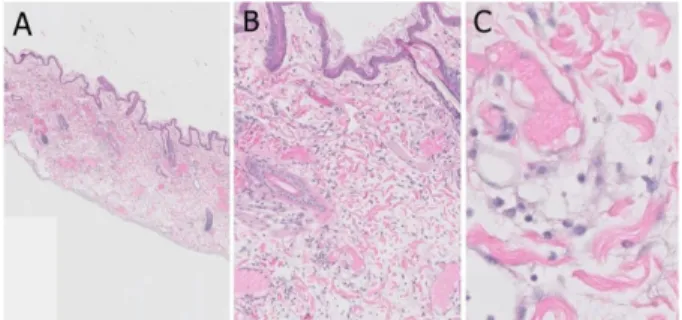
Actinic keratoses showed a trend towards increased expression in the basal layer compared with normal skin.. In actinic keratoses, Keratoacanthomas and seborrheic

Mean Value and Skin Improvement Effect that Prepare of 50 year-old Korean by Black Soybean Powder E xtract Applied to the Human Skin ……… 32 Fig.. Correlation

Serum uric acid levels and risk for vascular disease in patients with metabolic syndrome... Prevalence if the metabolic syndrome in a Turkish

tricuspidata on the production of proinflammatory cytokines in TNFα+IFNγ-stimulated HaCaT cells ...15 Fig.5: The cell viability of sub-fractions from 70% EtOH

In this study, it can be seen that sex and metabolic syndrome are important for biological factors, and occupation, smoking, and exercise are important

Objective: The purpose of this study was to investigate the expression of syndecan-1, E-cadherin, and beta-catenin in tissue sections of normal sun-damaged skin,

Herb Extract which exhibit several bene ficial effects including acne(pimple) and skin diseases was tested for antimicrobial activity and antiinflammation