상백피에 의한 MC/9 비만세포의 활성 억제 조절 연구
이기전1#, 김복규2, 길기정1*
1 : 중부대학교 한방제약과학과, 2 : 한국한의학연구원
Suppressive effects of
Morus alba
Linne Root Bark (MRAL) on activation of MC/9 mast cellsKi Jeon Lee
1#, Bok kyu Kim
2, Ki Jung Kil
1*1 : Department of Herbal Pharmaceutical Science, Joongbu University Geumsan 312-402, Korea 2 : Korea Institute of Oriental Medicine, Daejeon 305-811, Korea
ABSTRACT
Objective : Morus alba Linne Root Bark (MRAL) is a medicinal herb in Korean Medicine, known for its anti-inflammatory and anti-allergic properties. However, its mechanisms of action and the cellular targets have not yet been found and the study was developed to investigate the allergic suppressive effect of MRAL. The purpose of this study is to investigate the allergic suppressive effects of MRAL on activation of MC/9 mast cells.
Methods : Cytotoxic activity of MRAL (50, 100, 200, 400 µg/mL) on MC/9 mast cells measured using EZ-Cytox cell viability assay kit (WST reagent). The levels of interleukin-5 (IL-5), IL-13 and IL-4, IL-5, IL-6, IL-13 mRNA expression were measured by enzyme-linked immunosorbent assay (ELISA) and real-time PCR respectively. The expression of transcription factors such as GATA-1, GATA-2, NFAT, AP-1 and NF-κB p65 DNA binding activity were measured by western blot and electrophoresis mobility shift assay (EMSA).
Results : Our results indicated that MRAL (50 µg/mL, 100 µg/mL) significantly inhibited PMA/Ionomycin-induced production of IL-5 and IL-13 and the expression of IL-4, IL-5, IL-6 and IL-13 mRNA in MC/9 mast cells. Moreover, MRAL (50 µg/mL, 100 µg/mL) inhibited PMA/Ionomycin-induced GATA-1, GATA-2, NFAT-1, NFAT-2, c-Fos protein expression and NF-κB p65 DNA binding activity in MC/9 mast cells.
Conclusions : In conclusion, we suspect the anti-allergenic activities of MRAL, may be related to the regulation of transcription factors GATA-1, GATA-2, NFAT-1, NFAT-2, c-Fos and NF-κB p65 DNA binding assay causing inhibition of Th2 cytokines IL-5 and IL-13 in mast cells.
Key words : Morus alba Linne Root Bark (MRAL), MC/9 mast cell line, Anti-allergy, Transcription factor, Th2 cytokines
1)
서 론
알레르기성 질환으로는 기관지 천식, 비염, 아토피성 피부 염 등이 있는데, 각종 환경오염 등으로 인해 해마다 증가하고 있으며, 심각한 문제로 대두되고 있다.
알레르기 질환의 원인으로 작용하는 비만세포는 알레르기 반응기간 동안 생리적인 변화를 유발하는 중심인자로 인식되 고 있다
1).
최근에 발표된 많은 병리소견에서 만성 염증성 반응이 알 레르기 반응에서 공통적으로 발견되었고, 특히 후기 반응의
*교신저자 : 길기정, 충남 금산군 추부면 대학로 101번지 중부대학교 한방제약과학과 ·Tel : 041-750-6225 ·E-mail : [email protected]
#제1저자 : 이기전, 충남 금산군 추부면 대학로 101번지 중부대학교 한방제약과학과 ·Tel : 042-581-1475 ·E-mail : [email protected]
·접수:2012년 12월 9일 ·수정:2013년 1월 7일 ·채택:2013년 1월 16일
기전이 염증반응으로 이해되고 있기 때문에 비만세포로부터 유리되는 화학매체 외에 염증성 사이토카인인 종양괴사인자 알파 (tumor necrosis factor, TNF-α), 인터루킨-1 (IL-1)과 인터루킨-6 (IL-6)이 이러한 알레르기성 염증반응 의 유도에 결정적인 역할을 하는 것으로 알려져 있다
2).
비만세포는 과립내 화학매개체와 사이토카인을 분비하여 기관지 평활근 수축이나 점막 부종 등을 일으키며, 비만세포 에서 생산되는 Th2 사이토카인 IL-4, IL-5, IL-13 등은 Th2 면역 반응을 증가시키고, IgE 생산을 증가시켜 만성 알 러지 반응에 기여한다
3).
상백피 (桑白皮, Morus alba Linne)는 뽕나무과에 속한 낙엽교목인 뽕나무속 식물의 근피로써 예로부터 해열, 항경 련, 항알레르기, 항염증 작용과 더불어 이뇨촉진, 혈당 강하 작용, 항암, 항균, 미백 효과 등 다양한 효과가 있는 것으로 알려져 있다
4-6).
상백피에 대한 기존연구로는 천식 모델 흰쥐의 BALF 내 면역세포 및 혈청 IgE에 미치는 영향
7), 상백피가 항알러지 및 항염증반응에 미치는 영향
8)등이 보고되어 있으나, 알레르 기 발병 기작에서 중요하게 작용하는 비만세포에 대한 연구는 미흡한 실정이다.
이에 저자는 MRAL가 알레르기 염증 반응에 미치는 영향 을 알아보기 위해 MRAL가 비만세포와 Th2세포에서 어떤 신 호전달 기전을 제어함으로써 활성이 억제되어 IL-5와 IL-13 의 발현을 억제하는지를 실험적으로 연구하여 유의한 결과를 얻었기에 보고하는 바이다.
재료 및 방법
1. 재료
1) 약재본 실험에 사용한 상백피 ( Morus alba Linne, MRAL)는 금산약초시장에서 구입하여 중부대학교 한방제약과학과에서 감정한 후 정선하여 사용하였다.
2) 세포주
본 실험에 사용된 MC/9 murine mast 세포주는 American Type Culture Collection (ATCC, Rockville, MD, U.S.A.)에서 구매하여 사용하였다.
3) 시약 및 기기
Dulbecco’s Minimal essential medium (DMEM), Fetal bovine serum (FBS), Trypsin-EDTA, Antibiotics (penicillin, streptomycin)는 (Gibco-BRL, U.S.A.)제품을 사용하였고, Dulbecco's Phosphate Buffered Saline (D-PBS), Phorbol 12-myristate 13-acetate (PMA), Ionomycin, Phenylmethylsulfonyl fluoride (PMSF), Diethyl pyrocarbonate (DEPC), Chloroform, Isopropanol, Ethanol, magnesium chloride (MgCl₂), 2-mercaptoethanol는 (Sigma, U.S.A.)제품을 사용하였다.
Trizol은 (Ambion, U.S.A), EZ-Cytox kit는 (Daeil Lab.
Co., Korea), Deoxynucleoside triphosphate (dNTP)는 (TaKaRa, Japan), Moloey Murine Leukemia Virus
Reverse Transcriptase (M-MLV RT)와 RNase inhibitor 는 (Promega, U.S.A.)제품을 사용하였다. SYBR master mix는 (Applied Biosystems, U.S.A), IL-5 ELISA kit는 (BD bioscience, U.S.A.)제품을, IL-13 ELISA kit는 (Biosource, U.S.A), primary antibody (NFAT-1, NFAT-2, c-Jun, c-Fos, p-c-Jun, GATA-1, GATA-2, NF-κB p65)는 (Santa-Cruz, U.S.A), ECL-Hybond film은 (Amersham, U.S.A), SYBR master mix는 (Applied Biosystems, U.S.A)제품을 사용하였고, 기기는 열 탕추출기 (대웅, DWT-1800T, Korea), 감압 증류장치 (BUCHI B-480, Switzerland), 동결 건조기 (EYELA, FDU-540, Japan), CO
2배양기 (Forma scientific Co., U.S.A), 원심분리기 (한일과학, Korea), plate shaker (Lab-Line, U.S.A), spectrophotometer는 (Shimazue, Japan), Bio-freezer (Sanyo, Japan), Quantitative Real-Time PCR (Applied Biosystems, U.S.A), ELISA leader (Molecular Devices, U.S.A) 등을 사용하였다.
2. 방법
1) 추출물 제조상백피 100 g에 증류수 2,000 mL를 가하여 열탕 추출기 에서 2시간 동안 열수 추출하여 얻은 액을 흡입 여과하여 감 압증류장치 (rotary vaccum evaporator)로 농축하였다. 이 를 다시 동결건조기 (freeze dryer)를 이용하여 완전 건조한 상백피 추출물 (이하 MRAL) 14.5 g을 냉동 보관 (-84℃)하 면서 적당한 농도로 희석하여 사용하였다.
2) MC/9 비만 세포주 배양
MC/9 비만 세포주는 American Type Culture Collection에서 구매하였으며, DMEM with 10% fetal bovine serum, 100 µg/mL penicillin-streptomycin, 10%
T-stim, 0.05 mM 2-mercaptoethanol 그리고 2mM L-glutamine 첨가하여 37℃, 5% CO
2에서 10
5cells/mL 농도로 맞추어 96 well plate에 분주하였다.
3) 세포독성 (cytotoxicity)측정
MC/9 비만세포는 37℃, 5% CO₂배양기에서 1 시간 배 양한 후 MRAL (50, 100, 200, 400 µg/mL)을 48시간 동안 처리하였다. 배양종료 6시간 전에 EZ-Cytox WST reagent 10 µL씩 각 well에 가하고 실험 종료 시까지 배양하였다. 이 plate를 plate shaker에서 2,000 rpm으로 5분간 shaking하 고 ELISA LEADER에서 450㎚에서 흡광도를 측정하였다.
4) Real-time PCR
(1) RNA 추출
MC/9 세포를 6-well plate에 2.5×10
5cells/mL로 2
mL 씩 분주하고 24시간 동안 배양한 다음 MRAL을 100
µg/mL과 50 µg/mL로 처리하고 1시간 후에 PMA (50
ng/mL)와 ionomycin (0.5 µM)으로 자극한 뒤 3시간, 6시
간 후에 세포를 얻었다. 양성대조군으로는 cyclosporin
A(CsA) 10 µg/mL을 사용하였다. 배양종료 후 세포에 Trizol
1mL를 넣고 e-tube에 넣은 후 클로로포름 100 µL를 넣었
다. 얼음에 17분 동안 두었다가 13,000 rpm으로 15분 동안 원심분리 하였다. 층이 분리되면 e-tube에 상층액을 옮겨 담 고, 동량으로 이소프로판올을 넣어 얼음에 10분 정도 두었다 가 13,000 rpm으로 15분 동안 원심분리 하였다. 상층액은 버리고 80% 에탄올로 세척하고 13,000 rpm으로 10분 동안 원심분리 하였다. 상층액을 완전히 제거하고 투명해질 때까지 말린 후 DEPC water로 cell의 양에 따라 20∼30 µL를 넣어 녹였다.
(2) 역전사반응
역전사 (reverse transcription) 반응은 준비된 total RNA 3 µg을 75℃에서 5분 동안 변성 (denaturation)시키 고, 이에 2.5 µL 10 mM dNTPs mix, 1 µL random sequence hexanucleotides (25 pmole/ 25 µL), RNA inhibitor로서 1 µL RNase inhibitor (20 U/µL), 1 µL 100 mM DTT, 4.5 µL 5×RT buffer (250 mM Tris-HCl, pH 8.3, 375 mM KCl, 15 mM MgCl
2)를 가한 후, 1 µL의 M-MLV RT (200 U/µL)를 다시 가하고 DEPC 처리된 증류 수로서 최종 부피가 20 µL가 되도록 하였다. 이 20 µL의 반 응 혼합액을 잘 섞은 뒤 2,000 rpm에서 5초간 원심침강하여 37℃ 항온 수조에서 60분 동안 반응시켜 first-strand cDNA를 합성한 다음, 95℃에서 5분 동안 방치하여 M-MLV RT를 불활성화 시킨 후 합성이 완료된 cDNA를 polymerase chain reaction (PCR)에 사용하였다.
(3) cDNA PCR
Applied Biosystems 7500 Real-Time PCR system을 이용하여 수행하였다.
사용된 primers는 Table1과 같다.
Target gene Primer Sequences
IL-4 Forward 5'-cctcacagcaacgaagaaca-3'
Reverse 5'-tgcagctccatgagaacact-3'
IL-5 Forward 5'-agcctaaccctgttggaggt-3'
Reverse 5'-gtgatcggcttttcttgagc-3'
IL-6 Forward 5'-gctggagtcacagaaggagtggc-3'
Reverse 5'-ggcataacgcactaggtttgccg-3
IL-13 Forward 5'-gaagaatggcctgttacactca-3'
Reverse 5'-tttccggtttctagtttgacag-3' β-actin Forward 5'-agcagatgtggatcagcaag-3' Reverse 5'-aacagtccgcctagaagcat-3 Table 1. Primer sequence for Real-time PCR analysis
Mouse GAPDH probe set ; Endogenous Control (VIC® / MGB Probe, Probe limited) from Applied Biosystems (4352339E).
유전자 발현은 Taqman PCR Master mix (ABI)를 사용 하였고, internal standard를 G3PDH를 사용하였으며, primer의 최종 농도가 200 nM이 되게 반응시켰다. Real time quantitative PCR의 조건으로서: pre-denaturation은 50℃에서 2min, 94℃에서 10min, 그리고 40 cycles을 9 5℃에서 0.15 min, 60℃에서 1 min간 수행하였다. Target group의 RQ (relative quantitative)는 Quantitative PCR 로 아래와 같이 측정하였다.
y = x(1+e)n
x = starting quantity y = yield
n = number of cycles e = efficiency
5) ELISA
MC/9 cell을 48-well plate에 4×10⁵ cells/mL로 250 µL씩 분주하고 24시간 동안 배양한 다음 MRAL을 50 µg/mL, 100 µg/mL 그리고 CsA 10 µg/mL로 각 well에 처 리하고 1시간 후에 PMA (50 ng/mL)와 ionomycin (0.5 µM)으로 자극한 뒤 16시간 후에 상층액을 얻었다. Mouse IL-5, IL-13 ELISA kit 를 사용하여 제조사의 지시에 따라 코팅 antibody를 microwell에 100 mL 씩 분주하고 4℃에 서 16시간 두었다. 각 well을 wash buffer로 세척하고 Assay diluent를 200 µL 씩 넣어서 1시간 동안 well을 막은 후 실온에서 배양하였다. 표준품을 희석하고 상층액을 20배 희석한 후 microplate를 세척하고 각 표준품과 상층액을 100 µL 씩 넣었다. 2시간 동안 well을 막은 후 실온에서 배양하 였다. microplate를 세척하고 working detector를 만들어서 각 well에 100 µL 씩 넣고 1시간 동안 well을 막은 후 실온 에서 배양하였다. microplate를 세척하고 substrate solution을 만들어서 각 well에 100 µL 씩 넣고 30분 동안 어두운 곳에서 실온으로 배양하였다. stop solution을 각 well에 50 mL씩 넣고 microplate spectrophotometer에서 흡광도 450㎚로 측정하였다.
6) Western blot
MC/9 비만세포를 100×20 ㎜ plate에 4×10
5cells/mL 로 10 mL씩 분주하고 24시간 동안 배양한 다음 MRAL을 50 µg/mL과 100 µg/mL로 처리하고 1시간 후에 PMA (50 ng/mL와 ionomycin 0.5 µM)으로 자극한 뒤 6시간 후에 차 가운 PBS로 세척한 후 scraper로 세포를 얻고 원심 분리하 여 상층액을 버렸다. 양성대조군으로는 CsA 10 µg/mL을 사 용하였다.
Whole cell lysate는 Lysis buffer (RIPA buffer 980 µL + protease inhibitor cocktail 100 × 10 µL + PMSF 100 mM 10 µL) 100 µL로 얼음에서 15∼20분간 배양한 후 상층액을 얻었다. Nuclear extract lysate는 Nuclear extract kit (Active motif)를 사용하여 Hypotonic buffer를 넣고 얼음에서 15분간 배양한 후 원심분리 하여 상층액 (cytoplasmic fraction)을 모았다. 튜브에 남아있는 핵에 complete Lysis buffer를 넣고 30초 동안 얼음에서 배양한 후 원심 분리하여 상층액 (nuclear fraction)을 얻었다.
얻은 단백질은 BCA 단백질 정량법으로 정량하였다. 10%
SDS page gel을 만들어서 running buffer를 채운 뒤 20분 동안 pre-running시키고 그 사이에 단백질을 loading buffer로 희석하고 끓는 물에 5분 동안 끓여서 단백질이 꼬 인 것을 풀어주었다.
Gel의 첫 번째 빈칸은 띄우고 두 번째 칸에 단백질 마커를
넣고 그 다음 칸부터 샘플을 넣어 120 V로 내렸다. 다 내린
Gel을 크기에 맞게 자르고 20분 동안 transfer buffer에 담
가 놓고, 그 사이에 membrane을 Gel의 크기에 맞게 자르고 transfer buffer에 미리 담가 두었다. Transfer buffer로 가 득 채우고 뜨거워지지 않게 얼음을 넣은 후 150 V로 1시간 30분 동안 membrane으로 이동시켰다. Membrane을 크기에 맞게 자르고 5% skin milk (TBS/T buffer)로 1시간 동안 blocking 해주었다. Primary antibody(NFAT-1, NFAT-2, c-Jun, p-c-Jun, c-Fos, GATA 1, GATA 2, NF-kB p65, β-actin)로 4℃에서 하루 동안 반응시키고 다음날 secondary antibody를 1시간 동안 실온에서 반응시켰다. 암 실에서 ECL Detection 용액을 사용하여 필름에 옮기고 현상 용액에 담가 현상하고 고정액에 고정시켜 건조시켰다. 결과에 대한 밴드밀도 (band densities)는 YY-1과 비교하여 Image-Rab densitometer (Bio-Rad, Hercules, CA, USA)를 사용하여 측정 분석하였다.
7) Electromobility shift assay (EMSA)
EMSA를 위한 gel shift oligonucleotide 염기배열은 다음
과 같다. Annealing된 NF-κB;
5'-GGCAACTGCTCACTCTCCCTTT-3' probe 2 µL, polynucleatide kinase 2 µL, kinase buffer 1 µL, 32P γ -dATP 3 µL, D.W 12 µL를 넣고 혼합한 다음 37℃
heating block에서 1시간 방치한 후 짧게 원심 분리하였다.
원심분리 후 1x TE buffer 100 µL를 넣고 혼합하고, sephadax G50 column에서 1,800 rpm에서 5분간 원심 분 리하여 hot probe 100 µL의 hot probe를 얻어 probes로 사용하였다. DNA와 핵단백질과의 결합반응은 20 µL의 nuclear extract, 2 µL의 poly (dl-dC)가 들어있는 반응액 (10 mM Tris-Cl, pH 8.0, 100 mM KCl, 5 mM MgCl₂, 0.1 mM EDTA, 2 mM DTT, 250 µg/mL BSA)을 넣고 짧 게 원심분리한 후 실온에서 20분간 반응시켰다. 미리 준비한 hot-probe (0.5-1.0 ng, 100,000-2000,000 cpm)를 첨가 한 후 다시 20분간 실온에서 반응시켰다. 반응이 끝난 반응 액에 loading dye(10x EMSA dye) 2 µL를 넣고 6%
acrylamide gel의 각 well에 loading 한 다음, 110 V로 2 시간동안 전기영동 하였다. 6% polyacrylamide gel을 20분 간 prerun하여 반응액을 loading 한 다음 150 V, 10 mA로 3시간 동안 전기영동을 하였다.
Bromophenol blue (BPB) dye가 밑으로부터 1-2 cm정 도까지 이동하게 되면 전기영동을 멈추고 이를 vacuum gel dryer (Bio-Rad)에서 약 1시간 건조한 다음 X-ray film에 노출시켜 감광시켰다.
8) 통계 처리
각 실험군 결과 값 통계처리는 unpaired student's T-test를 하였으며, P<0.05 이하의 수준에서 유의성 검정을 실시하였다.
결 과
1. 세포독성 평가
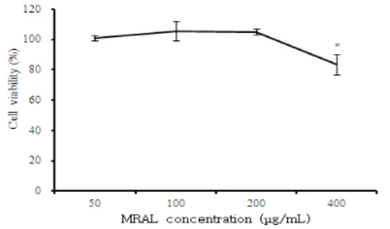
MC/9 비만세포에 대한 MRAL 추출물의 세포독성을 알아 보기 위하여 EZ-Cytox assay를 수행하였다(Fig. 1). MRAL
추출물을 농도별 (50, 100, 200, 400 µg/mL)로 처리한 결 과, 모든 농도에서 세포독성이 나타나지 않았다.
그러므로 MRAL 추출물의 알레르기 반응에 대한 효과를 평가할 때 독성이 없는 50, 100, 200 µg/mL의 농도를 선택 하여 실험을 진행하였다.
Fig. 1. Cytotoxic effects of MRAL on MC/9 mast cell line.
MC/9 mast cell line were pretreated with various concentration MRAL.
The results are expressed the mean±S.E. Statistically significant value compared with control group data by T-test.
2. Real -time PCR 분석
1) IL-4 mRNA 유전자 발현IL-4 mRNA 유전자 발현은 정상군은 0.013±0.012이었 고, PI만으로 자극한 대조군은 1.032±0.032로 정상군에 비 하여 유전자발현 증가가 나타났다. 양성대조군인 CsA 처리군 은 0.115±0.094로 대조군에 비하여 약 8.9배 정도 통계학 적으로 유의성 있게 (p<0.001)억제되었다. MRAL 처리 후 PI로 자극한 실험군의 IL-4 mRNA 유전자 발현은 50 µg/mL에서는 0.831±0.065로 약 1.2배 정도 유의성 있게 (p<0.01)억제되었으며, 100 µg/mL에서는 0.486±0.072로 대조군에 비하여 약 2.1배 정도 유의성 있게 (p<0.001)억제 되었다(Fig. 2).
Fig. 2. Suppressive effects of MRAL on IL-4 mRNA expression.
MC/9 mast cells were pretreated with CsA (10 µg/mL) or MRAL (50, 100 µg/mL) and then stimulated with PI. IL-4 mRNA expression was analyzed by real-time PCR. The results represent the mean±S.E. Statistically signigicant value was calculated by compared with PI-control group by student’s T-test. (** p<0.01,
*** p<0.001)
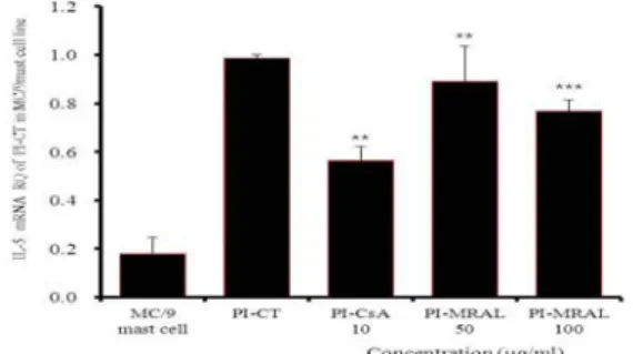
2) IL-5 mRNA 유전자 발현
IL-5 mRNA 유전자 발현은 정상군은 0.179±0.066이었고,
PI만으로 자극한 대조군은 0.986±0.015로 정상군에 비하여
유전자발현 증가가 나타났다. 양성대조군인 CsA 처리군은
0.566±0.058로 대조군에 비하여 약 42.5% 정도 통계학적으
로 유의성 있게 (p<0.01)억제되었다. MRAL 처리 후 PI로 자 극한 실험군의 IL-5 mRNA 유전자 발현은 50 µg/mL에서는 0.892±0.144로 약 9.5% 정도 유의성 있게 (p<0.01)억제되었 으며, 100 µg/mL에서는 0.768±0.046으로 대조군에 비하여 약 22.1% 정도 유의성 있게 (p<0.001)억제되었다(Fig. 3).
Fig. 3. Suppressive effects of MRAL on IL-5 mRNA expression.
MC/9 mast cells were pretreated with CsA (10 µg/mL) or MRAL (50, 100 µg/mL) and then stimulated with PI. IL-5 mRNA expression was analyzed by real-time PCR. The results represent the mean±S.E. Statistically signigicant value was calculated by compared with PI-control group by student’s T-test. (** p<0.01,
*** p<0.001)
3) IL-6 mRNA 유전자 발현
IL-6 mRNA 유전자 발현은 정상군은 0.087±0.059이었 고, PI만으로 자극한 대조군은 1.052±0.052로 정상군에 비 하여 유전자발현 증가가 나타났다. 양성대조군인 CsA 처리군 은 0.388±0.03로 대조군에 비하여 약 63% 정도 통계학적으 로 유의성 있게 (p<0.01)억제되었다. MRAL 처리 후 PI로 자 극한 실험군의 IL-6 mRNA 유전자 발현은 50 µg/mL에서는 0.943±0.122로 약 10.3% 정도 유의성 있게 (p<0.05)억제되 었으며, 100 µg/mL에서는 0.595±0.088로 대조군에 비하여 약 43.4% 정도 유의성 있게 (p<0.001)억제되었다(Fig. 4).
Fig. 4. Suppressive effects of MRAL on IL-6 mRNA expression.
MC/9 mast cells were pretreated with CsA (10 µg/mL) or MRAL (50, 100 µg/mL) and then stimulated with PI. IL-6 mRNA expression was analyzed by real-time PCR. The results represent the mean±S.E. Statistically signigicant value was calculated by compared with PI-control group by student’s T-test. (*p<0.05, **
p<0.01, *** p<0.001)
4) IL-13 mRNA 유전자 발현
IL-13 mRNA 유전자 발현은 정상군은 0.062±0.017이었 고, PI만으로 자극한 대조군은 0.987±0.013으로 정상군에 비 하여 유전자발현 증가가 나타났다. 양성대조군인 CsA 처리군 은 0.113±0.005로 대조군에 비하여 약 88.5% 정도 통계학적 으로 유의성 있게 (p<0.001)억제되었다. MRAL 처리 후 PI로 자극한 실험군의 IL-13 mRNA 유전자 발현은 50 µg/mL에서
는 0.797±0.082로 약 19.2%정도 유의성 있게 (p<0.05)억제 되었으며, 100 µg/mL에서는 0.427±0.105로 대조군에 비하 여 약 56.7% 정도 유의성 있게 (p<0.001)억제되었다(Fig. 5).
Fig. 5. Suppressive effects of MRAL on IL-13 mRNA expression.
MC/9 mast cells were pretreated with CsA (10 µg/mL) or MRAL (50, 100 µg/mL) and then stimulated with PI. IL-13 mRNA expression was analyzed by real-time PCR. The results represent the mean±S.E. Statistically signigicant value was calculated by compared with PI-control group by student’s T-test. (*p<0.05,
*** p<0.001)
3. ELISA
1) IL-5 단백질 생성
MRAL이 비만세포에서 IL-5 단백질 생성에 미치는 영향 을 관찰하기 위하여, MC/9 세포에 CsA와 다양한 농도의 MRAL을 처리한 후 PI로 자극한 뒤 24시간 후 ELISA로 IL-5 생산량을 측정하였다. IL-5 생산량은 정상군은 121±6.0 pg/mL이었고, 대조군은 1946.6±24.5 pg/mL로 정상군에 비해 약 16배 정도 증가하였다. 실험군의 IL-5 생 산량은 50 µg/mL에서는 2062.3±43.1 pg/mL로 대조군과 비슷한 수준으로 나타났고, 100 µg/mL에서는 1255.9±21.9 pg/mL로 대조군에 비하여 약 1.5배 정도 유의성 있게 (p<0.001)억제되었고, 200 µg/mL에서는 834.5±134.9 pg/mL로 대조군에 비하여 약 2.3배 정도 유의성 있게 (p<0.001)억제되었다(Fig. 6).
Fig. 6. Suppressive effects of MRAL on IL-5 production.
MC/9 mast cells were pretreated with MRAL (50, 100, 200 µg/mL) and then stimulated with PI. IL-5 production levels were measured by ELISA. The results represent the mean±S.E.
Statistically signigicant value was calculated by compared with PI-control group by student’s T-test. (*** p<0.001)
2) IL-13 단백질 생성
IL-13 단백질 생산량은 정상군은 90±3.9 pg/mL이었고,
대조군은 3211.0±112.1 pg/mL로 정상군에 비해 약 35배
정도 증가하였다. 실험군의 IL-13 생산량은 50 µg/mL에서
는 3163.0±65.3 pg/mL로 대조군과 비슷한 수준으로 나타
났고, 100 µg/mL에서는 2361.0±96.0 pg/mL로 대조군에 비하여 약 1.4배 정도 유의성 있게 (p<0.001)억제되었고, 200 µg/mL에서는 1746.0±63 pg/mL로 대조군에 비하여 약 1.8배 정도 유의성 있게 (p<0.001)억제되었다(Fig. 7).
Fig. 7. Suppressive effects of MRAL on IL-13 production.
MC/9 mast cells were pretreated with MRAL (50, 100, 200 µg/mL) and then stimulated with PI. IL-13 production levels were measured by ELISA. The results represent the mean±S.E.
Statistically signigicant value was calculated by compared with PI-control group by student’s T-test. (*** p<0.001)
4. Western blot analysis
1) GATA-1과 GATA-2 발현GATA-1의 단백질 발현은 대조군에서 GATA-1 densities (이하 DT)가 1로 나타났고 (Fig. 8, GATA-1 band lane 1), 양성대조군의 DT는 0.25로 대조군에 비하여 약 4배 정도 억제되었다(Fig. 8, GATA-1 band lane 2).
실험군의 GATA-1의 단백질 발현은 대조군에 비하여 50 µg/mL에서 DT는 0.78, 100 µg/mL에서 DT는 0.18로 각각 약 1.2배 및 5.5배 정도로 농도의존적으로 단백질 발현이 억 제되었다(Fig. 8, GATA-1 band lane 3-4). GATA-2의 단백질 발현은 대조군에서 GATA-2 densities (이하 DT)가 1로 나타났고(Fig. 9, GATA-2 band lane 1), 양성대조군 의 DT는 0.18로 대조군에 비하여 약 5.5배 정도 억제되었다 (Fig. 9, GATA-2 band lane 2). 실험군의 GATA-2의 단 백질 발현은 대조군에 비하여 50 µg/mL에서 DT는 0.64, 100 µg/mL에서 DT는 0.48로 각각 약 1.5배 및 2배 정도로 농도의존적으로 단백질 발현이 억제되었다(Fig. 9, GATA-2 band lane 3-4).
Fig. 8. Effect of MRAL on GATA-1 signal events in MC/9 line cells induced with PI.
MC/9 mast cells were pretreated with CsA (10 µg/mL) or MRAL (100, 50 µg/mL) for 1 hour, and then stimulated with PI for 6 hours. After adding lysis buffer, one part of the lysates was subjected to western blot with GATA-1 proteins. Another part of the lysates was directly subjected to SDS-PAGE and
immunoblotting with the indicated antibodies. The levels of GATA-1 proteins were selected as the positive controls for Anti-GATA-1 respectively. Band densities were compared with YY-1 and measured using an Image-Rab densitometer.
Fig. 9. Effect of MRAL on GATA-2 signal events in MC/9 mast cells induced with PI.
MC/9 mast cells were pretreated with CsA (10 µg/mL) or MRAL (100, 50 µg/mL) for 1 hour, and then stimulated with PI for 6 hours. After adding lysis buffer, one part of the lysates was subjected to western blot with GATA-2 proteins. Another part of the lysates was directly subjected to SDS-PAGE and immunoblotting with the indicated antibodies. The levels of GATA-2 proteins were selected as the positive controls for Anti-GATA-2 respectively. Band densities were compared with YY-1 and measured using an Image-Rab densitometer.
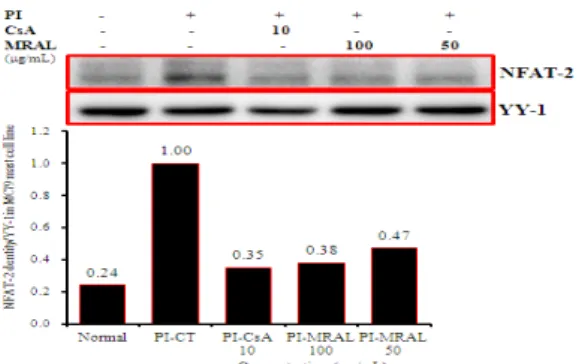
2) NFAT 발현
NFAT-1의 단백질 발현은 정상군에서 NFAT-1 densities (이하 DT)가 0.02로 나타났고(Fig. 10, NFAT-1 band lane 1), 대조군에서 DT가 1로 정상군에 비하여 현저하게 증가하였으 며(Fig. 10, NFAT-1 band lane 2), 양성대조군의 DT는 0.18 으로 대조군에 비하여 약 5.5배 정도 억제되었다(Fig. 10, NFAT-1 band lane 3). 실험군의 NFAT-1의 단백질 발현은 대조군에 비하여 50 µg/mL에서 DT는 0.64, 100 µg/mL에서 DT는 0.48로 각각 약 1.5배 및 2배 정도로 농도의존적으로 억 제되었다 (Fig. 10, NFAT-1 band lane 4-5). NFAT-2의 단 백질 발현은 정상군에서 DT가 0.24로 나타났고(Fig. 11, NFAT-2 band lane 1), 대조군에서 DT가 1로 정상군에 비하 여 현저하게 증가하였으며(Fig. 11, NFAT-2 band lane 2), 양 성대조군의 DT는 0.35로 대조군에 비하여 약 2.8배 정도 억제 되었다(Fig. 11, NFAT-2 band lane 3). 실험군의 NFAT-2의 단백질 발현은 대조군에 비하여 50 µg/mL에서 DT는 0.47, 100 µg/mL에서 DT는 0.38로 각각 약 2.1배 및 2.6배 정도로 농도의존적으로 억제되었다(Fig. 11, NFAT-2 band lane 4-5).
Fig. 10. Effect of MRAL on NFAT-1 signal events in MC/9 mast cells induced with PI.
MC/9 mast cells were pretreated with CsA (10 µg/mL) or MRAL (100,
50 µg/mL) for 1 hour, and then stimulated with PI for 6 hours. After adding lysis buffer, one part of the lysates was subjected to western blot with NFAT-1 proteins. Another part of the lysates was directly subjected to SDS-PAGE and immunoblotting with the indicated antibodies. The levels of NFAT-1 proteins were selected as the positive controls for Anti-NFAT-1 respectively. Band densities were compared with YY-1 and measured using an Image-Rab densitometer.
Fig. 11. Effect of MRAL on NFAT-2 signal events in MC/9 mast cells induced with PI.
MC/9 mast cells were pretreated with CsA (10 µg/mL) or MRAL (100, 50 µg/mL) for 1 hour, and then stimulated with PI for 6 hours. After adding lysis buffer, one part of the lysates was subjected to western blot with NFAT-2 proteins. Another part of the lysates was directly subjected to SDS-PAGE and immunoblotting with the indicated antibodies. The levels of NFAT-2 proteins were selected as the positive controls for Anti-NFAT-2 respectively. Band densities were compared with YY-1 and measured using an Image-Rab densitometer.
3) AP-1 발현
c-Jun의 단백질 발현은 정상군에서 c-Jun densities (이 하 DT)가 0.75로 나타났고(Fig. 12, c-Jun band lane 1), 대조군에서 DT가 1로 정상군에 비하여 증가하였으며(Fig.
12, c-Jun band lane 2), 양성대조군의 DT는 0.86로 대조 군에 비하여 억제되었다(Fig. 12, c-Jun band lane 3). 실 험군의 c-Jun 단백질 발현은 대조군에 비하여 50 µg/mL에 서 DT는 1.04, 100 µg/mL에서 DT는 0.98로 대조군에 비 하여 차이가 나타나지 않았다(Fig. 12, c-Jun band lane 4-5). c-Fos의 단백질 발현은 정상군에서 c-Fos densities (이하 DT)가 0.33으로 나타났고(Fig. 13, c-Fos band lane 1), 대조군에서는 DT가 1로 정상군에 비하여 현저하게 증가 되었으며(Fig. 13, c-Fos band lane 2), 양성대조군의 DT 는 0.52로 대조군에 비하여 약 1.9배 정도 억제되었다(Fig.
13, c-Fos band lane 3). 실험군의 c-Fos 단백질 발현은 대조군에 비하여 50 µg/mL서 DT는 0.78, 100 µg/mL에서 DT는 0.61로 억제되었다(Fig. 13, c-Fos band lane 4-5).
Fig. 12. Effect of MRAL on AP-1 proteins (c-Jun) in MC/9 mast cells induced with PI.
MC/9 mast cells were pretreated with CsA (10 µg/mL) or MRAL
(100, 50 µg/mL) for 1 hour, and then stimulated with PI for 6 hours. After adding lysis buffer, one part of the lysates was subjected to western blot with AP-1 proteins. Another part of the lysates was directly subjected to SDS-PAGE and immunoblotting with the indicated antibodies. The levels of AP-1 proteins were selected as the positive controls for anti-c-Jun respectively. Band densities were compared with YY-1 and measured using an Image-Rab densitometer.
Fig. 13. Effect of MRAL on AP-1 proteins(c-Fos) in MC/9 mast cells induced with PI.
MC/9 mast cells were pretreated with CsA (10 µg/mL) or MRAL (100, 50 µg/mL) for 1 hour, and then stimulated with PI for 6 hours. After adding lysis buffer, one part of the lysates was subjected to western blot with AP-1 proteins. Another part of the lysates was directly subjected to SDS-PAGE and immunoblotting with the indicated antibodies. The levels of AP-1 proteins were selected as the positive controls for anti-c-Fos respectively. Band densities were compared with YY-1 and measured using an Image-Rab densitometer.
5. NF-κB 신호전달경로에 대한 효과
NF-κB p65 DNA binding activity는 정상군 (Fig. 14, NF-κB p65 band lane 1) 보다도 대조군에서 NF-κB p65 DNA binding activity가 현저하게 증가되었고(Fig. 14, NF- κB p65 band lane 2), 양성대조군의 NF-κB p65 DNA binding activity는 대조군에 비하여 크게 억제되었다(Fig.
14, NF-κB p65 band lane 3). 실험군의 NF-κB p65 DNA binding activity은 100 µg/mL에서 대조군에 비하여 현저하게 억제되었다(Fig. 14, NF-κB p65 band lane 4).
Fig. 14. Effect of MRAL on DNA-binding activity of NF-κB p65 signal events in MC/9 cells induced with PI.
MC/9 mast cells were pretreated with CsA (10 µg/mL) or MRAL (100 µg/mL) for 1 hour, and then stimulated with PI for 6 hours.
Nuclear extract from MC/9 mast cells were incubated with a 32P-labeled DNA probe and analyzed by 6% polyacrylamide gels.
Electrophoresis mobility shift assay by NF-κB binding activity.
고 찰
비만세포는 천식이나 알레르기성 비염과 같은 알레르기 반 응을 매개하는 중요한 세포로 활성화 되면 탈과립되고 또한 아라키돈산 대사물질과 염증반응을 유발하는 다양한 사이토카 인이 분비된다
2).
이에 저자는 MRAL가 알레르기 염증 반응에 미치는 영향 을 알아보기 위해 MC/9 비만세포를 이용하여 Real-time PCR, ELISA, 그리고 EMSA로 비만세포에서 발현하는 IL-4, IL-5와 IL-13 유전자 발현을 분석하였고, western blot으로 GATA-1, GATA-2, NFAT-1, NFAT-2, c-Fos, c-Jun, 전사인자의 발현을 억제하는지 분석하였으며, NF- κB p65는 EMSA법으로 DNA binding activity를 측정하였 다. 또한 Th2 세포에서 발현하는 cytokine IL-5와 IL-13의 변화를 관찰하였다.
IL-4는 B-세포를 활성화하여 IgE 항체를 생성하며, Th2 세포의 분화를 촉진시키므로 천식과 같은 알레르기 질환에서 중요한 역할을 하는 사이토카인이다. 정상인의 경우는 같은 알레르겐에 대해서 IFN-γ를 분비하는 Th1 세포의 기능을 갖고 있으나, 천식 환자의 기관지 생검, 아토피 환자의 피부 생검, 알레르기성 비염 환자의 비점막에서는 IL-4, IL-5, IL-9 등과 같은 Th2 세포가 분비하는 사이토카인의 양상이 관찰된다
9).
MRAL 추출물을 투여하였을 때 IL-4 mRNA 유전자 발현 이 유의적으로 감소된 것으로 보아 MRAL에는 세포가 Th2 세포의 기능을 획득하는 것을 억제함으로써 항알레르기 작용 이 있을 것이라 생각된다.
알레르기반응 중 유리되는 IL-3, IL-5, 그리고 GM-CSF 등은 호산구를 생성을 활성화시키고 세포자멸에 관여하며, 이 들의 길항제나 항체를 이용하여 알레르기 염증반응을 억제하 려는 시도가 이루어지고 있다
10). 본 실험에서 MRAL 추출물 이 IL-5 mRNA의 발현을 효과적으로 억제시키는 것으로 보 아 항알레르기 작용이 있을 것으로 사료된다.
TNF-α는 비만세포에서 본질적으로 발현되어지지만, IL-6는 자극제에 의해서 새롭게 합성된다
11). 따라서 IL-6 mRNA 유전자 발현정도를 확인한 결과 MRAL이 비만세포에 서 IL-6 mRNA 발현을 억제한 것으로 보아 IgE 유도성 비 만세포 활성화를 억제한다고 생각된다.
최근에 알레르기 질환과 관련된 많은 종류의 세포들 (비만 세포, 호중성백혈구, 림프구, 호산성백혈구 등)로부터 여러 종 류의 매개물질들 (leukotriene, 혈소판 활성인자(PAF), 히스 타민 등)과 사이토카인들 (Cytokines ; TNF-α, IL-4, IL-5, IL-9, IL-12, IL-13 등)이 유리되고, adhesion molecules (ICAM-12, VCAM-1 등)과 transcription factor (nuclear factor kappa B, AP-1, NF-AT, STAT 등)의 발현이 중요한 역할을 하며, 이들의 상호 관련성에 관 한 광범위한 많은 연구가 진행되고 있고
12), 이러한 알레르기 질환의 병태생리에 중요한 역할을 하는 비만세포로부터 분비 되는 많은 물질들 (histamine, cytokines, interleukins, PAF, leukotrienes 등)의 유리를 유발하거나 억제하는 인자 들에 대한 연구를 통하여, 비만세포 안정화물질 (sodium cromoglycate, mast cell stabilization compound; MSC 등)을 이용하여 알레르기 질환을 예방하거나 치료할 수 있는
방법을 개발하는 차원에서 볼 때 MRAL의 IL-13과 같은 비 만세포 활성화 인자의 발현 정도를 보아 억제효능 도출은 큰 의미를 갖는다고 생각된다.
비만세포에서 분비된 IL-4, IL-13은 B cell을 활성화하여 IgE 항체를 만들게 하며, IL-5는 호산구를 모으고 활성화시 키는 역할을 하고, 활성화된 호산구 역시 IL-5를 분비하는 데, 이렇게 분비된 IL-5 또한 호산구 활성화와 그를 통한 매 개물질 생성을 촉진하는 것으로 알려져 있다
13). MRAL이 비 만세포에서 IL-5와 IL-13 단백질 생산을 억제하여 항알레르 기 효능을 갖는다고 생각된다.
비만세포에서 IL-4와 IL-13의 발현은 선택적으로 발현되 는 GATA-1, GATA-2, PU.1과 같은 인트론 부위에 결합하 는 전사인자
14)및 NF-AT를 포함하는 여러 전사인자와의 협 동에 의해 조절된다
15). FcεRI의 교차결합 반응으로 여러 가 지 어댑터 분자와 키나아제의 보충과 활성화는 NF-AT 및 NF-κB의 핵전이와 c-Jun 아미노 말단 키나아제와 같은 단 백질 키나아제에 의한 AP-1의 활성화에 이르게 한다. 많은 연구자들이 AP-1 (c-jun, c-fos)와 GATA 전사인자 모두 IL-13 유전자의 transactivation을 위한 필수 신호전달 단백 질로 알려져 있다
14). 비만세포에서 IL-13의 발현 기전이 아직 까지 명확하게 밝혀지진 않았지만 다수의 전사인자가 IL-13의 프로모터에 작용하여 조절되는 것으로 확인되고 있다.
IL-13의 생산은 NF-AT, GATA, AP-1을 포함하는 여러 전사인자와의 협동에 의해 조절된다. NF-AT2는 GATA와 협 력하여 IL-13 전사 조절에 수반되는 가장 중요한 전사인자이 다. GATA-2는 IL-13에 결합하는 AP-1 결합을 유도할 뿐 만 아니라 비만세포에서 IL-13 생성을 증가시킨다
16).
NF-κB는 면역기능, 염증반응, 혈관 내피세포의 활성화, 세포성장 등에 관여하는 전사인자로서 거의 모든 세포에 존재 한다. NF-κB는 구조상 같은 계열 (Rel family)에 속한 단 백인 heterodimer나 혹은 homodimer 형태로 존재하는데 가 장 대표적인 것이 p50과 p65의 heterodimer 형태이다. 기저 상태에서 IκB와 결합하여 세포질 내에 비활성화 상태로 존 재하는 NF-κB는 바이러스 감염, 내독소, TNF-α와 IL-1 β 등의 염증매개 cytokine에 의해 활성화된다. 즉, 외부 자 극에 의해 IκB가 분해되면 세포질에 존재하던 NF-κB가 핵 속으로 이동되어 여러 유전자의 κB element에 결합하여 그 유전자의 전사를 가져온다.
특히 IKK는 최근 들어서 IKKα와 IKKβ로 이루어진 복 합체라는 것이 발견되었으며, IKK는 IκB의 인산화와 전사인 자인 NF-κB의 활성화를 촉진시키는 kinase라는 것이 밝혀 졌다
17). 또한 IKK 중 IKKβ 단백질이 염증성 싸이토카인 발 현에 관여하는 NF-κB의 활성화에 필수적인 요소라는 것이
밝혀졌다
18-19). 전사인자 NF-κB는 TNF-α와 IL-6 유전자
내에 결합부위가 존재하여 TNF-α와 IL-6 유전자의 전사 인자로써 역할을 한다
20).
본 연구에서 MRAL은 활성화된 비만세포와 Th2 세포에서
발현하는 IL-5, IL-13의 생성을 억제함으로써 알레르기 염
증 반응을 제어하는 것으로 판단되며, GATA-1, GATA-2,
NFAT-1, NFAT-2, c-Fos 전사인자의 활성을 억제함으로
써 GATA, NF-AT, AP-1 신호전달 기전 및 IL-6 억제 기
전인 NF-κB 전사인자를 차단하여 IL-5, IL-13의 발현을
억제함으로써 항알레르기 효능을 나타내는 것으로 사료된다.
결 론
상백피 추출물(MRAL)이 비만세포와 Th2 세포의 cytokine IL-4, IL-5, IL-6 및 IL-13유전자 발현 및 단백질과 GATA-1, GATA-2, NFAT 및 AP-1발현 그리고 NF-KBp65 신호전달에 미치는 영향을 실험적으로 규명한 결 과 다음과 같다.
1. MRAL은 MC/9 세포주에서 모든 농도에서 세포독성이 나타나지 않았다.
2. MRAL은 MC/9 비만세포에서 IL-4, IL-5, IL-6와 IL-13 mRNA 유전자 발현을 억제하였다.
3. MRAL은 MC/9 비만세포에서 IL-5와 IL-13 단백질 생산을 억제하였다.
4. MRAL은 MC/9 비만세포에서 전사인자인 GATA-1, GATA-2, NFAT-1, NFAT-2 그리고 c-Fos 단백질 발현을 억제하였다.
5. MRAL은 MC/9 비만세포에서 전사인자인 binding activity of NF-kB을 억제하였다.
참고문헌