Available at http://www.formulastudy.com
HFS
Original Article / 원저
C2C12 근아세포의 산화적 손상에 대한 고삼, 감초 및 백선피 복합 추출물의 보호효과
최은옥
1,2, 황보현
1,2, 김민영
1,2, 손다희
2, 정진우
1,2, 박 철
3, 홍수현
2, 김민주
4, 이지영
4, 신수진
4, 최영현
1,2*1
동의대학교 항노화연구소,
2동의대학교 한의과대학 생화학교실,
3
동의대학교 자연생활과학대학 분자생물학과,
4㈜리오엘리
Protective Effects of Ethanol Extract Mixtures of Sophora flavescens, Glycyrrhiza uralensis and Dictamnus dasycarpus against Oxidative
Stress-induced Damage in C2C12 Murine Myoblasts
Eun Ok Choi
1,2, Hyun Hwang-Bo
1,2, Min Young Kim
1,2, Da Hee Son
2, Jin Woo Jeong
1,2, Cheol Park
3, Su Hyun Hong
2, Min Ju Kim
4, Ji Young Lee
4, Su Jin Shin
4, Yung Hyun Choi
1,2*1
Anti-Aging Research Center, Dong-Eui University
2
Department of Biochemistry, College of Korean Medicine, Dong-Eui University
3
Departments of Molecular Biology, College of Natural Sciences, Dong-Eui University
4
Lioele Cosmetic Co., LTD.
ABSTRACT
Objectives : Increased oxidative stress by reactive oxygen species (ROS) has been suggested as a major cause of muscle fatigue. Although several studies have demonstrated the various biological properties of Sophora flavescens Aiton, Glycyrrhiza uralensis Fischer and Dictamnus dasycarpus Turcz, but the antioxidative potentials have not been clearly demonstrated. The present study was designed to investigate the protective effects of their water and ethanol extract mixtures (medicinal herbal mixtures, MHMIXs) on hydrogen peroxide (H2O2)-induced cell damage and apoptosis in C2C12 myoblasts.
ⓒ 2017 The Korean Medicine Society For The Herbal Formula Study
This paper is available at http://www.formulastudy.com which permits unrestricted non-commercial use, distribution, and reproduction in any medium, provided the original work is properly cited.
Methods : Cytotoxicity was assessed by an MTT assay. Quantitative evaluation of apoptosis induction and ROS production was evaluated by flow cytometry analysis. Expression levels of apoptosis regulatory and DNA-damage proteins were detected by Western blotting.
Result : The inhibition of H2O2-induced cell proliferation was effectively blocked in extracts of 3: 1: 1 (EMHMIXs-1) or 2: 2: 1 (EMHMIXs-2) of S. flavescens, G. uralensis and D. dasycarpus Turcz, ethanol extracts from various complex extracts in C2C12 myoblasts. EMHMIXs-1 and EMHMIXs-2 also effectively attenuated H2O2-induced C2C12 cell apoptosis, which was associated with the restoration of the upregulation of Bad and death receptor 4, and downregulation of XIAP and cIAP-1 induced by H2O2. In addition, these herbal mixtures significantly blocked the H2O2‑induced ROS generation and phosphorylation of p-γH2A.X, which suggests that they can prevent H2O2‑induced cellular DNA damage.
Conclusions : The results suggest that EMHMIXs-1 and EMHMIXs-2 could block the DAN damage and apoptosis of C2C12 myoblasts by oxidative stress through blocking ROS generation.
Key words : C2C12 myoblasts, medicinal herbal mixtures, oxidative stress, DNA damage, apoptosis
Ⅰ. 서론
6)활성산소종(reactive oxygen species, ROS)은 미 토콘드리아 내에서 발생하는 산소의 환원 대사산물로 서, 과도한 스트레스, 방사선 및 화학물질 등에 노출 되거나 항산화 시스템의 기능이 저하되어 체내 산화 -환원 대사의 균형이 파괴될 경우 비정상적으로 생 성될 수 있다. 증가된 ROS에 의해 산화적 스트레스 (oxidative stress)가 유도되면 세포 내 DNA, 단백 질, 지질의 심한 손상이 초래된다1-3). 이러한 산화적 스트레스 및 ROS는 다양한 기전을 통하여 노화, 세 포의 돌연변이, 뇌질환, 심장질환, 동맥경화 등 다양 한 질병의 발생과 진행을 촉진하는 것으로 알려져 있다
4-6).
체내 산화적 스트레스를 저감하는 전통 약물 유래 기능성 소재를 발굴하기 위하여 다양한 세포주 모델 이 이용되고 있는데 C2C12 세포는 근육으로 분화되 기 전단계인 근아세포(myoblast)이다7). 최근 골격근 세포 손상의 주요한 기전으로 산화적 스트레스의 중 요성이 인식되면서8-10), C2C12 세포는 근기능 손상을 보호하기 위한 다양한 약물의 발굴을 위한 모델로서 널리 사용되고 있다11-13).
고삼(苦蔘, Sophora flavescens Aiton)과 감초(甘草, Glycyrrhiza uralensis Fisch)는 쌍떡잎식물 장미목
콩과에 속하는 다년생 초본식물이며, 백선피(白鮮皮) 는 운향과에 속하는 다년생 초본식물인 백선(Dictamnus dasycarpus Turcz.)의 뿌리껍질이다. 고삼의 뿌리는 인삼과 유사한 효능을 가지면서 소화불량, 신경통, 간염, 황달 등에 효과가 있고, 감초의 뿌리는 통증완 화, 혈압강하, 담즙분비 촉진, 근육경련과 다양한 염 증성 질환 억제 등에 효과가 있으며, 백선피는 백선 의 뿌리를 의미하며, 습진, 피부가려움증, 두드러기, 황달, 감기 등에 효과가 있을 뿐만 아니라 해열작용 과 피부진균 억제작용이 있어 오래 전부터 전통 의학 에서 많이 사용되어 왔다14-16). 본 연구실의 선행 연 구에서 3가지 약재 추출물을 이용한 다양한 비율로 조성된 복합 추출물의 항아토피성 효능을 조사하기 위하여 실험 동물에 아토피 피부염을 유발하여 몇 가 지 아토피 지표, 혈청 내 IgE와 cytokine 양의 변화 를 관찰한 결과, 각각의 단일 추출물과 복합 추출물 에서 항아토피성 조절제로서의 발굴 가능성이 있음을 확인하였다17). 특히, 고삼과 감초 및 백선피 추출물 이 3:1:1로 혼합된 복합 추출물 처리군에서 각각의 단일 추출물에 비하여 더 우수한 효과가 있음을 보고 한 바 있다17). 또한, lipopolysaccharide (LPS)로 자 극된 대식세포에서 항염증 효과를 검증하기 위하여 실시한 일산화질소(nitric oxide, NO) 및 염증성 cytokine의 생성량과 이들 조절 유전자들의 발현 변
* Corresponding author : Yung Hyun Choi, Department of Biochemistry, College of Korean Medicine, Dong-Eui University, 52-57, Yangjeong-ro, Busanjin-gu, Busan, 47227, Republic of Korea.
Tel:+82-51-850-7413, Fax:+82-51-853-4036, E-mail : [email protected]
∙ Received : May 16, 2017 / Revised : May 29, 2017 / Accepted : May 29, 2017
화 조사에서도 고삼, 감초 및 백선피 추출물이 3:1:1 로 혼합된 복합 추출물 처리군에서 한약재 단일 추출 물의 함량에 비해 각각의 한약재 단일 추출물의 함량 이 낮음에 비하여 더 효과적이거나 유사한 결과를 얻 었다18). 따라서 본 연구에서는 복합 처리 시 상승효 과를 볼 수 있을 것으로 기대되는 고삼, 감초 및 백선피 의 각 열수 및 에탄올 추출물의 혼합 비율을 달리한 복합 추출물(medicinal herber mixtures, MHMIXs)을 이용 하여 안전하면서 산화적인 스트레스에 대한 보호효능 이 우수한 혼합의 비율을 발굴하고자 하였다. 이를 위 하여 C2C12 세포를 이용하여 H2O2로 유도된 산화적 손상의 차단효과를 다양한 지표를 통하여 평가하였다.
Ⅱ. 재료 및 방법
1. 고삼, 감초 및 백선피의 열수 및 에탄올 추출물과 복합물의 준비
본 연구에 사용된 고삼, 감초 및 백선피는 ㈜서경한 방약업사(Busan, Republic of Korea)에서 파쇄된 상 태로 구입하였다. 고삼 열수 추출물(water extract of S.
flavescens, WESF), 감초 열수 추출물(water extract of G. uralensis, WEGU) 및 백선피 열수 추출물(water extract of D. dasycarpus, WEDD)을 얻기 위하여 약재 무게의 10배에 해당하는 증류수로 100℃에서 3 시간 동안 추출하였다. 또한, 고삼 에탄올 추출물 (ethanol extract of S. flavescens, EESF), 감초 에탄 올 추출물(ethanol extract of G. uralensis, EEGU) 및 백선피 에탄올 추출물(ethanol extract of D.
dasycarpus, EEDD)을 얻기 위하여 각 약재 100 g을 2 L의 30% 에탄올을 가하여 초음파추출(ultrasonification extraction)로 추출물을 제조하였다. 이를 위해 추출 용기에 한약재와 30% 에탄올을 혼합한 후 초음파 수 조(Power Sonic 405, SJ BioLab., Anyang, Korea) 바닥에 닿지 않도록 하여 40 KHz 초음파를 가하여 2시간 동안 추출하였다. 각각의 추출물은 여과지 (Whatman No. 3 filter paper, Whatman International Ltd., Maidstone, England)로 여과한 후 용매를 Rotary evaporator (Eyela, A-1000, Tokyo Rikakikai Co., Tokyo, Japan)를 이용하여 농축하고, 각 농축물은 동결 건조하여 분말화시켰다. 이후 열수 추출물은 멸 균 증류수에 100 mg/ml의 농도로 녹이고, Minisart®
Syringe filter (0.2 μm, Sartorius AG, Weender Landstr.
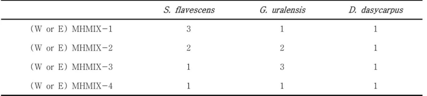
Germany)로 거른 후 사용 전까지는 –20℃에서 보관 하였고, 30% 에탄올 추출물은 dimethyl sulfoxide (DMSO, Sigma-Aldrich Chemical Co., St. Louis, MO, USA)를 이용하여 100 mg/ml로 stock solution을 만들어 실 험에 따라 적절하게 희석하여 사용하였다. 추출된 각 추출물은 Table 1에 제시된 비율로 혼합하여 추출혼 합물(MHMIXs)을 조성한 후 용매와 조성에 따른 추 출물의 효능을 비교하였다.
2. 세포 배양
본 실험에 사용된 C2C12 근아세포, HaCaT 피부각 질세포, C6 신경교세포는 American Type Culture Collection (Manassas, MD, USA)에서 분양받았으며, 세포의 배양을 위해 10% 우태아혈청(fetal bovine serum, FBS, WELGENE, Daegu, Republic of Korea) 및 1%의 penicillin/streptomycin이 포함된 Dulbecco's Modified Eagle's Medium (DMEM, WELGENE)을 사용하여, 37℃, 5% CO2 조건 하에서 배양하였다.
세포의 증식에 따른 과밀도 현상을 해소하기 위하여 매 48 시간마다 0.05% trypsin-0.02% ethylene- diaminetetracetic acid (EDTA, Sigma-Aldrich Chemical Co.)를 이용하여 세포를 부유시킨 후 적정수의 세포 를 유지하였다.
3. MTT assay에 의한 세포 생존율 조사
복합 추출물 처리가 세포생존율에 미치는 영향과 H2O2처리에 따른 산화적 스트레스에 의한 세포 손상 보호효과를 확인하기 위하여 MTT assay를 이용하였 다. 이를 위하여 세포 배양용 6 well plate에 해당 세포를 1×10⁵ cells/well로 분주하고 24시간 동안 안 정화시킨 후 적정 농도의 복합 추출물을 1시간 전처 리한 후 H2O2 (Sigma-Aldrich Chemical Co.)를 처 리하였다. 24시간 배양 후, 3-(4,5-dimethyl-2 thiazolyl)-2,5-diphnyl-2H-tetrazolium bromide (MTT, Sigma-Aldrich Chemical Co.)를 0.5 mg/ml 농도로 희석하여 처리한 후 37oC에서 2 시간 동안 다시 반응시켰다. 반응이 끝난 다음 MTT 시약을 제 거하고 DMSO를 이용하여 각 well에 생성된 formazin을 모두 녹인 후 96 well plate에 200 μ l씩 옮겨서 enzyme- linked immunosorbent assay (ELISA) reader (Molecular Devices, Sunnyvale, CA, USA)로 540 nm에서 흡 광도를 측정하였다.
4. Annexin V-FITC 염색에 의한 apoptosis의 정량 적 측정
C2C12 세포에서 H2O2 처리에 의한 apoptosis 유 발 및 복합 추출물에 의한 차단 효과의 정량적 분석 을 위해 준비된 세포들을 모은 다음 2,000 rpm으로 5분간 원심분리하여 상층액을 제거하였다. 이들 세포 를 phosphate buffer saline (PBS)를 이용하여 2~3 회 정도 세척하고 10 mM HEPES/NaOH, pH 7.4, 140 mM NaCl 및 2.5 mM CaCl2가 포함된 annexin V binding buffer (Becton Dickinson, San Jose, CA, USA)에 부유시킨 다음 annexin V-fluorescein isothiocyanate (FITC, Becton Dickinson) 및 propidium iodide (PI, Sigma-Aldrich Chemical Co.)를 처리하 여 암실에서 15분 동안 반응을 시켰다. 반응이 끝난 후 35-mm mesh를 이용하여 단일세포로 분리하고 flow cytometer (FACS Calibur, Becton Dickinson)를 적용시켜 apoptosis가 유발된 세포(V+/PI–)를 형광반 응에 따라 분석하였다.
5. 단백질의 분리, 전기영동 및 Western blot analysis 단백질 발현 검출을 위하여 준비된 세포에 적당량의 lysis buffer [25 mM Tris-Cl (pH 7.5), 250 mM NaCl, 5 mM EDTA, 1% Nonidet-P40 (NP-40), 1 mM phenymethylsulfonyl fluoride (PMSF), 5 mM dithiothreitol (DTT)]를 첨가하여 4℃에서 1시간 이 상 반응시킨 후, 14,000 rpm으로 30분간 원심 분리 하여 상층액에 있는 단백질을 분리하였다. 상층액의 단백질 농도는 Bio-Rad 단백질 정량 시약(Bio-Rad, Hercules, CA, USA)과 그 사용방법에 따라 정량 한 다음 동량의 Laemilni sample buffer (Bio-Rad)와 혼합하여 sodium dodecyl sulphate (SDS)-polyacrylamide gel을 이용하여 전기영동을 실시하였다. 이를 다시 nitrocellulose membrane (Schleicher and Schuell, Keene, NH, USA)으로 electroblotting에 의해 전이 시키고 분리된 단백질이 전이된 membrane에 5%
skim milk를 1 시간 처리하여 비특이적인 단백질들 에 대한 blocking을 실시하였다. 그리고 적정 1차 항 체를 처리하여 상온에서 2 시간 이상 또는 4℃에서 over night 반응시킨 다음 PBS-T (PBS with Tween 20) 로 세척하고 1차 항체에 맞는 2차 항체를 사용하여 상온에서 1 시간 정도 반응시켰다. 반응이 끝난 후 암실에서 enhanced chemiluminoesence (ECL) solution
(Santa Cruz Biotechnology Inc., Santa Cruz, CA, USA)을 적용시킨 다음 X-ray film에 감광시켜 특정 단백질의 발현 변화를 분석하였다. 본 실험에서 단백 질 분석을 위하여 사용된 항체들(Table 2)은 Santa Cruz Biotechnology 및 Abcam (Danvers, MA, USA)에 서 구입하였다. Immunoblotting을 위해 2차 항체로 사용된 horseradish peroxidase (HRP)-conjugated anti-mouse 및 anti-rabbit 항체는 Santa Cruz Biotechnology에서 구입하였다.
6. ROS 생성 변화 측정
세포 내 ROS 생성 변화를 확인하기 위하여 C2C12 세포에 적정 농도의 복합 추출물을 1시간 전처리한 후 H2O2를 재처리하였다. 30분 후, 세포를 모아 fluorescent probe 인 2’,7’-di-chlorodihydrofluorescein diacetate (DCF-DA, Molecular Probes, Leiden, Netherlands) 용액(10 μM)으로 20분간 염색하였다.
반응이 끝난 후 35-mm mesh를 이용하여 단일세포 로 분리하고 flow cytometer를 적용시켜 ROS 값의 변 화를 분석하였다.
7. 통계 분석
시료로부터 얻어진 실험 결과들의 유의성을 검정하 기 위하여 분산분석(ANOVA)을 실시한 후 p < 0.05 수준에서 Duncan's multiple range tests를 실시하 였으며, 그 결과는 평균(mean) ± 표준편차(standard deviation, SD)로 표시하였다. 이때 사용한 모든 통 계분석은 SPSS 17.0 (IBM SPSS Inc, New York, USA) 통계 프로그램을 이용하여 처리하였다.
Ⅲ. 결과
1. C2C12 세포에서 H2O2로 유도된 산화적 스트레스 에 대한 한약재 복합 추출물의 보호 효과
C2C12 세포에서 H2O2 처리에 의한 산화적 세포손 상에 각 한약재 복합 추출물의 보호 효과를 조사하기 위하여, C2C12 세포에 50% 전후의 생존율을 보여준 H2O2처리 농도 0.5 mM를 산화적 스트레스 처리 조 건으로 설정하였으며(data not shown), 열수와 에탄 올 복합 추출물(0.25~1.0 mg/ml)을 1시간 전처리 한 후 H2O2를 처리하여 24시간 동안 배양한 뒤 세포 생 존율을 확인하였다. Fig. 1의 결과에 의하면, H2O2를
단독 처리 하였을 때 감소하였던 세포 생존율이 열수 복합 추출물(WMHMIX)을 처리하였을 때 보호 효과 가 없거나 고농도 처리군에서는 오히려 더 감소하는 것으로 나타났다. 그러나 H2O2를 단독 처리 하였을 때 감소하였던 세포 생존율이 에탄올 복합 추출물 (EMHMIX) 중 EMHMIX-1과 EMHMIX-2를 처리한 군에서는 농도 의존적으로 산화적 스트레스에 의한 생 존율 저하를 차단한다는 것을 확인하였으나, EMHMIX-3 과 EMHMIX-4 처리군에서는 이러한 보호 효과가 관 찰되지 않았다(Fig. 2). 따라서 여러 종류의 WMHMIX 및 EMHMIX 중 고삼의 비율이 상대적으로 높은 EMHMIX-1 과 EMHMIX-2 처리군이 C2C12 세포에서의 산화적 스트레스에 대한 보호 효과를 가지는 것을 알 수 있 었다.
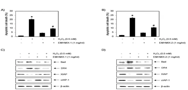
2. C2C12 세포에서 산화적 스트레스에 의해 유도된 apoptosis에 미치는 한약재 복합 추출물의 영향 다음으로 세포보호 효과가 관찰된 EMHMIX-1과 EMHMIX-2가 H2O2에 의하여 유발된 apoptosis 억 제 효능을 나타내는지를 조사하기 위하여 annexin V-FITC/PI 염색을 이용한 flow cytometer 분석을 실시하였다. 결과에서 볼 수 있듯이, H2O2단독 처리 된 배지에서 24시간 동안 배양된 C2C12세포에서는 약 20~22%에 해당되는 세포에서 apoptosis가 유발 되었으나, EMHMIX-1와 EMHMIX-2가 전처리된 세 포에서는 각각 9.4와 10.3% 정도로 나타났으므로 H2O2에 의하여 유발된 apoptosis가 억제된다는 것을 알 수 있었다(Fig. 3A 및 B). 또한, Western blot analysis를 통해 apoptosis 관련 단백질 발현의 변화 를 확인한 결과, H2O2가 단독 처리된 배지에서 배양된 C2C12세포에서는 apoptosis 유발과 연관이 있는 Bad 및 death receptor 4 (DR4)의 발현증가와 apoptosis 차 단에 관여하는 XIAP 및 cIAP-1의 발현 감소가 나타 났으나, EMHMIX-1와 EMHMIX-2의 전처리에 의하 여 이러한 현상들이 회복된다는 것을 확인 할 수 있 었다(Fig. 3C 및 D). 이상의 결과를 살펴볼 때 EMHMIX-1과 EMHMIX-2가 산화적 스트레스에 의한 C2C12세포의 apoptosis를 효율적으로 억제하였으며, 이 는 apoptosis 관련 유전자들의 조절에 따른 세포 생존 율 회복과 연관성이 있음을 알 수 있었다.
3. C2C12 세포에서 산화적 스트레스에 의해 유도된 γH2A.X 단백질의 인산화에 미치는 한약재 복합 추출물의 영향
γH2A.X 단백질은 DNA 이중 나선이 산화적 스트 레스에 의하여 절단되었을 경우 인산화형의 발현이 증가하기 때문에 DNA 손상 여부를 확인할 수 있는 바이오 마크로서 활용되는 지표 중 하나이다19,20). 따 라서 EMHMIX-1과 EMHMIX-2에 의한 C2C12 세포 의 산화적 스트레스에 의한 보효효과가 DNA 손상 차 단 효과와 연관성이 있는지를 조사하기 위하여 γH2A.X 단백질의 인산화 여부에 미치는 영향을 조사하였다.
이를 위하여 Fig. 3과 동일한 조건에서 배양된 세포 에서 단백질을 분리하여 Western blot analysis를 실시한 결과, C2C12세포에 H2O2를 단독 처리하였을 경우 증가되었던 인산화형의 γH2A.X의 발현이 EMHMIX-1 과 EMHMIX-2가 전처리에 의해 현저히 억제되는 것 으로 나타났다(Fig. 4). 이러한 결과를 살펴볼 때 산 화적 스트레스에 대한 C2C12 세포에서의 EMHMIX-1과 EMHMIX-2의 보호 효과는 DNA 손상 차단과 직접 적인 연관성이 있음을 의미하는 것이다.
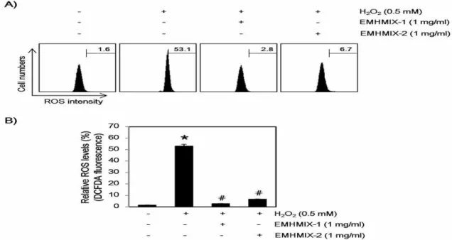
4. C2C12 세포에서 산화적 스트레스에 의해 유도된 ROS 생성에 미치는 한약재 복합 추출물의 영향 H2O2에 의한 apoptosis 유발은 mitochondria에서의 비정상적인 ROS 생성에 의한 mitochondria 기능 손상과 관련이 있다21,22). 따라서 EMHMIX-1과 EMHMIX-2에 의한 C2C12 세포의 apoptosis 유발 억제 효과가 ROS 생 성의 차단에 의한 것인지 조사하기 위하여 H2O2 처 리에 의한 ROS의 생성 차단 여부를 DCF-DA 염색을 통한 flow cytometry 분석을 통하여 조사하였다.
Fig. 5에 나타낸 바와 같이, 0.5 mM의 H2O2가 30분 동안 처리된 C2C12 세포에서 ROS의 생성이 대조군 의 1.6%에 비하여 53.1%까지 증가되었지만 EMHMIX-1 과 EMHMIX-2가 전처리된 조건에서 H2O2가 처리된 세포에서의 ROS의 생성은 각각 2.8과 6.7%로 나타 나 거의 완벽한 ROS 생성 차단 효과를 보였다. 따라 서 EMHMIX-1과 EMHMIX-2는 C2C12 세포에서 산 화적 스트레스를 유발하는 H2O2에 대한 강력한 ROS 소거능이 있음을 알 수 있었고, 이는 H2O2 처리에 의 한 EMHMIX-1과 EMHMIX-2의 apoptosis 유발 억 제가 ROS 생성의 차단과 연관성이 있음을 제시하는 것이다.
5. 다양한 정상 세포의 생존율에 미치는 한약재 단일 추출물과 복합 추출물의 영향
C2C12 세포에서 산화적인 스트레스에 의하여 유도 되는 apoptosis 및 DNA 손상에 대하여 보호효과를 나타낸 EMHMIX-1과 EMHMIX-2를 비롯하여 복합 추출물을 이루고 있는 EESF, EEGU 및 EEDD의 단 일 추출물과 일정한 비율로 구성된 복합 추출물의 세 포독성 여부를 C2C12 세포를 포함한 HaCaT 세포 및 C6 세포에서도 추가로 확인하였다. 이를 위하여 다양 한 농도의 3가지 한약재 단일추출물과 4가지 복합 추 출물이 함유된 배지에서 24시간 동안 배양된 3가지 세포주를 대상으로 MTT assay를 실시하였다. Fig. 6 의 결과에서 알 수 있듯이 C2C12와 HaCaT 세포에서 3가지 단일 추출물 및 4가지 복합 추출물이 모두 약 95% 이상의 생존율을 유지하였다. 하지만 C6 세포에 서는 오히려 세포증식을 유도하여 최고농도(1 mg/ml)에 서는 150% 이상의 세포증식이 나타나 세포 성장을 촉진시키는 작용을 하는 것으로 생각되며 이에 대한 추가적인 연구가 필요할 것으로 생각된다. 이를 통하 여 C2C12 세포에서 산화적인 스트레스로 인해 유도 되는 apoptosis와 DNA 손상에 대해 보호효과를 나 타낸 EMHMIX-1과 EMHMIX-2는 세포독성이 없는 범위에서 산화적 스트레스를 차단할 수 있음을 재확 인하였다.
Ⅳ. 고찰
인체 내 대사과정에서 생성되는 산화적 대사 부산 물인 ROS는 흡연, 음주, 각종 환경오염물질, 알코올, 약물, 자외선, 그리고 과도한 스트레스와 같은 외적 요인에 의해서도 생성될 수 있으며, 과잉 생성될 경 우 산화적 스트레스를 유발시켜 세포 내 여러 구성성 분인 핵산, 지질 및 단백질의 산화를 유도하며, DNA 손상과 더불어 세포사멸을 촉진하고 암, 당뇨병, 동 맥경화, 치매, 류마티스 관절염 및 알레르기 등의 여 러 가지 질환의 원인이 된다23-27). 따라서 산화적 스 트레스에 대항하는 방어기전 향상을 통한 질병의 예 방과 치료를 목적으로 하는 항산화제 발굴을 위하여 부작용과 독성이 낮은 천연물에 존재하는 항산화 성 분들에 의한 세포 내 산화적 손상 억제와 보호효과에 대한 연구가 필요한 실정이다.
본 연구실에서는 선행 연구로서 고삼, 감초, 백선
피 추출물 및 다양한 비율로 조성된 복합 추출물의 항아토피성 효능과 LPS로 자극된 대식세포 모델을 이용한 항염증 효능을 확인한 결과, 한약재 단일 추 출물보다 복합 추출물에서 더욱 효과적이라는 결과를 보고한 바 있다17,18). 이는 아마도 단일 추출물 각 함 유 성분들의 효능이 더 복합적으로 나타난 현상일 것 으로 추측되며, 본 연구에서는 C2C12 세포 모델을 대상으로 H2O2에 의하여 유발된 산화적 손상에서 복 합 처리 시 상승효과를 볼 수 있을 것으로 기대되는 3가지 한약재의 열수 및 에탄올 추출물의 혼합비율을 달 리한 복합 추출물(medicinal herber mixtures, MHMIXs) 을 전처리하였을 경우 나타나는 세포 보호효과를 통 한 산화적 손상 억제 효능을 평가하였다.
이를 위하여 복합 추출물이 산화적 스트레스에 의해 감소된 C2C12 세포의 생존율을 회복시킬 수 있는지 조사한 결과, 열수 및 에탄올 복합 추출물 중 에탄올 복합 추출물인 EMHMIX-1과 EMHMIX-2를 전처리 하였을 경우, H2O2처리에 의한 세포 생존 억제 현상 이 유의적으로 차단되었음을 확인하였다(Fig. 1 및 2). 이를 통하여 EMHMIX-1과 EMHMIX-2가 산화 적 스트레스에 대한 C2C12 세포의 세포보호 효과를 가지고 있음을 확인하였다. 또한 EMHMIX-1과 EMHMIX-2가 유발하는 H2O2에 의한 산화적 스트레 스 의존적인 세포생존 억제에 대한 차단 효과가 apoptosis 유도 차단과 직접 연관성이 있다는 것을 flow cytometer 분석을 통하여 확인할 수 있었다 (Fig. 3A 및 B). 열수 추출물보다 에탄올 추출물이 산화적 스트레스에 대한 보호 효과가 뛰어난 것은 아 마도 단순 열수보다도 에탄올에 의하여 더 다양한 물 질들이 침출되어 나오기 때문이라 생각하며, 이에 대한 추가적인 지표 물질을 포함한 성분 분석이 요구된다.
일반적으로 apoptosis는 세포막에 존재하는 DR과 해 당 DR 특이적 death legend와의 결합으로 개시되는 DR-mediated extrinsic apoptosis (extrinsic pathway) 경로와 mitochondria를 중심으로 apoptosis가 활성 화되는 mitochondria-mediated intrinsic apoptosis (intrinsic pathway) 경로로 구분된다28-30). 이러한 extrinsic 및 intrinsic pathway는 공통경로인 caspase-3의 활성화와 표적 단백질인 poly(ADP-ribose) polymerase (PARP)의 절단을 유도하여 apoptosis를 야기한다21,31). 본 연구에서는 apoptosis 유발 과정에서 extrinsic pathway를 경유하는 과정에서 중요한 역할을 하는
DR4와 intrinsic pathway 조절에 중요한 역할을 하 는 Bcl-2 family인 Bad의 유전자의 발현에 미치는 영향을 관찰한 결과, EMHMIX-1와 EMHMIX-2는 H2O2로 유도된 Bad와 DR4의 발현 증가를 유의적으 로 감소시켰다(Fig. 3C 및 D).
한편 다양한 종류의 caspase는 세포가 정상적으로 성장 및 생존할 경우 불활성 상태로 존재하고 있지만 세포 내, 외부의 다양한 자극에 의하여 활성화되면 기질 단백질들을 분해함으로서 apoptosis를 유도하게 된다32,33). 이러한 caspase는 inhibitors of apoptosis protein (IAP) family에 속하는 단백질들과의 직접적 인 결합을 통하여 활성이 억제되는 것으로 알려져 있 다34). 특히 IAP family 중 활성화된 caspase와 높은 친화력을 가지는 것으로 알려진 XIAP는 caspase-9 및 -3과 결합하여 apoptosis를 직접 억제하는 것으로 알려져 있으며, cIAP-1도 각각 다양한 caspase와의 결합을 통하여 apoptosis를 억제하는 것으로 알려져 있다35). 따라서 XIAP와 cIAP-1의 유전자의 발현에 미치는 영향을 관찰한 결과 EMHMIX-1와 EMHMIX-2는 H2O2로 감소된 XIAP와 cIAP-1의 발현 증가를 유의 적으로 증가시켰다(Fig 3C 및 D). 이러한 결과로서 EMHMIX-1와 EMHMIX-2에 의한 C2C12 세포의 apoptosis 보호 효과는 extrinsic 및 intrinsic pathway 유전자 발현의 동시 조절과 연관성이 있음을 알 수 있었다.
산화적 스트레스는 DNA 손상을 유도하는데 histone H2Ab family 중 하나인 H2A.X는 DNA double-strand break가 일어나면 H2A.X의 C-terminal tail에 존재 하는 Ser 139이 인산화되어 p-ɤH2A.X 형태로 바뀌 게 되어 DNA 손상 지표로서 사용된다19,20). 따라서 본 연구에서 EMHMIX-1과 EMHMIX-2에 의한 C2C12 세포의 산화적 스트레스에 의한 보효효과가 DNA 손 상 차단 효과와 연관성이 있는지를 조사하기 위하여 γH2A.X 단백질의 인산화 여부에 미치는 복합 추출 물의 영향을 조사하였다. 이를 위하여 동일한 조건에 서 배양된 세포의 단백질을 분리하여 Western blot analysis를 실시한 결과, C2C12세포에 H2O2를 단독 처리하였을 경우 γH2A.X 단백질의 인산화가 매우 증가되었으나, EMHMIX-1과 EMHMIX-2의 전처리에 의 해 현저히 억제되었다(Fig. 4). 따라서 산화적 스트레스 에 대한 C2C12 세포에서 EMHMIX-1과 EMHMIX-2의 보호 효과는 DNA 손상 차단과 직접적인 연관성이 있
음을 알 수 있었다.
한편 산화적 스트레스로 인해 발생된 ROS의 증가 는 apoptosis를 유도하며, H2O2에 의한 apoptosis 유발은 mitochondria에서의 비정상적인 ROS 생성에 의한 mitochondria 기능 손상과 연관이 있다고 알려 져 있다22,36). 따라서 EMHMIX-1과 EMHMIX-2에 의한 apoptosis 유발 억제가 ROS 생성 억제와 연관 되어 있는지를 조사한 결과 H2O2 처리에 의하여 증 가된 ROS의 생성이 EMHMIX-1과 EMHMIX-2에 의 하여 현저하게 감소되었다(Fig. 5). 이러한 결과는 H2O2 처리에 의한 EMHMIX-1과 EMHMIX-2의 apoptosis 유발 억제가 ROS 생성의 차단과 연관성이 있음을 의 미한다. 아울러 복합 추출물에 의한 효능 상승은 각 단일 추출물이 가지는 다양한 약리 작용의 복합적 작 용에 의해 나타날 가능성이 높기 때문에 복합 추출물 의 활용에 대한 접근은 유의적인 의미를 가질 수 있 을 것이다.
V. 결론
본 연구에서는 고삼, 감초와 백선피의 열수 및 에 탄올 추출물의 혼합비율을 달리한 복합 추출물의 산 화적 스트레스에 의한 세포 보호 효과를 조사하였다.
이를 확인하기 위해 C2C12 세포에서 H2O2에 대한 복 합 추출물의 생존율 억제 차단 효과와 apoptosis 및 DNA 손상 억제 그리고 ROS 생성 억제 여부 등을 확인하였다. 그 결과, 복합 추출물 중 EMHMIX-1와 EMHMIX-2를 전처리하였을 때 H2O2에 의한 세포생 존율 저해가 회복되었고 H2O2로 유발된 apoptosis도 현저히 감소되었다. 이러한 현상은 Bad, DR4, XIAP, cIAP-1, γH2A.X와 같은 apoptosis 및 DNA 손상 관련 단백질의 발현 변화 억제와 연관성이 있었다. 또한 H2O2에 의해 세포 내 증가된 ROS가 EMHMIX-1와 EMHMIX-2의 전처리에 의해 감소되어 EMHMIX-1 와 EMHMIX-2의 이러한 산화적 스트레스에 대한 보호 효과와 DNA 손상 억제 및 apoptosis 억제 효능은 ROS 생성 억제를 통해 일어남을 확인 할 수 있었다.
감사의 글
본 연구는 산업통상자원부와 한국산업기술진흥원의
“지역특화산업육성사업(KIAT, R0003671)” 및 한국연
구재단(2015R1A2A2A01004633)의 지원을 받아 수행 된 연구임
References
1. Selman C, Blount JD, Nussey DH, Speakman JR. Oxidative damage, ageing, and life-history evolution: where now? Trends Ecol Evol.
2012;27(10):570-7.
2. Finsterer J, Drory VE. Wet, volatile, and dry biomarkers of exercise-induced muscle fatigue.
BMC Musculoskelet Disord. 2016;17:40.
3. Rani V, Deep G, Singh RK, Palle K, Yadav UC.
Oxidative stress and metabolic disorders:
Pathogenesis and therapeutic strategies. Life Sci. 2016;148:183-93.
4. Sultana R, Butterfield DA. Role of oxidative stress in the progression of Alzheimer's disease.
J Alzheimers Dis. 2010;19(1):341-53.
5. Johnston-Carey HK, Pomatto LC, Davies KJ.
The immunoproteasome in oxidative stress, aging, and disease. Crit Rev Biochem Mol Biol.
2015;51(4):268-81.
6. Mikhed Y, Daiber A, Steven S. Mitochondrial oxidative stress, mitochondrial DNA damage and their role in age-related vascular dysfunction.
Int J Mol Sci. 2015;16(7):15918-53.
7. Kang YS, Park C, Han MH, Hong SH, Hwang HJ, Kim BW, et al. Ethanol extract of Schisandra chinensis (Turcz.) Baill. reduces AICAR-induced muscle atrophy in C2C12 myotubes. J Life Sci.
2015;25(3):293-98.
8. Jackson MJ. Skeletal muscle aging: role of reactive oxygen species. Crit Care Med. 2009;
37(10 Suppl):S368-71.
9. Renjini R, Gayathri N, Nalini A, Srinivas Bharath MM. Oxidative damage in muscular dystrophy correlates with the severity of the pathology: role of glutathione metabolism.
Neurochem Res. 2012;37(4):885-98.
10. Sun S, Wong S, Mak A, Cho M. Impact of oxidative stress on cellular biomechanics and
rho signaling in C2C12 myoblasts. J Biomech.
2014;47(15):3650-6.
11. Choi YH. Berberine hydrochloride protects C2C12 myoblast cells against oxidative stress- induced damage via induction of Nrf-2- mediated HO-1 expression. Drug Dev Res. 2016;
77(6):310-8.
12. Lee MH, Han MH, Lee DS, Park C, Hong SH, Kim GY, Hong SH, et al. Morin exerts cytoprotective effects against oxidative stress in C2C12 myoblasts via the upregulation of Nrf2-dependent HO-1 expression and the activation of the ERK pathway. Int J Mol Med.
2017;39(2):399-406.
13. Han MH, Park C, Lee DS, Hong SH, Choi IW, Kim GY, et al. Cytoprotective effects of esculetin against oxidative stress are associated with the upregulation of Nrf2-mediated NQO1 expression via the activation of the ERK pathway. Int J Mol Med. 2017 ;39(2):380-6.
14. Woo ER, Kwak JH, Kim HJ, Park H. A new prenylated flavonol from the roots of Sophora flavescens. J Nat Prod. 1998;61(12):1552-4.
15. Kim HJ, Seo JY, Suh HJ, Lim SS, Kim JS.
Antioxidant activities of licorice-derived pre- nylflavonoids. Nutr Res Pract. 2012;6(6):491-8.
16. Kim H, Kim M, Kim H, Lee GS, An WG, Cho SI. Anti-inflammatory activities of Dictamnus dasycarpus Turcz., root bark on allergic contact dermatitis induced by dinitrofluorobenzene in mice. J Ethnopharmacol. 2013;149(2):471-7.
17. Lee MH, Han MH, Yoon JJ, Song MK, Kim MJ, Hong SH, et al. Medicinal herb extracts attenuate 1-chloro-2,4dinitrobenzene-induced development of atopic dermatitis-like skin lesions. J Life Sci. 2014;24(8):851-9.
18. Han MH, Lee MH, Hong SH, Choi YH, Moon JS, Song MK, et al. Comparison of anti-inflammatory activities among ethanol extracts of Sophora flavescens, Glycyrrhiza uralensis and Dictamnus dasycarpus, and their mixtures in RAW 246.7 murine macrophages. J Life Sci. 2014;24(3):
329-35.
19. Rogakou EP, Pilch DR, Orr AH, Ivanova VS, Bonner WM. DNA double-stranded breaks induce histone H2AX phosphorylation on serine 139. J Biol Chem. 1998;273(10):5858-68.
20. Mah LJ, El-Osta A, Karagiannis TC. gammaH2AX:
a sensitive molecular marker of DNA damage and repair. Leukemia. 2010;24(4):679-86.
21. Franklin JL. Redox regulation of the intrinsic pathway in neuronal apoptosis. Antioxid Redox Signal. 2011;14(8):1437-48.
22. Bauer G. Targeting extracellular ROS signaling of tumor cells. Anticancer Res. 2014;34(4):
1467-82.
23. Balavoine GG, Geletii YV. Peroxynitrite scavenging by different antioxidants. Part I: convenient assay. Nitric Oxide. 1999;3(1):40-54.
24. Wickens AP. Ageing and the free radical theory.
Respir Physiol. 2001;128(3):379-91.
25. Valko M, Rhodes CJ, Moncol J, Izakovic M, Mazur M. Free radicals, metals and antioxidants in oxidative stress-induced cancer. Chem Biol Interact. 2006;160(1):1-40.
26. Dias V, Junn E, Mouradian MM. The role of oxidative stress in Parkinson's disease. J Parkinsons Dis.
2013;3(4):461-91.
27. Navarro-Yepes J, Burns M, Anandhan A, Khalimonchuk O, del Razo LM, Quintanilla- Vega B, et al. Oxidative stress, redox signaling, and autophagy: cell death versus survival.
Antioxid Redox Signal. 2014;21(1):66-85.
28. Gupta S. Molecular signaling in death receptor
and mitochondrial pathways of apoptosis (Review). Int J Oncol. 2003;22(1):15-20.
29. Fulda S, Debatin KM. Extrinsic versus intrinsic apoptosis pathways in anticancer chemotherapy.
Oncogene. 2006;25(34):4798-811.
30. Burz C, Berindan-Neagoe I, Balacescu O, Irimie A. Apoptosis in cancer: key molecular signaling pathways and therapy targets. Acta Oncol.
2009;48(6):811-21.
31. Agarwal A, Mahfouz RZ, Sharma RK, Sarkar O, Mangrola D, Mathur PP. Potential biological role of poly (ADP-ribose) polymerase (PARP) in male gametes. Reprod Biol Endocrinol. 2009;
7:143.
32. Boatright KM, Salvesen GS. Mechanisms of caspase activation. Curr Opin Cell Biol. 2003;15(6):
725-31.
33. Mazumder S, Plesca D, Almasan A. Caspase-3 activation is a critical determinant of genotoxic stress-induced apoptosis. Methods Mol Biol.
2008;414:13-21.
34. Dubrez-Daloz L, Dupoux A, Cartier J. IAPs: more than just inhibitors of apoptosis proteins. Cell Cycle. 2008;7(8):1036-46.
35. Hunter AM, LaCasse EC, Korneluk RG. The inhibitors of apoptosis (IAPs) as cancer targets. Apoptosis.
2007;12(9):1543-68.
36. Fang J, Nakamura H, IyerAK. Tumor-targeted induction of oxystress for cancer therapy. J Drug Target. 2007;15(7-8):475-486.
Figure Legends
Fig. 1. Effects of WMHMIXs on the H2O2-induced growth inhibition in C2C12 myoblasts cells. Cells were pre-treated with or without (A) WMHMIX-1 (0.25 ~ 1 mg/ml), (B) WMHMIX-2 (0.25 ~ 1 mg/ml), (C) WMHMIX-3 (0.25 ~ 1 mg/ml), and (D) WMHMIX-4 (0.25 ~ 1 mg/ml) for 1 h, then treated with H2O2 (0.5 mM). After 24 h, the cell viability was measured by MTT assay. The results are the mean ± SD values obtained in three independent experiments (*P<0.05 compared with the untreated control group).
Fig. 2. Effects of EMHMIXs on the H2O2-induced growth inhibition in C2C12 myoblasts cells. Cells were pre-treated with or without (A) EMHMIX-1 (0.25 ~ 1 mg/ml), (B) EWMHMIX-2 (0.25 ~ 1 mg/ml), (C) EMHMIX-3 (0.25 ~ 1 mg/ml), and (D) EMHMIX-4 (0.25 ~ 1 mg/ml) for 1 h, then treated with or without H2O2 (0.5 mM). After 24 h, the cell viability was measured by MTT assay. The results are the mean
± SD values obtained in three independent experiments (*P<0.05 compared with the untreated control group; #P<0.05 compared with H2O2‑treated group).
Fig. 3. Effects of EMHMIXs on the H2O2-induced apoptosis in C2C12 myoblasts cells. Cells were pre-treated with or without EMHMIX-1 (1 mg/ml), EWMHMIX-2 (1 mg/ml) for 1 h, then treated with or without H2O2 (0.5 mM) for 24h. (A and B) The cells were stained with FITC-conjugated Annexin-V and PI for flow cytometry analysis. The percentages of apoptotic cells were determined by counting the percentage of Annexin V-positive cells. Each point represents the mean ± SD of three independent experiments (*P<0.05 compared with the untreated control group; #P<0.05 compared with H2O2‑treated group). (C and D) The cells were lysed and then equal amounts of cell lysates were separated on SDS-polyacrylamide gels and transferred to nitrocellulose membranes. The membranes were probed with specific antibodies against Bad, DR4, XIAP and cIAP-1, and the proteins were visualized using an ECL detection system. β-actin was used as an internal control.
Fig. 4. Effects of EMHMIXs on H2O2-induced phosphorylation of γH2A.X in C2C12 myoblasts cells. Cells were pre-treated with or without EMHMIX-1 (1 mg/ml), EWMHMIX-2 (1 mg/ml) for 1 h, then treated with or without H2O2 (0.5 mM) for 24h. (A and B) The cells were lysed and then equal amounts of cell lysates were separated on SDS-polyacrylamide gels and transferred to nitrocellulose membranes.
The membranes were probed with specific antibodies against p-γH2A.X, and the proteins were visualized using an ECL detection system. β-actin was used as an loading control. (C and D) The relative expression of p-γH2A.X represent the average densitometric analyses as compared with β -actin (*P<0.05 compared with the untreated control group; #P<0.05 compared with H2O2‑treated group).
Fig. 5. Effects of EMHMIXs on H2O2-induced ROS generation in C2C12 myoblasts cells. (A and B) The cells were pre-treated with the indicated concentration of EMHMIXs for 1 h and then stimulated with or without 0.5 mM H2O2 for 30 min. The cells were incubated at 37˚C in the dark for 20 min with a culture medium containing 10 μM DCFH-DA to monitor ROS production. ROS generation was measured by flow cytometry (*P<0.05 compared with the untreated control group; #P<0.05 compared with H2O2‑treated group).
Fig. 6. Effects of EESF, EEGU, EEDD and EMHMIXs on the cell viability in various normal cell lines.
C2C12 myoblasts (A), HaCaT keratinocyte (B) and C6 glial (C) cells (1x10⁵ cells/ml) were treated with the indicated concentrations of EESF, EEGU, EEDD (0.2~1.0 mg/ml) and EMHMIXs (0.2~1.0 mg/ml).
After 24 h, cell viability was assessed by MTT assays and the results are expressed as mean ± SD values obtained from three independent experiments. Results are representative of those obtained from three independent experiments.
Table 1. Ratio of medicinal herbs water or 30% ethanol extracts mixtures.
S. flavescens G. uralensis D. dasycarpus
(W or E) MHMIX-1 3 1 1
(W or E) MHMIX-2 2 2 1
(W or E) MHMIX-3 1 3 1
(W or E) MHMIX-4 1 1 1
Table 2. Antibodies used in the present study
Antibody Cat. No. Company
Bad sc-8044 Santa Cruz Biotechnology
DR4 sc-7863 Santa Cruz Biotechnology
XIAP sc-11426 Santa Cruz Biotechnology
cIAP-1 sc-7943 Santa Cruz Biotechnology
p-γH2A.X Ab76026 abcam
β-actin sc-1616 Santa Cruz Biotechnology Goat anti-rabbit IgG-HRP sc-2004 Santa Cruz Biotechnology Goat anti-mouse IgG-HRP sc-2005 Santa Cruz Biotechnology