흰쥐 흑질내 수산화도파민 주입으로 유도된 파킨슨병 모델에서 흑질과 선조체의 신경교세포 반응
가톨릭대학교 의과대학 신경외과학교실, 해부학교실
*양경원·성재훈·김문찬·이문용*·이상원·최승진·박춘근·강준기
= Abstract =
Neuroglial Reaction in the Substantia Nigra and Striatum of 6-Hydroxydopamine Induced Parkinson’s Disease Rat Model
Kyung Won Yang, M.D., Jae Hoon Sung, M.D., Moon Chan Kim, M.D., Moon Yong Lee, M.D.,* Sang Won Lee, M.D., Seung Jin Choi, M.D.,
Choon Keun Park, M.D., Joon Ki Kang, M.D.
Department of Neurosurgery and Anatomy,* Catholic University Medical College, Seoul, Korea
bjectives:Parkinson’s disease is a well-known neurodegenerative disease characterized by dopaminergic cell death in the substantia nigra. The reactive gliosis by activated astrocytes and microglias is no more regarded as a simple sequel of neuronal cell death. Microglial activation takes place in a stereotypic pattern with graded morphologic and functional(resting, activated and phagocytic) changes. In Parkinson’s disease animal model, the degree of microglial activation along the nigro-striatal dopaminergic tract has not been studied intensively. The purpose of this study was to elucidate the characteristics of microglial reaction and to grade its degree of activation at substantia nigra and corpus striatum using 6-hydroxydopamine induced rat model of Parkinson’s disease.
Methods:Using Sprague-Dawley rat, parkinsonian model was made by 6-hydroxydopamine(OHDA) induced destruction of medial and lateral substantia nigra(SN). The rat was sacrificed 3-, 5-, 7-, 14- and 21-day-after operation. For control group, we injected saline with same manner and sacrificed 3-day after operation. With im- munohistochemistry, we examined dopaminergic neuronal cells and microglial expression using tyrosine hydroxylase (TH) and OX-42 antibodies, respectively. Also we performed in situ hybridization for osteopontin, a possible marker of subset in activated microglia.
Results:
1) In lesioned side of substantia nigra and corpus striatum, the TH immunoreactivity was markedly decreased in whole experimental groups.
2) Using optical densitometry, microglia induced immunoreactivity of OX-42 was counted at SN and corpus striatum. At SN, it was increased significantly on the lesioned side in control and all time-dependent experimental groups. At striatum, it was increased significantly in post lesion 3-day group only(p <0.05). Compared to control group, immunoreactivity of OX-42 on lesioned side was increased in groups, except post lesion 21-day group, at SN.
Only post lesion 3-day group showed significance at striatum(p <0.05). Compared to SN region, immunoreactivity of OX-42 was much weaker in striatum.
3) Microscopically, the microglias showed typically different activation pattern. At SN, numerous phagocytic micro- glias were found at pars compacta and reticularis of lesion side. At striatum, no phagocytic form was found and the intensity of staining was much weaker.
4) At SN, the immunoreactivity of osteopontin showed definite laterality and it was markedly increased at pars compacta of lesion side with relatively short duration time. At striatum, however, it was not detected by in situ hybridization technique.
OOOO
Conclusion:The nigral 6-OHDA induced rat model of Parkinson’s disease revealed several characteristic patterns of microglial reaction. At SN, microglias was activated shortly after direct neuronal damage and maintained for about three weeks. In contrast, despite of sufficient dopaminergic insufficiency at striatum, activation of microglias was trivial, and distinguished 3 day later. Antegrade slow neuronal degeneration is major pathophysiology in striatal dopa- minergic deficiency. So, the acuteness of neuronal damage and consequential degree of neuronal degeneration may be important factor for microglial activation in neurodegenerative diseases such as Parkinson’s disease. Additionally, osteopontin may be a possible marker for several subsets of activated microglia, possibly the phagocytic form.
KEY WORDS:6-hydroxydopamine・Microglia・Osteopontin・Parkinson’s disease・Reactive gliosis・Animal model.
서 론
중추신경계는 감염, 염증, 외상, 허혈, 출혈, 종양 및 퇴 행성 신경질환등 다양한 종류의 손상에 접할 수 있고 이로 인한 신경의 사멸이나 신경전도로의 장애가 발생하였을 경 우 이들을 처리하고 복구시키려면 신경교세포(neuroglia) 의 활성이 필수적이다. 성상교세포(astrocyte)와 미교세포 (microglia)는 뇌 손상에 대하여 반응성 활성을 보이는 대 표적인 교세포이고, 이중 성상교세포는 뇌 안에서 가장 많 은 비율을 차지하고 있어 다양한 생리, 병리학적 과정에 관 여하며 미교세포는 극히 미미한 주변환경 변화에도 가장 신 속하게 반응하는 일종의 항원 제공 세포(antigen presen- ting cell)로서 손상의 강도에 따라 휴식형(resting), 활성 형(activated) 및 탐식형(phagocytic)으로 형태가 변하면 서 면역 및 탐식기능을 수행하는 특징이 있다
21). 활성화된 미교세포가 분비하는 여러 효소, cytokine, neurotrophic factor, 면역표지물등은 손상된 신경계의 복구와 재생과정 에 관여하기도 하지만 역으로 신경계 손상을 악화시키는 작용도 있음이 밝혀져 더욱 흥미를 끌고 있다
14). Banati 등
2)은 미교세포의 방어적 기능에 중점을 두어 형태별 변형 단계에 따라 중추신경계 내에서 담당하는 역할이 분담되어 있을 것이라는 가설을 제시하면서 휴식형은 감독과 감시 (surveillance)기능을, 활성형은 병리적 환경에 대한 중재 (intervention)와 방어 기능을, 탐식형은 세포독성(cytoto- xicity) 및 탐식을 통한 항원제거 기능을 담당한다고 하였다.
특히 파킨슨병은 외상이나 허혈과 달리 발병원인도 다양 하고 지속적인 진행 및 악화가 특징적인 병이므로 병인의 기전규명을 위하여 도파민성 신경세포의 사멸 과정에 대한 연구 못지 않게 이에 뒤이은 신경교세포 반응의 특성에 관 심이 집중되고 있다
20)24). Osteopontin은 원래 골수에서 발 견된 물질로 신체 내 여러 기관에서 발현되어 손상이후의 탐식세포 활성을 통한 창상치유과정에 관여하는 물질로 알
려져 있다
4). 발현되는 세포 또한 파골세포(osteoclast), 섬 유모세포(fibroblast), T 임파구, 탐식세포 및 상피세포에 이르기까지 다양하며 염증반응에 의한 cytokine과 성장인 자와 연관하여 작용한다
12). 중추신경계에서는 내재된 탐식 세포인 미교세포와 osteopontin과의 관련성이 보고되고 있
으나
15)22)이에 대한 연구는 주로 혈액-뇌 관문이 파손된
뇌 허혈모델에 국한되어 이루어졌기 때문에 퇴행성 뇌질환 에 의해 진행되는 점진적인 미교세포 반응과는 차이가 있 으리라 생각된다. 또한 도파민을 합성하는 신경세포에 선 택적 독성을 주는 6-hydroxydopamine을 이용한 파킨슨 병 동물모델에서 신경교세포의 발현을 관찰한 기존의 보고 들은 성상교세포에 국한하여 시차 변동만을 관찰하거나
29), 약물을 투여한 흑질부에 국한하여 미교세포의 주 조직적합 성 복합체(major histocompatibility complex:MHC) 항 원의 발현정도만을 관찰하였을 뿐이다
1).
이에 저자는 흰쥐 흑질에 6-hydroxydopamine을 주입 한 파킨슨병 모델에서의 흑질과 선조체에서 미교세포의 반 응양상을 OX-42 항체를 이용한 면역세포화학법으로 밝히 고, osteopontin probe를 이용한 in situ 보합반응을 이용 하여 미교세포의 상동적(stereotypic) 변화상의 특성 및 시차에 따르는 추이를 비교 분석함으로써 파킨슨병에서 미 교세포 반응의 특징을 알아보고자 한다.
대상 및 방법
1. 흰쥐를 이용한 뇌 정위적 병소제작(파킨슨병 흰쥐 모델 수술)
36마리의 체중 200~250g Sprague-Dawley계 숫 흰 쥐를 철저한 환경조절 하에 사육, 적응시킨 뒤 실험에 사 용하였다. 생리 식염수에 6-hydroxydopamine(OHDA;
Sigma-Aldrich, St. Louis, MO, U.S.A.)을 0.2% 농도로
용해시킨 뒤 자가산화를 방지하기 위하여 0.02% 농도로
ascorbic acid 용액을 추가한 실험 약물을 준비하였다. 뇌
정위적 실험직전에 제조한 뒤, 차광 및 냉동보관 하였으며 주입직전 해동하여 사용하였다. 뇌 정위적 좌표는 중뇌 흑 질의 내측 및 외측 모두를 정확히 적중시키기 위해 기존의 보고
5)28)30)에서 사용한 좌표점을 Paxinos와 Watson
27)의 흰쥐 뇌 정위적 좌표점과 비교 분석 후 일부 수정하여 적용 하였으며, 이는 apomorphine(Sigma-Aldrich, 2.5mg/kg) 을 이용한 행동검사를 통하여 모델의 신뢰성을 충분히 높인 예비실험 후에 얻어졌다. 7% choral hydrate를 0.4mg/g의 용량으로 복강내 주입하여 마취하고 Kopf 뇌 정위 기구 (900 Small Animal Stereotaxic, David Kopf Instruments, Tujunga, CA, U.S.A.)를 두개골에 고정한 후 내측 흑질에 도달하기 위해 전정(bregma)에서 후방으로 5.3mm, 우측 으로 1.3mm 되는 점을 설정하여 전기드릴을 이용하여 두 개골을 천공한 뒤, 26G Hamilton syringe의 주사침을 직 상방에 직각 방향으로 고정하고, 노출된 경막으로부터의 깊이가 7.5mm 되는 목표점까지 천천히 삽입, 고정하였다.
6-OHDA용액 4μl(8μg)를 분당 0.5μl의 속도로 천천히 주입한 후 3분간 기다렸다가, 분당 1mm의 속도로 천천히 제거하였다. 외측 흑질에 도달하기 위해서는 전정에서 후방 으로 5.3mm, 우측으로 2.3mm, 경막으로부터의 깊이 6.8 mm되는 목표점을 설정하여 같은 방법으로 6-OHDA를 주입하였다. 수술후 7일 이후 희생시키는 군에 대하여는 apomorphine(Sigma-Aldrich, 2.5mg/kg)을 복강내 주입 한 후 Photobeam Rotometer System(San Diego Instru- ments, San Diego, CA, U.S.A.)을 이용하여 수술한 반대 편을 향하여 유의한 편향 회전운동을 보이는 동물만을 선 택하였다. 대조군 설정을 위해 6-hydroxydopamine 대신 같은 양의 생리식염수를 동일한 방법으로 주입한 모델을 만들어 시술 3일째 희생시켰다.
2. 뇌 적출 및 조직처리
파킨슨병 모델 흰쥐는 수술 후 3, 5, 7, 14 및 21일째에 각각 5마리씩 희생시켜 조직을 준비하였다. urethane(10 mg/kg)을 복강내 주입하여 마취시킨 후 흉곽을 열어 심장 을 통하여 0.1M 인산 완충용액(pH 7.4)을 충분히 관류시 켜 혈관 내 혈액성분을 제거하고, 4% paraformaldehyde 고정액 300ml를 관류시켰다. 약 30분 후에 경부를 절단하 고 두개골을 제거하며 조심스럽게 뇌를 적출하여 같은 고 정액에서 4시간동안 재고정 하였다. 30% sucrose 용액에 16시간 이상 담구어 동결시 발생할 수 있는 결빙구의 발생 을 방지하였다. 뇌조직을 OCT compound에 포매한 뒤 액 체질소로 급속 냉동시켰다.
3. 면역조직화학 염색
본 실험에 사용한 1차 항체는 도파민성 세포 표지자인 anti-TH(tyrosine hydroxylase, 1:5000, Sigma-Al- drich)항체와 미교세포 표지자인 OX-42(1:1000, Che- micon International, Temecula, CA, U.S.A.)항체였다. 뇌 조직을 Cryostat(Leica CM1850, Leica Microsystems, Wetzlar, Germany)으로 -16℃에서 선조체와 흑질부가 노 출될 때까지 절단하였다. 목표부위를 확인한 다음에는 25 μm의 두께로 절단한 후 4% paraformaldehyde 용액에 15분간 고정한 뒤 PBS로 5분씩 3회 세척하였다. 비특이 적 면역반응을 제거하기 위하여 10% 정상 염소 혈청을 1시간동안 적용시킨 다음 면역염색을 시행하였다. 4℃에서 24~48시간 각각 반응시켰다. 이후 인산 완충용액으로 세 척한 다음, 2차 항체는 peroxidase가 표지된 goat anti- mouse IgG(Jackson ImmunoResearch Laboratories, Philadelphia, PA, U.S.A.)였으며, 3차 시약으로 avidin- biotin-peroxidase(ABC)를 사용 30분전에 미리 1:100으 로 희석혼합하여 실온에서 1시간 동안 반응시킨 후, 0.05%
3,3′ -diaminobenzidine-tetrahydrochloride-0.01% H
2O
2혼합액에 넣어 10분간 발색하였다.
4. in situ 보합반응
Osteopontin(OPN) 발현에 대하여만 시행하였다.
1) Probe의 제작
Reverse transcription, PCR(polymerase chain reac- tion), transformation, 그리고 cloning 과정을 거쳐 제작 한 osteopontin cDNA(580bp)를 사용하였고 digoxigenin labeled riboprobe를 얻기 위하여 DIG RNA Labeling Kit (Roche Diagnostics, Indianapolis, IN, U.S.A.)를 사용하 였다. riboprobe를 사용한 in situ 보합반응에서는 RNase 의 오염을 막기 위하여 사용하는 기구는 170℃ oven에 전 처리하거나 DEPC 처리하며, 사용하는 모든 용액을 DEPC 처리하였다.
2) in situ 보합반응 조직화학염색
조직절편을 2×standard citrate solution(SSC)으로 3
번 세척한 후 57℃에서 2시간 prehybridization을 시행하
였다. 이때 사용한 prehybridization solution은 50% for-
mamide, 250μm/ml denatured salmon sperm DNA,
100μg/ml yeast tRNA, 0.05 M sodium phosphate(pH
7.0), 4×SSC, 5% dextran sulphate 및 1×denhardt’ s
solution이었다. OPN probe를 1:1000으로 희석시킨
hybridization용액에 57℃에서 16시간 hybridization을 시
행한 다음 67℃에서 2×SSC, 2×SSC/50% formamide, 0.1×SSC/50% formamide 및 0.1×SSC용액으로 각각 30분씩 세척하였고, 이후 tris-buffered saline(TBS, 0.15 M NaCl, 0.1M Tris, pH 7.5)에 세척한 조직절편을 block- ing solution에서 1시간 처리한 후 alkaline phosphate가 표지된 sheep anti-digoxigenin antibody(Roche Diag- nostics)를 1:2000으로 희석시켜 16시간 반응시켰다.
TBS/50mM MgCl
2용액에 녹인 4-nitroblue tetrazolium chloride(0.35mg/ml)와 5-bromo-4-chloro-3-indolyl phosphate(0.18mg/ml)혼합용액으로 빛을 차단한 상태에서 6시간 정색시킨 다음, 대물유리에 붙여 말린 후 Kaiser’s gelatin액을 사용하여 밀봉하였다.
5. 관 찰
OX-42 항체를 이용하여 면역조직 화학법으로 발색된 슬라이드를 단위면적(1mm
2)으로 격자화된 현미경(대물렌 즈 10배, 접안렌즈 20배)에 위치시키고 흑질 치밀부와 선 조체 배외측부에서 뇌 정위적 시술을 시행한 환측과 반대편 건측에서 단위면적별로 각각 5군데씩을 선택한 뒤 optical densitometry로 흡광도(absorbance)를 비교하기 위해 image analysis system(BMI-PLUS;Bummi Universe
Co., Ansan, Korea)을 이용하여 숫자로 환산하였다.
6. 통계처리
검사 결과는 시술을 시행한 환측과 시술을 시행하지 않은 건측에서 계측된 수치를 평균값±표준편차로 표시한 뒤 환 측값의 변동이 건측값과 비교하여 유의한가는 paired t- test를 통해 검정하였다. 실험군에서의 각 시간별 변동 추 이가 대조군과 비교하여 유의한가는 one-way ANOVA (SPSS for Windows, version 8.0, SPSS Inc., USA) 및 Bonferroni와 Duncan의 post-hoc test로 검정하였다. 통 계학적인 의의는 95% 신뢰구간을 사용하여 p <0.05 일 때 를 유의한 것으로 판단하였다.
결 과
1. 흑질(substantia nigra)과 선조체(striatum)의 도파민성 신경세포 퇴행양상
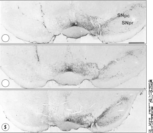
흑질에서 6-OHDA에 의해 직접 손상을 받은 환측의 TH양성 면역반응성은 건측에 비해 수술 3일부터 현저하 게 감소되어 거의 발현되지 않았으며, 모든 실험군에서 동 일한 소견을 보였다(Fig. 1). 선조체에서도 환측의 TH양성
Fig. 1. Light photomicrographs of tyrosine hydroxylase(TH) immun- ocytochemically stained coronal section of the midbrain of a rat 3(A), 7(B) and 21(C) days after unilateral injection of 6-hydroxydo- pamine(OHDA) into the substantia nigra . Note the substantia nigra of the lesion side(left column) is almost devoid of TH-positive neur- ons in whole experimental periods.
SNpc, substantia nigra pars comp- acta;SNpr, substantia nigra pars reticularis. Scale bar, 25μm.
A A A A
C C C
C
B B
B B
면역반응성은 시술 3일 이후 발현이 현저히 감소하여 7일 과 21일째까지 그 차이가 명확하였다(Fig. 2). 생리 식염수 를 주입한 대조군은 양측에서 TH 양성 세포를 모두 관찰 할 수 있었으며 면역반응성에 차이를 보이지 않았다.
2. 미교세포의 활성도 관찰(OX-42 및 osteopontin발현양상) 1) 흑질 부위
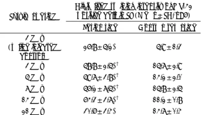
흑질에서 수술 3일 후부터 미교세포의 활성에 따라 환측 의 OX-42 항체의 발현이 급속히 증가하여 발현정도를 optical densitometry로 측정하여 건측과 비교한 결과, 환 측과 건측간의 발현은 생리 식염수를 투여한 대조군을 포 함하여 모두 유의하게 관찰되었다. 또한 환측의 발현강도 변동을 대조군을 기준으로 검정한 결과 3, 5, 7 및 14일에 유의하게 증가하였다. 21일의 유의성 결여는 대조군의 기 계적 손상에 의한 미교세포 활성때문으로 판단된다(Table 1). 면역조직화학 염색 소견상 건측의 미교세포는 나뭇가 지를 뻗은 듯한 길고 가느다란 주위돌기를 가진 방추모양 의 휴식형 미교세포의 모습으로 드물게 발현되는데 반해, 환측에서는 둥근(ameboid) 모양의 탐식성 상태로 강한 염
색을 보이며 활성화된 미교세포가 다수 관찰되었으며 주변 의 돌기 또한 두꺼워진 모양을 보이면서 그 수가 증가한 양상을 보였다. 3일군의 환측에서 주로 흑질 치밀부에서 보
Table 1. Time dependent changes of OX-42 immunoreactivity, measured and digitalized by image analyzer, in the rat subst- antia nigra(SN) examined after nigral 6-hydroxydopamine lesion Digitalized staining intensity of OX-42
positive cells in SN(Mean±SD, n=6) Survival period
Lesion side Contralateral side 3 day
(saline injected
control) 26.8±5.2* 5.9±1.3
3 day 58.8±2.5*† 15.7±2.9
5 day 59.7±3.8*† 14.2±2.0
7 day 56.2±7.5*† 15.8±2.5
14 day 64.3±3.7*† 11.2±0.8
21 day 30.6±3.4* 14.7±0.4
Asterisk*:denotes significant increase in the digitalized staining intensity of OX-42 in SN of lesion side, compared to that in contralateral side(p <0.05, paired t-test).
Cross†:indicates significant increment of absorbance in lesion side, compared to that of saline injected control group, by Bonferroni’s & Duncan’s post-hoc test, following ANOVA (p <0.05).
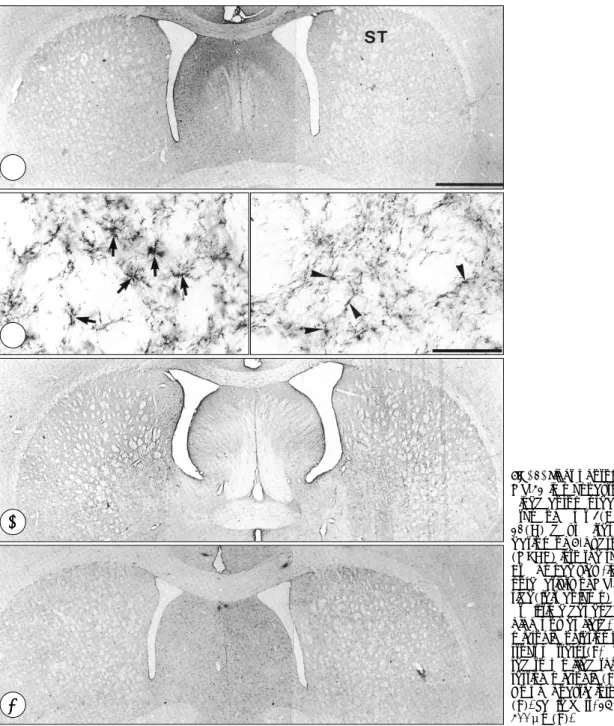
Fig. 2. Light photomicrographs of tyrosine hydroxylase(TH) immun- ocytochemically stained coronal section of the striatum of a rat 3(A), 7(B) and 21(C) days after unilateral injection of 6-hydroxyd- opamine(OHDA) into the substa- ntia nigra. TH immunoreactivity of the lesion side(left column) is ma- rkedly decreased in whole expe- rimental periods. ST, striatum. Scale bar, 40μm.
B B B B
C
C C
C
A
A A
A
였던 탐식형 미교세포는 7일군에서 흑질 망상부까지 증식, 이동한 소견을 보였다(Fig. 3). osteopontin은 in situ 보합 반응에서 정상적으로 발현되는 적핵(red nucleus)과 일부 흑질 망상부에서는 양측모두에서 발현되었으며 흑질 치밀 부를 비교해보니 환측에서만 발현되었고 건측에서는 발현 되지 않았다. 환측에서의 발현은 손상초기에 강하게 관찰 되었으며 21일군에서는 거의 발현되지 않았다(Fig. 4).
2) 선조체 부위
전반적인 활성강도가 상당히 감소되어 있었으며 optical densitometry로 측정하여 건측과 비교한 결과 수술 3일군
에서만 의의있게 상승하였다. 또한 대조군에서도 거의 반응 이 관찰되지 않아, 대조군을 기준으로 한 상승변동을 검정 해봐도 역시 3일군에서만 유의성이 관찰되어 흑질의 발현 양상(Table 1)과 비교해 볼 때 반응강도가 낮음은 물론 손 상초기에 국한된 일과성 변화를 보였다(Table 2). 면역조직 화학 염색상 미교세포는 흑질부에서 관찰되는 두꺼워진 돌 기를 가진 비후된 모양의 활성형만 관찰될 뿐, 크고 둥글며 강하게 발현되는 탐식형은 관찰되지 않아 활성의 강도가 낮 을 뿐 아니라 그 기능상에서도 차이가 있음을 확인할 수 있 었다(Fig. 5). 흑질과 달리 osteopontin은 발현되지 않았다.
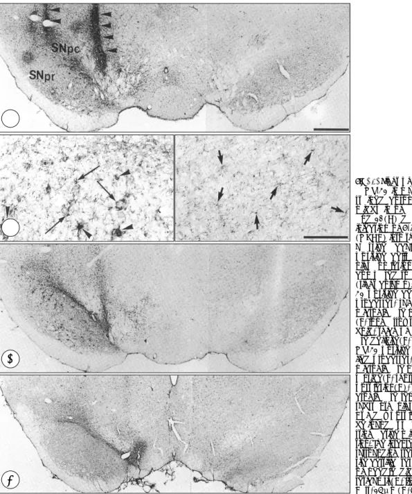
Fig. 3. Light photomicrographs of a OX-42 immunocytochemically stained coronal section of the midbrain of a rat 3(A, B), 7(C) and 21(D) days after unilateral injection of 6-hydroxydopamine (OHDA) into the substantia nigra.
Massive accumulation of OX-42 positive cells in the substantia nigra on lesion side(left column), compared to contralateral side (right column). Hypertrophic OX- 42 positive cells with thick stout processes, suggesting activated microglia are noted at lesion side (B, long arrows). Moreover, ame- boid, thick phagocytic microglia are definite(B, arrow heads). Thin OX-42 positive sells bearing rami- fied processes, suggesting resting microglia are noted at contralate- ral side(B, short arrows). At 3 day post lesion(A), the phagocytic mi- croglia are relatively confined to substantia nigra pars compacta.
At 7 day post lesion(C), they can be found at pars reticularis, sugge- sting active migration or prolifera- tion. The intense microglial reaction surrounding stereotactic operation site certificates that the end point of needle did not invade pars reticularis(A, linear arrows). Scale Bars, 25μm(A, C, D);400μm(B).
B B B B A A A A
C C C C
D D D
D
고 찰
파킨슨병은 대표적인 퇴행성 신경계 질환으로, 흑질 치밀
부(substantia nigra pars compacta)에서 도파민성 신경 세포의 점진적인 사멸에 기인하는 것으로 알려져 있다. 파 킨슨병이 있는 흑질부에서는 철 농도의 상승, glutathione 의 감소 및 미토콘드리아 기능의 감소 등이 확인되고 있어 흑질 치밀부에 가해지는 지속적인 산화성 자극(oxidative stress)으로 인한 생화학적 변성이 세포 사멸의 한 원인으 로 인정되고 있다
6)17).
파킨슨병에서 뇌간부에 존재하는 모든 도파민성 신경세 포가 균등하게 손상되는 것이 아니고 부위에 따라 손상의 정도가 다른 선택성이 있음이 알려지면서 신경세포와 교세 포의 상호작용에 대한 관심이 높아졌다. 즉 선조체로 투사 하는 도파민성 신경원이 변연계로 투사하는 신경원보다 손 상에 민감하며, 파킨슨병을 앓았던 환자의 부검연구상 도 파민성 신경세포가 분포하는 뇌간부위중 흑질 치밀부의 소 실이 가장 심하여 약 70%에 다다르고, 흑질 외측부(pars lateralis)에서는 약 30%, 배측 피개부(ventral tegmental area)에서는 약 50%의 소실이 관찰됨이 보고되고 있다
18). 신경세포 소실과정에 있어 선택성이 나타나는 원인을 Da- mier
10)는 흑질에서 도파민성 신경세포의 소실정도와, 그 주 변의 성상교세포의 밀도사이에 역상관 관계가 있음을 들어
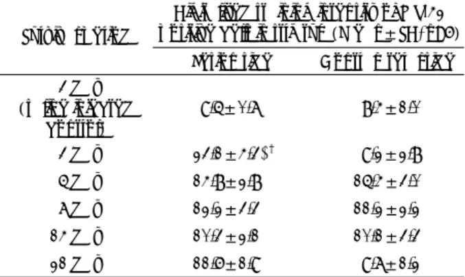
Table 2. Time dependent changes of OX-42 immunoreactivity, measured and digitalized by image analyzer, in the rat striatum examined after nigral 6-hydroxydopamine lesion
Digitalized staining intensity of OX-42 positive cells in striatum(Mean±SD, n=6) Survival period
Lesion side Contralateral side 3 day
(saline injected
control) 9.5±0.7 8.4±1.0
3 day 23.1±4.3*† 9.2±2.8
5 day 14.8±2.8 15.4±3.0
7 day 12.2±3.3 11.2±2.2
14 day 10.3±2.1 10.1±3.3
21 day 11.6±1.9 9.7±1.2
Asterisk*:denotes significant increase in the digitalized staining intensity of OX-42 in striatum of lesion side, compared to that in contralateral side(p <0.05, paired t-test).
Cross†:indicates significant increment of absorbance in lesion side, compared to that of saline injected control group, by Bonferroni’s & Duncan’s post-hoc test, following ANOVA (p <0.05).
Fig. 4. Light photomicrographs of osteopontin(OPN) in situ hybridi- zation in coronal section of the midbrain of a rat 3(A), 7(B) and 21(C) days after unilateral inj- ection of 6-hydroxydopamine (OHDA) into the substantia nigra.
At SNpc area, a number of OPN mRNA labelled microglia are seen at lesion side(left column). No la- belled cell exists at contralateral side(right column). The time course is rapid and OPN labelled cell can not be found at 21 day post lesion.
Red nucleus and some portion of SNpr also have OPN immunore- active cells. SNpc, substantia nigra pars compacta. SNpr, substantia nigra pars reticularis. RN, red nu- cleus. Scale bar, 25μm.
A A A A
B B B B
C
C
C
C
교세포가 신경세포의 선택적인 소실과정에 어떠한 역할을 할 것이라 제시하였다. 즉 성상교세포가 많은 부위에 존재 하는 신경세포는 적은 부위의 그것보다 질환의 이환에 덜 민감함을 밝혔다.
6-hydroxydopamine등의 신경독소를 이용하여 흑질내 도파민성 신경세포를 손상시켰을 경우 흑질에서 뿐만아니라 선조체에서도 성상교세포의 대표적인 표지물질인 GFAP (glial fibrillary acidic protein)의 발현이 증가됨은 여러 보 고들로 입증되고 있으며, 이러한 GFAP 발현증가는 손상이 일어난 후 일정기간동안 지속되는 양상을 보인다고 알려져
있다
8). 즉 성상교세포의 활성은 도파민성 신경세포의 손상 시기에 맞추어 손상 초기부터 발현이 상승되어 비교적 지 속적으로 발현되어 도파민성 신경세포의 손상에 잇따르는 반응형 신경교증 현상이다. 증가한 GFAP는 기존의 성상교 세포의 크기와 숫자가 증가해서 나타난 것이며 새로운 증 식때문은 아닌 것으로 생각된다. GFAP는 새로운 성상교세 포 돌기가 뻗어나가는데 필수적인 세포골격 단백질이므로 아마도 손상후 연접재생 과정에 도움을 줄 것으로 생각되 고 있다
7). 현재로서 GFAP의 증가는 손상에 반응하여 활 성화된 성상교세포의 지표로서의 의미를 가지며 그 정확한
Fig. 5. Light photomicrographs of OX-42 immunocytochemically st- ained coronal section of the stri- atum of a rat 3(A, B), 7(C) and 21(D) days after unilateral inj- ection of 6-hydroxydopamine (OHDA) into the substantia nigra.
At a glance view, increased immu- noreactivity of OX-42 of the lesion side(left column) is not evident at all time dependent groups. On high power field, the activated microglia of lesion side show more stout patterns(B, arrows) compa- red to ramified thin contralateral resting microglia(B, arrowheads).
No phagocytic form can be found (B). Scale bars, 25μm(A, C, D);
400μm(B).
A A A A
C C C C B B B B
D D D
D
생리학적 기능을 밝히기 위한 연구가 지속되고 있다.
활성화된 성상교세포는 매우 다양한 역할을 수행한다. 손 상된 신경세포로부터 차단된 목표점까지의 축삭 발아(spr- outing)가 원활히 이루어 질 수 있도록 glial cell derived neurotrophic factor(GDNF) 및 basic fibroblast growth factor 등을 분비한다. 반면에 중추신경계의 일부영역에서 는 반응성 신경교증에 의한 물리적 반흔이 재생과정의 장애 물이 되거나 탐식작용 및 neuronal protease와 cytokine 의 분비를 통해 염증반응 유발, nitric oxide(NO) 합성, iron 및 calcium 항상성의 저해, apoptosis 유도등의 작용 을 통해 도파민성 신경세포의 파괴에 관여하여 손상을 촉 진시키는 반대작용을 나타내기도 한다
3)20). 따라서 파킨슨 병에서 나타나는 반응성 신경교증은 단순한 손상후 이차적 변화가 아닌 질환 진행은 물론 재생, 복구과정에도 연관될 수 있는 매우 중요한 병태 생리 현상임을 알 수 있다.
전체 교세포의 약 20%를 차지하는 미교세포는 중추신경 계내 면역반응을 시작하고 유지하는데 중요한 역할을 하는 교세포이다
16). 미교세포는 여러 뇌손상, 즉 허혈, 축삭절단, kainic acid에 의한 경련유도 및 염증 등에 의하여 활성화 되는 것으로 알려져 있다
25). 활성화된 미교세포는 증식, 변 형과 같은 형태학적 변화를 보이면서 기능적으로는 항원 제공 세포(antigen presenting cell)로 작용하여 주 조직적 합성 복합체(major histocompatibility complex:MHC) 항원 유도, 다양한 면역매개 물질의 분비, 탐식작용 발현, 손상부위 쪽으로의 세포동원 및 세포독성 물질의 합성분비 와 같은 비교적 상동(常同)적인(stereotypic)반응을 보이 는 특징이 있다
13). McGeer등
24)에 의해 파킨슨병의 세포사 멸과정에 활성형 미교세포에 의한 염증반응이 관여함이 처 음 보고된 이래 미교세포에 의해 유도되는 interleukin-1 및 interleukin-6등이 파킨슨병 환자의 뇌척수액에서 증가 해 있음이 입증되었다. 이처럼 미교세포에서 분비되는 여 러 물질들은 신경재생에 도움을 주기도 하고, 역으로 신경 독소로도 작용하는 면을 보인다.
미교세포는 형태학적 변화와 기능적 활성간에 관련이 있 다. 정상상태에서는 일정하게 분산되어 배치되면서 가는 나 뭇가지를 뻗은 모양(ramified)을 보이면서 기능적으로 정 지 상태를 나타내는 휴식기 미교세포(resting microglia)상 태로 존재하다가, 뇌 조직의 손상이나 질환에 이환될 경우 두꺼운 나뭇가지 모양을 보이는 활성형(activated microglia) 으로 변화하여 여러 신경물질을 분비하면서 이동, 증식하 며, 최종적으로 탐식기능을 보일때는 동그랗고(ameboid) 뚱뚱한 전형적인 탐식세포 모양을 보이는 탐식형 미교세포 (phagocytic microglia)로 변형된다. 신경손상에 대해 미
교세포의 반응이 단순한 보수에 국한되는지 신경세포의 재 생에 관여하는지를 규명하기 위한 안면신경 절단모델이 잘 알려져 있다. 즉 안면신경을 중추신경계인 안면신경핵(facial nucleus)과 먼 경상유돌기 공(stylomastoid foramen) 근 처에서 절단함으로써 혈관에서 기인한 탐식세포나 단핵세 포가 중추신경계로 침윤되는 것을 사전에 봉쇄함으로써 중 추신경계내에 이미 존재하고 있는 실질적인 미교세포의 반 응만을 관찰함이 목적이다. 이에 따르면 안면신경을 절단 한지 3~4일 이후에 신경핵내에서는 미교세포의 활성이 관 찰되며
18), 이들 미교세포에서는 탐식작용이 관찰되지 않는 것으로 보아 탐식을 통한 손상부 퇴행보다는 신경영양인자 (neurotrophic factor) 분비를 통한 복구와 관련이 있음이 알려졌다. 그러나 이와 달리 안면신경에 유해한 독소인 ricin을 주사할 경우에는 ricin이 안면신경 핵으로 역행성 (retrograde) 이동하여 단백합성을 저해함으로써 신경핵 의 괴사를 유도하고 이때 활성화되는 미교세포는 탐식형 미교세포로 변화하여 탐식작용을 나타낸다. 신경독소를 뇌 에 주사할 경우 신경전도 경로를 따라 주 조직적합성 복합 체 1군 또는 2군 양성인 미교세포가 침윤되면서 손상된 신 경세포의 탐식이 이루어진다. 이처럼 미교세포가 탐식세포 로 변형되는 것은 신경에 가해진 손상의 정도와 이로 인한 신경세포의 생존여부와 관련이 있는 것으로 생각된다
26).
본 실험에서는 미교세포의 활성강도의 차이를 규명하기
위해 미교세포에 공통적으로 존재하여 가장 민감도가 높다
고 알려진 CR3 complement 수용체 항원에 대한 OX-42
항체와 활성형 이상상태의 미교세포와 반응할 것으로 보고
된 바 있는 osteopontin 항체
22)를 사용하였다. osteopontin
(OPN)은 처음 골격계에 존재하는 단백질로 알려졌으나
다양한 조직에서 존재하며 그 기능에 대하여는 정확하게
알려져 있지 않으나 창상치유과정에 관여하리라 여겨지고
있다
22). 최근 국소뇌허혈후 활성형 미교세포에서 OPN이
발현되며, OPN receptor인 αvβ3의 발현이 증가되어
OPN의 chemotactic role의 가능성이 제시되었으며, 또한
OPN과 이들 수용체간의 결합이 세포내 칼슘이온의 분비
를 촉진시켜 성상교세포의 GFAP filament network의 재
조합을 유발할 수 있을 것으로 보고되어 OPN이 중추신경
계통 손상에 잇따르는 창상치유 과정에도 관여할 것이라
생각되고 있다
15). 뇌허혈 모델에서 탐식형 미교세포는 2주
까지 관찰되나 OPN 발현은 2~3일에 절정에 달한후 5일
이후 에서는 관찰되지 않았다. 따라서 OPN이 미교세포 활
성과정의 조절인자로 작용할 것으로 생각된다. 심근괴사후
OPN이 괴사조직의 탐식을 촉진시킨다는 보고가 있으나, 중
추신경 계통내 OPN의 기능에 대한 연구는 미흡하며, OPN
발현을 보이는 미교세포가 반응형 미교세포중 일부에서만 관찰된다는 보고
15)22)는 있으나 특히 신경퇴행성 질환에서 OPN 발현을 보이는 반응형 미교세포의 특성에 대한 연구 는 아직 없다.
본 실험결과 흑질에서는 활성형 미교세포의 발현이 증가 하였으나 선조체에서는 tyrosine hydroxylase의 발현이 떨 어져 있음에도 불구하고 미교세포의 발현이 미약함은 흥미 로운 사실이다. 저자들은 도파민성 신경세포의 사멸을 유도 하기 위해 6-hydroxydopamine을 뇌 정위적으로 직접 흑 질에 주입하였다. 또 다른 도파민성 신경세포 독소인 MPTP (N-methyl-4-phenyl-1,2,3,6-tetrahydropyridine)
11)는 뇌 정위적 수술로 도파민성 신경세포에 도달시킬 필요 없이 복강내 주입만 하여도 뇌 전반에 걸친 도파민성 신경 세포를 직접 사멸시키므로 파킨슨병 동물모델을 만들 때 흔히 사용되는 약물이다. MPTP를 이용한 생쥐 파킨슨병 모델에서는 흑질은 물론 선조체에서도 강력한 미교세포 반 응이 관찰된다
9).
6-OHDA를 이용할 경우 흑질의 도파민성 세포는 직접적 으로 사멸되지만 선조체는 이로인한 전향성 변성(antegr- ade degeneration)으로 인한 점진적 결손이 오는 것이므 로 파킨슨병과 같은 신경퇴행성 질환에서의 미교세포의 활 성에는 신경세포에 가해지는 손상의 신속성과 치명도 및 이로 인한 세포 사멸의 정도가 중요한 요인이 될 것으로 판단되며 더디거나 미약한 손상에 대해서는 활성형 미교세 포가 탐식형까지는 전환되지 못하도록 제어하는 세포내 신 호경로가 존재하거나 또는 이러한 종류의 손상이 가져오는 각각의 세포외 미세환경 변화가 영향을 미칠 것이라는 가 설을 제시할 수 있다. 아울러 흑질에서는 osteopontin이 건 측에서는 전혀 발현되지 않지만, 환측에서는 발현되고 있는 것으로 보아, osteopontin이 활성형 단계 이상의 미교세포 를 감지하는데 유용함을 알 수 있었다. 면역조직화학 염색 상 흑질에서는 활성형과 탐식형이 혼재하고 있는데 반해 선조체에서는 짧은 시간동안 활성형 단계까지만 발현되는 것으로 보아 osteopontin은 활성형 미교세포 가운데 존재 하는 탐식형 미교세포를 감지하는 지표라 생각된다.
결 론
이상의 결과로 파킨슨병의 병태생리에 신경교세포가 중 요한 역할을 할 것으로 생각되며 동물모델을 선정할 때 도 파민성 신경세포의 사멸에 잇따르는 신경교세포 반응의 강 도 또한 중요한 요인으로 고려되어야 할 것이다. 특히 태아 중뇌세포 이식 연구시 성상교세포나 미교세포와 같은 신경
교세포 반응의 강도가 이식편의 생존에 영향을 미칠 수 있 음을 감안하여야 할 것이다.
•
논문접수일:2001년 1월 27일•
심사완료일:2001년 5월 8일•
책임저자:성 재 훈442-723 경기 수원시 팔달구 지동 93-6 가톨릭대학교 의과대학 성빈센트병원 신경외과학교실 전화:031) 249-7190, 전송:031) 245-5208 E-mail:[email protected]
References
1) Akiyama H, McGeer PL:Microglial response to 6-hydoxy- dopamine-induced substantia nigra lesions. Brain Res 489: 247-253, 1989
2) Banati RB, Graeber MB:Surveillance, intervention and cy- totoxicity:is there a protective role of microglia? Dev Neurosci 16:114-127, 1994
3) Benveniste EN:Inflammatory cytokines within the central nervous system:sources, function, and mechanism of action.
Am J Physiol 263(Cell Physiol 32):C1-C16, 1992 4) Butler WT:The nature and significance of osteopontin. Con-
nect Tissue Res 23:123-136, 1989
5) Carman LS, Gage FH, Shults CW:Partial lesion of the sub- stantia nigra:lesion between extent of lesion and rotational behavior. Brain Res 553:275-283, 1991
6) Cassarino DS, Bennett JPJ:An elevation of the role of mitoch- ondria in neurodegenerative disease:mitochondrial mutations and oxidative pathology, protective nuclear responses, and cell death in neurodegeneration. Brain Res Rev 29:1-25, 1999 7) Chen WJ, Liem RK:Reexpression of glial fibrillary acidic
protein rescues the ability of astrocytoma cells to form proce- sses in response to neurons. J Cell Biol 127:813-823, 1994 8) Cheng HW, Jiang T, Brown SA, Pasinetti GM, Finch CE,
McNeill TH:Response of striatal astrocyte to neuronal dea- fferentation:An immunohistochemical and ultrastructural study. Neuroscience 62:425-439, 1994
9) Czlonkowska A, Kohutnicka M, Kurkowska-Jastrzebska I, Czlonkowski A:Microglial reaction in MPTP(1-methyl-4- phenyl-1,2,3,6-tetrahydropyridine) induced Parkinson’s disease mice model. Neurodegeneration 5:137-143, 1996
10) Damier P, Hirsch EC, Zhang P, Agid Y, Jabid-Agid F:Gluta- thione peroxidase glial cells and Parkinson’s disease. Neuro- science 52:1-6, 1993
11) Davis GC, Williams AC, Markey SP, Ebert MH, Caine ED, Reichert CM, et al:Chronic parkinsonism secondary to in- travenous injection of meperidine analogues. Psychiat Res 1:249-254, 1979
12) Denhardt DT, Guo X:Osteopontin:A protein with diverse functions. FASEB J 7:1475-1482, 1993
13) Dickson DW, Mattiace LA, Kure K, Hutchins K, Lyman WD,
Brosnan CF:Microglia in human disease, with an emphasis on the acquired immune deficiency syndrome. Lab Invest 64: 135-156, 1991
14) Eddleston M, Mucke L:Molecular profiles of reactive astro- cytes - implications for their role in neurologic disease. Neu- roscience 54:15-36, 1993
15) Ellison JA, Velier JJ, Spera P, Jonak ZL, Wang X, Barone F, et al:Osteopontin and its integrin receptor v3 are upregulated during formation of the glial scar after focal stroke. Stroke 29:1698-1707, 1998
16) Gehrmann J, Matsumoto Y, Kreutzberg GW:Microglia: intrinsic immunoeffector cell of the brain. Brain Res Rev 20: 269-287, 1995
17) Gotz ME, Kunig G, Riederer P, Youdim MB:Oxidative stress:free radical production in neural degeneration. Phar- macol Therap 63:37-122, 1994
18) Graeber MB, Streit WJ, Kreutzberg GW:Axotomy of the rat facial nerve leads to increased CR3 complement receptor expression by activated microglial cells. J Neurosci Res 21: 18-24, 1988
19) Hirsch EC, Graybiel AM, Agid Y:Melanized dopaminergic neurons are differentially susceptible to degeneration in Par- kinson’s disease. Nature 334:345-348, 1988
20) Hirsch EC, Hunot S, Damier P, Faucheux B:Glial cells and inflammation in Parkinson’s disease:A role in neurodegene- ration? Ann Neurol 44(3 Suppl 1):115S-120S, 1998 21) Kato H, Walz W:The initiation of the microglial response.
Brain Pathol 10:137-143, 2000
22) Lee MY, Shin SL, Choi YS, Kim EJ, Cha JH, Chun MY, et al:Transient upregulation of osteopontin mRNA in hippo- campus and striatum following global forebrain ischemia in rats. Neuroscience Lett 271:81-84, 1999
23) McGeer PL, Itagaki S, Boyes BE, McGeer EG:Reactive microglia are positive for HLA-DR in the substantia nigra of Parkinson’s disease and Alzheimer disease brains. Neurology 38:1285-1291, 1988
24) McGeer PL, McGeer EG:The inflammatory response system of brain:implications for therapy of Alzheimer and other neurodegenerative diseases. Brain Res Rev 21:195-218, 1995 25) Mitchell J, Sundstrom LE, Wheal VH:Microglial and astro- cytic cell responses in the rat hippocampus after an intracere- broventricular kainic acid injection. Expl Neurol 121:224- 230, 1993
26) Nakajima K, Kohsaka S:Functional roles of microglia in the brain. Neurosci Res 17:187-203, 1993
27) Paxinos G, Watson C:The rat brain in stereotaxic coordinates.
4th ed. San Diego:Academic Press, 1998
28) Perese DA, Elman J, Viola J, Ewing SE, Bankiewicz KS:A 6-hydroxydopamine induced selective parkinsonian rat model.
Brain Res 494:285-293, 1989
29) Sheng JG, Shirabe S, Nishiyama N, Schwartz JP:Alteration in striatal glial fibrillary acidic protein expression in response to 6-hydroxydopamine-induced denervation. Exp Brain Res 95:450-456, 1993
30) Ungerstedt U:Stereotaxic mapping of the monoamine path- ways in the rat brain. Acta Physiol Suppl 367:1-48, 1971