Prevalence of Human Papillomavirus by DNA Chip Test in Women
Jae-Woo Kim, Yun-Tae Kim1, Dae-Sik Kim2 and Seok-Cheol Choi*
Department of Clinical Laboratory Science, College of Health Sciences, Catholic University of Pusan, Busan 609-757, Korea
1Seoul Medical Science Institute (SMSI), Seoul 140-809, Korea
2Department of Clinical Laboratory Science, Dongnam Health College, Suwon 440-714, Korea Received November 5, 2008 /Accepted December 24, 2008
We determined the prevalence of human papillomavirus (HPV) by DNA chip test in 549 women and cytologic diagnosis. 237 of 549 women (43.17%) subjected with HPV DNA Chip examination were found positive for HPV. 210 (88.60%, High group) were infected with high-risk HPV types. 17 (7.17%, Low group) were infected with low-risk HPV types (6, 11, 40, 44, 70) and 17 (7.17%, Mixed group) were infected with mixed types. According to age, in their twenties, thirties, forties, fifties and over sixties, the prevalence of infection with high-risk HPV types were 1.26% (3/237), 15.61% (37/237), 31.65% (75/237), 23.21% (55/237), and 13.92% (33/237), respectively. In the Low and Mixed group, percentages of infection with HPV were significantly lower than that of the High group. On the com- parison of cytologic diagnosis (224 women) by Pap smear and DNA chip positive (237 women) for HPV, 132 out of 194 cases in the High group (68.04%) suffered cervical lesions with ASCUS (atypical squamous cells of undetermined significance, 7.22%), LSIL (low grade squamous intraepithelial lesion, 15.98%), HSIL (high grade SIL, 23.20%) and ICC (invasive cervical cancer, 21.65%). The Low group (14/224 women) showed 1 case of ASCUS and 6 cases of LSIL, whereas the Mixed group (4/224 women) had only 2 cases of ASCUS. According to the HPV subtypes, the high-risk types 16 and 18 induced 26 and 7 cases of ICC, respectively, whereas other HPV subtypes induced lower or no ICC incidence. In conclusion, the present data imply that the prevalence of HPV was 43.17%, high-risk HPV type 16 is a major factor, which causes precancerous and/or cervical cancer in woman and that HPV DNA chip is an accurate and useful tool for detecting HPV.
Key words : Human paillomavirus, cervical cancer, DNA chip, Pap smear
*Corresponding author
*Tel:+82-51-510-0564, 0569, Fax:+82-51-510-0568
*E-mail : [email protected]
Introduction
The development of the Pap smear by Papanicolaou has contributed to reduce morbidity and mortality in patients with cervical cancer. It is unquestionable that the Pap smear is the best example of a successful cervical cancer screening [12,13]. However, the Pap smear test has a limitation with false negative although it is an excellent screening test.
Sherman et al. [15] showed that ASCUS (atypical squamous cells of undetermined significance) cytology reports are not reproducible, even by expert cytopathologists. Several stud- ies demonstrated that even though the majority of ASCUS findings by Pap smear represents a benign reactive process, 5 to 10% of the women with ASCUS have potential for HSIL (high grade squamous intraepithelial lesion) [4,9,17]. Human papillomavirus, which is sexually transmitted, is a strong agent of cervical cancer [18]. HPV can infect basal epithelial
cells of the skin or inner lining of tissues and are classified as cutaneous type and mucosal type. Cutaneous types are epidermitrophic and target the skin of the hands and feet, while mucosal types infect the lining of the mouth, throat, respiratory tract, or genital epithelium. Methods for detect- ing HPV include PCR, in situ hybridization, Hybrid-Capture II assay, and HPV DNA chip test. Although HAP DNA chip test has merits such as identification for 22 to 24 genotypes of HPV, it has been less used compared with Hybrid- Capture II assay. However, we do not have enough data for studies of HPV DNA chip in Korea.
This study was carried out to investigate the prevalence of human papillomavirus (HPV) by DNA chip test in 549 women and cytologic diagnosis.
Materials and Methods Patients
In 2006, 549 women were tested for human papil- lomavirus (HPV) by DNA chip. 237 women having tested positive on the HPV DNA chip test were subjected to this
study. A cytologic test was carried out for 224 out of 237 women. Finally, the data of 224 patients were analyzed in the present study. Cytologic diagnosis was determined by Pap smear.
Cytologic diagnosis
Specimens were collected by scraping the endocervix and ectocervix with a brush.
The specimens were smeared on a slide glass and imme- diately immersed in 95% alcohol for test. The Pap stain pro- cedure was the following : the specimen on the slide glass was fixed in 95% alcohol for 30 min and washed in tap water. The Nucleus in the specimen was stained with Harris' hematoxylin (Sigma, America), decolorized with 1% HCL-al- cohol and neutralized in tap water for 10 min. The Cytoplasm was stained with orange G (Young Dong Diagnostics., Korea) for 2 min and with EA 50 (Young dong Co., Korea) for 2 min. The specimen was hydrated in abso- lute alcohol and mounted with Synthetic Mountant (Shandon, England).
Pathologists interpreted the results of the specimen and classified them with TBS (the Bethesda system).
HPV DNA chip test
A specimen was taken by cytobrush of HPV DNA chip sampler (BioMed Lab., Korea) from the endocervix and ecto- cervix and inserted into a transfer medium. The HPV DNA chip test was performed as follows.
DNA isolation
The specimen was isolated from cytobrush by strong vor- texing and inserted into a microcentrifuge tube of 1.5 ml.
It was centrifuged with 8,800 rpm for 10 min and the super- natant was removed. The pellet with 1 ml of 1× washing buffer (10× washing buffer + DW) was mixed in vortex, cen- trifuged with 8,800 rpm for 5 min and the supernatant was removed. 200 μl of DNA extraction buffer was mixed, in- cubated into water bath of 50oC for over 3 hrs and boiled at 90-100oC for 20 min. It was centrifuged with 14,000 rpm for 10 min and 100-150 μl of supernatant was prepared for PCR analysis.
Polymerase chain reaction (PCR) analysis
PCR procedures consisted of three steps. The first step of PCR was as follows; 6.5 μl of PCR I, 0.3 μl of Taq polymer- ase and 12.7 μl of DW were infused into PCR tube and 5 μl of DNA extractive was added into it. This mixed DNA
was amplified as follows; initial denaturation at 50oC for 3 min followed by 1 cycle of 95oC for 15 min. And, procedures such as 95oC for 30 sec, 55oC for 30 sec, 35 cycles of 72oC for 30 sec, and a final cycle of 72oC for 3 min were employed.
The second PCR procedures were the following; 6.5 μl of PCR II, 0.3 μl of Taq polymerase and 16.2 μl of DW were infused into PCR tube and 2 μl of the first PCR product was added into it. One cycle of 95oC for 5 min and proce- dures such as 95oC for 30 sec, 55oC for 40 sec, 20 cycles of 72oC for 30 sec, and a final cycle of 72oC for 3 min were performed.
Hybridization
10 μl of the 2nd product was denaturated at 95oC for 5 min. Frozen hybridization buffer was incubated at 43oC 10 min until it was utilized. Denatured 10 μl of the 2nd product combined with 30 μl of hybridization buffer were distributed into a well of HPV DNA Chip slide under appropriate humidity. After hybridization at 43oC for 2 to 3 hr, the hy- bridization seal was removed from the slide. It was re- spectively washed two times with washing buffer I [2× SSC (sodium citrate, sodium chloride), 0.1% SDS (sodium do- decyl sulfate)] for 5 min, two times with buffer II [0.2× SSC]
for 5 min, and one time with buffer III [0.1× SSC]. The HPV DNA chip includes 24 type specific probes; 16 types are high-risk group (HPV-16, HPV-18, HPV-31, HPV-33, HPV-35, HPV-39, HPV-45, HPV-51, HPV-52, HPV-53, HPV-54, HPV-56, HPV-58, HPV-59, HPV-66, HPV-68) and 8 types are low-risk group (HPV-6, HPV-11, HPV-34, HPV-40, HPV-42, HPV-43, HPV-44, HPV-70). Positive and negative control were utilized in every test.
Interpretation
The HPV type was determined by scanning (ScanArray Gx, PerkinElmer, America) after air drying at room temperature.
Results and Discussion Cytologic diagnosis
Table 1 shows the cytologic diagnosis according to age in 224 HPV positive women. 70 women (35.27%, Fig. 1) were normal, ASCUS was detected in 19 out of 224 women (8.48%) consisting of 1 case in the age group 20 years (5.26%), 4 cases in the age group 30 years (21.05%), 7 cases in the age group 40 years (36.84%), 5 cases in the age group 50
Table 1. Finding of cytologic diagnosis according to age group Cytologic diagnosis
(case, %)
Age (year)
20-29 (n=4) 30-39 (n=40) 40-49 (n=84) 50-59 (n=60) >60 (n=36) Total (n=224) Normal (79)
ASCUS (19) LSIL (39) HSIL (45) Invasive Ca (42)
1(1.26%) 1(5.26%)
0 2(4.44%)
0
12(15.19%) 4(21.05%) 4(10.26%) 15(33.33%) 5(11.90%)
33(41.77%) 7(36.84%) 19(48.72%) 14(31.11%) 11(26.19%)
22(27.85%) 5(12.82%) 9(23.08%) 9(20.00%) 15(35.71%)
11(13.92%) 2(10.53%) 7(17.95%) 5(11.11%) 11(26.19%)
79(35.27%) 19(8.48%) 39(17.41%) 45(20.09%) 42(18.75%) Abbreviations: ASCUS, atypical squamous cells of undetermined significance; LSIL, low grade squamous intraepithelial lesion; HSIL, high grade squamous intraepithelial lesion; Ca, cervical carcinoma.
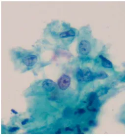
Fig. 1. Normal superficial (eosinophilic) and intermediate (basophilic) cells by cytologic finding with Pap stain.
Abundant cytoplasm has a polygonal form. It is thin and transparent. Eosinophilic cytoplasm may possess thin and/or folded cytoplasm. The round nucleus of superficial cell is smaller than a red blood cell (RBC) and karyopyknotic. The nucleus of an intermediate cell, which is larger than a RBC, is round to oval and vesicular with finely divided, evenly distributed chromatin.
years (26.32%), and 2 cases in the age group 60 years and older (10.53%) (Fig. 2). LSIL was detected in 39 out of 224 women (17.41%) consisting of 4 cases in the age group 30 years (10.26%), 19 cases in the age group 40 years (48.72%), 9 cases in the age group 50 years (23.08%), and 7 cases in the age group 60 years and older (17.95%) (Fig. 3). HSIL was found in 45 out of 224 women (20.09%) with 2 cases in the age group 20 years (4.44%), 15 cases in the age group 30 years (33.33%), 14 cases in the age group 40 years (31.11%), 9 cases in the age group 50 years (20.00%), and 5 cases in the age group 60 years and older (11.11%) (Fig.
4). Invasive cervical carcinoma (ICC) was detected in 42 out of 224 women (17.72%) with 5 cases in the age group 30
Fig. 2. Finding of ASCUS (Atypical squamous cells of un- determined significance). The nuclei are 2 to 3 times the size of that of intermediate cells. These cells have slight- ly increased nuclear/cytoplasmic ratio. It shows a smooth membrane or very limited irregularity with mild hyperchromatin and evenly distributed chromatin without granularity. The cytoplasm is usually mature superficial/intermediate type.
years (11.90%), 11 cases in the age group 40 years (26.19%), 15 cases in the age group 50 (35.71%), and 11 cases in the age group 60 years and older (26.19%) (Fig. 5). Overall, the prevalence of ASCUS and LSIL was highest in the age group 40 years, and that of HSIL was highest in age group 30 years with vigorous sexual activity. The patients in the age group 50 years suffered invasive cervical carcinoma because they may have a menopausal disorder or they may be going through menopause.
Our findings were different from previous studies.
Hong et al. [5] reported lower prevalence of abnormal findings than our results. They investigated subjects with and without HPV infection. However, Hwang [6] showed similar results in his study on women with HPV infection.
Fig. 3. Finding of LSIL (low-grade squamous intraepithelial le- sion). The nuclei are at least 3 times the size of that of intermediate cells. They have increased nuclear/cyto- plasmic ratio, binucleation or multinucleation. Nuclear membranes are slightly irregular, and show hyper- chromasia with uniform chromatin distribution, chroma- tin degeneration and smudging. The cytoplasm has ir- regular perinuclear clearing (halo).
Fig. 4. Finding of HSIL(high-grade squamous intraepithelial le- sion). nucleoli are enlarged in range of LSIL but dimin- ished amount of cytoplasm and marked increase in nu- clear/cytoplasmic ratio. Nuclear membranes are irregular. They have hyperchromasia, fine to coarse, and evenly distributed chromatin. Nucleoli are generally ab- sent and the cytoplasm is lacy and delicate.
HPV DNA test
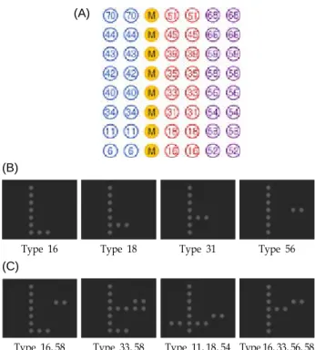
If patients were infected with HPV, a fluorescent color can be seen (Fig. 6). 237 of 549 women (43.17%) subjected to the HPV DNA Chip examination were found positive for HPV.
Fig. 5. Finding of invasive cervical carcinoma. Nuclei show a marked variation of size/shape, numerous dense, opa- que forms chromatin, which is coarsely granular and ir- regularly distributed. Parachromatin is clear and macro- nucleoli are present. The cytoplasm is frequently dense and orangeophilic.
(A)
(B)
Type 16 Type 18 Type 31 Type 56
(C)
Type 16, 58 Type 33, 58 Type 11, 18, 54 Type 16, 33, 56, 58 Fig. 6. (A) Interpretation of HPV DNA chip test. M-markers
are control. Left circles are low-risk HPV subtypes. Right circles indicate high-risk HPV subtypes. (B) Single in- fection cases of HPV subtypes. (C) Multiple infection cases of HPV subtypes.
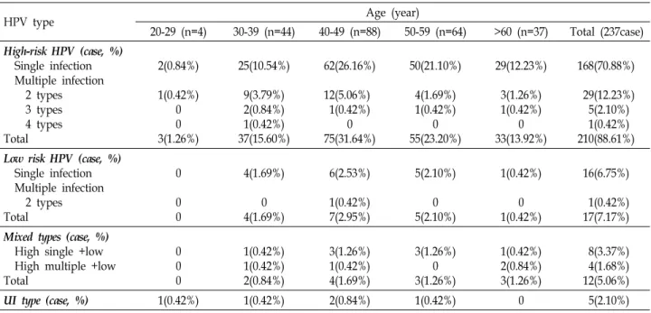
Incidences of HPV infection were summarized in Table 2. Among 237 cases, 203 cases (85.65%) were high-risk HPV infections and were comprised of single infections (with one subtype), infections with two, three, and four subtypes. In
Table 2. Incidences of HPV infection by DNA chip according to age group
HPV type Age (year)
20-29 (n=4) 30-39 (n=44) 40-49 (n=88) 50-59 (n=64) >60 (n=37) Total (237case) High-risk HPV (case, %)
Single infection Multiple infection 2 types 3 types 4 types Total
2(0.84%) 1(0.42%)
0 0 3(1.26%)
25(10.54%) 9(3.79%) 2(0.84%) 1(0.42%) 37(15.60%)
62(26.16%) 12(5.06%) 1(0.42%)
0 75(31.64%)
50(21.10%) 4(1.69%) 1(0.42%)
0 55(23.20%)
29(12.23%) 3(1.26%) 1(0.42%)
0 33(13.92%)
168(70.88%) 29(12.23%) 5(2.10%) 1(0.42%) 210(88.61%) Low risk HPV (case, %)
Single infection Multiple infection 2 types Total
0 0 0
4(1.69%) 0 4(1.69%)
6(2.53%) 1(0.42%) 7(2.95%)
5(2.10%) 0 5(2.10%)
1(0.42%) 0 1(0.42%)
16(6.75%) 1(0.42%) 17(7.17%) Mixed types (case, %)
High single +low High multiple +low Total
0 0 0
1(0.42%) 1(0.42%) 2(0.84%)
3(1.26%) 1(0.42%) 4(1.69%)
3(1.26%) 0 3(1.26%)
1(0.42%) 2(0.84%) 3(1.26%)
8(3.37%) 4(1.68%) 12(5.06%)
UI type (case, %) 1(0.42%) 1(0.42%) 2(0.84%) 1(0.42%) 0 5(2.10%)
Abbreviation: UI, unidentified.
Table 3. Incidence of HPV infection according to cytologic diagnosis
HPV type (224 cases) Cytologic diagnosis
normal ASCUS LSIL HSIL Invasive
High-risk HPV (case, %) Single
2 types 3 types 4 types Total (n=194)
51 10 1 0 62(31.96%)
11 2 1 0 14(7.22%)
27 3 1 0 31(15.98%)
36 8 1 0 45(23.20%)
36 4 1 1 42(21.65%) Low-risk HPV (case, %)
Single 2 types Total (n=14)
7 0 7(50.0%)
1 0 1(7.14%)
5 1 6(42.86%)
0 0 0
0 0 0 Mixed types (case, %)
High single+low High multiple+low Total (n=12)
5 3 8(66.67%)
2 0 2(16.67%)
1 1 2(16.67%)
0 0 0
0 0 0 UI tupes (case, %)
Total (n=4)
2 2(50%)
2 2(50%)
0 0
0 0
0 0 single and two-types infections, patients in their forties had
the highest incidence, in three- and four-type infections pa- tients in their thirties had the highest prevalence. These data suggest that age group 30 and 40 years with energetic sexual activity may be exposed to more risk of HPV infection. On the other hand, 17 cases (7.17%) were identified as low-risk HPV infections. The highest incidence was in patients in their forties. Mixed types (with single infections high-risk and low-risk HPV or multiple infections high-risk and low-risk HPV) and unidentified type infections were re-
spectively found in 12 (5.06%) and 5 (2.10%) cases. In all cases, age group 40 years had the highest prevalence of HPV infections. These results were similar to previous studies.
Muñoz et al. [11] demonstrated the highest prevalence of both groups in women in their 40s with and without squ- amous-cell cervical cancer. Hong et al. [5] explained that pa- tients aged between 35 to 54 years were highly infected with HPV. Furthermore, Kim et al. [8] found that women in the 40 years age group had the highest HPV detection and HSIL.
However, Lazacano-Ponce et al., [10] whom investigated on
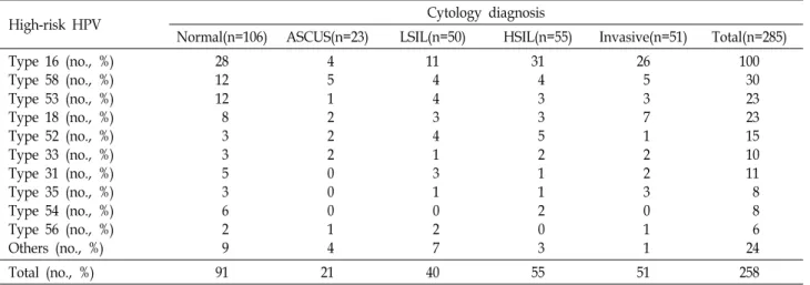
Table 4. Comparison and analysis between cytology diagnosis and high-risk HPV subtypes
High-risk HPV Cytology diagnosis
Normal(n=106) ASCUS(n=23) LSIL(n=50) HSIL(n=55) Invasive(n=51) Total(n=285) Type 16 (no., %)
Type 58 (no., %) Type 53 (no., %) Type 18 (no., %) Type 52 (no., %) Type 33 (no., %) Type 31 (no., %) Type 35 (no., %) Type 54 (no., %) Type 56 (no., %) Others (no., %)
28 12 12 8 3 3 5 3 6 2 9
4 5 1 2 2 2 0 0 0 1 4
11 4 4 3 4 1 3 1 0 2 7
31 4 3 3 5 2 1 1 2 0 3
26 5 3 7 1 2 2 3 0 1 1
100 30 23 23 15 10 11 8 8 6 24
Total (no., %) 91 21 40 55 51 258
Abbreviation: Others, HPV subtypes 39, 45, 51, 59, 66 and 68.
Table 5. Comparison and analysis between cytology diagnosis and low-risk HPV subtypes
Low-risk HPV type Cytology diagnosis
Normal(n=106) ASCUS(n=23) LSIL(n=50) HSIL(n=55) Invasive(n=51) Total(n=285) Type 70 (no., %)
Type 6 (no., %) Type 11 (no., %) Others (no., %)
7 5 1 2
1 1 0 0
5 0 5 0
0 0 0 0
0 0 0 0
13 6 6 2
Total (n=27) 15 2 10 0 0 27
Abbreviation: Others, HPV subtypes 40 and 44.
the epidemiology of HPV infection among Mexican women with normal cervical cytology, reported a rate of HPV in- fection of 16.7% in the age group under 25 years, which de- clined to 3.7% in the age group 35-44 years, then increased progressively to 23% among women 65 years and older.
Also, Stoler [16] described that age group 20 years in American women had the highest prevalence of HPV infection.
These discrepancies among several studies may be attrib- uted to social or sexual cultural backgrounds.
Comparison of cytologic diagnosis with prevalence of HPV infection
Relationships between high- and low-risk HPV subtypes are described in Tables 3, 4, and 5. When comparing the cytologic diagnosis (224 women) by Pap smear and DNA chip positive (237 women) for HPV, 62 out of 194 women with high-risk HPVs (31.96%), 7 of 14 women with low-risk HPVs (50.0%), 8 of 12 women infected with mixed types, and 2 of 4 women with unidentified HPV subtypes (50.0%) had a normal cytology. In high-risk HPV-infected cases (194
of 224 women), 132 out of 194 women (68.04%) suffered cer- vical lesions with ASCUS (14 patients, 7.22%), LSIL (31 pa- tients, 15.98%), HSIL (45 patients, 23.20%) and ICC (42 pa- tients, 21.65%). In low-risk HPV-infected cases (14 of 224 women), 7 women showed 1 case of ASCUS and 6 cases of LSIL, whereas 12 women infected with mixed subtypes (12/224 women) had 2 cases of ASCUS and 2 cases of LSIL, respectively. 2 of 4 women infected with unidentified HPV subtypes had ASCUS (Table 3).
In high-risk HPV, patients with ASCUS showed the high- est prevalence of subtype 58 infection (5/23 cases, 21.74%), whereas those with LSIL (11/50 cases, 22.00%), HSIL (31/55 cases, 56.36%), or ICC (26/51 cases, 50.98%) had more in- fections of subtype 16 than other subtypes, which were 58, 53, 18, 52, 33, 31, 35, 54, 56, 39, 45, 51, 59, 66, and 68, re- spectively (Table 4). On the other hand, low-risk HPV cases with LHIL showed the highest incidence of subtype 70 (10%) (Table 5).
The results of the present study agree with previous stud- ies, in the fact that high-risk HPV subtype 16 causes cervical cancer. An [1], Hong et al. [5], Hwang [6], Kahng and Lee
[7], Kim et al. [8] and Muñoz et al. [10] demonstrated a con- sistent finding that women with various cervical lesions in- cluding invasive squamous cancer had the highest preva- lence of HPV subtype 16 infection. Rozendaal et al. [14] re- vealed that women with normal cervical smears containing high-risk HPV genotypes were 116 times more at risk of de- veloping CIN (cervical intraepithelial neoplasia) III com- pared with women without high-risk HPV. Although the prevalence of HPV DNA associated with invasive cervical carcinoma differs among countries, the most common HPV types in invasive cervical cancer were, in order of decreasing prevalence, HPV 16, 18, 45, 31, 33, 58, 52, 35, 59, 56, 6, 51, 68, 39, 82, 73, 66 and 70. In squamous cell carcinoma, HPV subtype 16 was the predominant type (46-63%) followed by HPV subtype 18 (10-14%), 45 (2-8%), 31 (2-7%) and 33 (3-5%) in all regions except Asia, where HPV subtypes 58 (6%) and 52 (4%) were more frequently detected. In adeno- and ad- enosquamous-carcinoma, the HPV prevalence was sig- nificantly lower (76.4%) than in squamous cell carcinoma (87.3%), and HPV subtype 18 was predominant in every re- gion (37-41%), followed by subtypes 16 (26-36%) and 45 (5-7%) [3]. Bosch et al. [2] found that 80-95% of patients suf- fering cervical cancer were infected with 5 subtypes such as HPV 16, 18, 31, 33, and 45. We support Bosch group's report by observing similar results.
In summary, the prevalence of HPV was 43.17%. Women with high-risk HPV subtype, especially type 16 infection, may be exposed to higher risk of cervical lesions and thus regularly routine HPV test with DNA Chip for women will appear to be useful for protection and prognosis of cervical cancer.
References
1. An, H. J., N. H. Cho, S. Y. Lee, I. H. Kim, C. Lee, S. J. Kim, M. S. Mun, S. H. Kim and J. K. Jeong. 2003. Correlation of cervical carcinoma and precancerous lesions with human papillomavirus (HPV) genotypes detected with the HPV DNA chip microarray method. Cancer 97, 1672-1680.
2. Bosch, F. X., A. Lorincz, N. Muñoz, C. J. L. M. Meijier and K. V. Shak. 2002. The causal relation between human papil- lomavirus and cervical cancer. J. Clin. Pathol. 55, 244-265.
3. Clifford, G. M., J. S. Smith, M. Plummer, N. Muñoz and S. Franceschi. 2003. Human papillomavirus types in in- vasive cervical cancer worldwide: a meta-analysis. Br. J.
Cancer 88, 63-73.
4. Cox, J. T., A. T. Lorincz, M. H. Schiffman, M. E. Sherman, A. Cullen and R. J. Kurman. 1995. Human papillomavirus
testing by hybrid capture appears to be useful in triaging women with a cytologic diagnosis of atypical squamous cells of undetermined significance. Am. J. Obstet. Gynecol.
172, 946-954.
5. Hong, S. H., D. H. Lee and H. R. Shin. 2004. Prevalence of human papillomavirus infection in women in South Korea. The Korean Journal of Cytopathology 15, 17-27.
6. Hwang, T. S. 1999. Detection and typing of human papil- lomavirus DNA by PCR using consensus primers in various cervical lesions of Korea women. J. Korean Med. Sci. 14, 593-599.
7. Kahang, J. M. and H. J. Lee. 2008. Clinical efficacy of HPV DNA chip test in the era of HPV vaccination: 1,211 cases, a single institution study. Korean J. Lab. Med. 28, 70-78.
8. Kim, S. R., S. Y. Song, D .S. Kim, J. W. Lee, C. S. Park, D. S. Bae, H. J. Lee, K. T. Kim, O. J. Kwon, E. S. Song, H. J. Joo and G. W. Ahn. 2004. Evaluation of self-collected pad sampling for the detection of HPV in cervicovaginal secretion. The Korean Journal of Pathology 38, 258-264.
9. Kinney, W. K., M. M. Manos, L. B. Hurley and J. E. Ransely.
1998. Where's the high-grade cervical neoplesia? the im- portance of minimally abnormal Papanicolaou diagnoses.
Obstet. Gynecol. 91, 973-976.
10. Lazacano-Ponce, E., R. Herrero and N. Muñoz. 2001.
Epidemiology of HPV infection among Mexican women with normal cervical cytology. Int. J. Cancer 91, 412-420.
11. Muñoz, N., X. Bosch, S. de Snajosé, R. Herrero, X.
Castellsagué, K. V. Shah, P. J. F. Sanijders and C. J. L. M.
Meijer. 2003. Epidemiologic classification of human papil- lomavirus types associated with cervical cancer. N. Engl. J.
Med. 348, 518-527.
12. National Institutes of Health Consensus Development Conference statement on cervical cancer, 1997. Gynecol.
Oncol. 66, 351-361.
13. Recommendations for cervical screening 1997. Members of the Working Party on Cervical Screening. 1998. N. Z. Med.
J. 111, 94-98.
14. Rozendaal, L., J. M. Walboomers, J. C. van der Linden, F.
J. Voorhorst, P. Kenemans, T. J. Helmerhorst, M. van Ballegooijen and C. J. Meijer. 1996. PCR-based high-risk HPV test in cervical cancer screening gives objective risk assessment of women with cytomorphologically normal cervical smears. Int. J. Cancer 68, 766-769.
15. Sherman, M. E., M. H. Schiffman and A. T. Lorincz. 1994.
Toward objective quality assurance in cervical cytopathol- ogy: correlation of cytopathologic diagnoses with detection of high-risk human papillomavirus types. Am. J. Clin. Pathol.
102, 182-187.
16. Stoler, M. H. 2000. Advances in Cervical Screening Technology. Mod. Pathol. 3, 275-284.
17. Wright, T. C., X. W. Sun and J. Koulos. 1995. Comparison of management algorithms for the evaluation of women with low-grade cytologic abnormalities. Obstet. Gynecol. 85, 202-210.
18. Zur Hausen, H. 1988. Papillomavirus in human cancers. Mol.
Carcinog. 1, 147-150.
초록:여성에 있어 DNA 칩검사에 의한 인유두종바이러스 감염률의 조사
김재우․김윤태1․김대식2․최석철*
(부산가톨릭대학교 보건과학대학 임상병리학과, 1서울의과학연구소, 2동남보건대학 임상병리과)
우리는 549명의 여성을 대상으로 HPV DNA Chip을 이용하여 자궁경부암의 주요 원인인 HPV 감염률을 조사하 고 Pap 도말 염색법을 이용한 세포학적 검사를 실시하였다. 전체 대상여성 549명 중 237명 HPV DNA Chip 검사에 서 양성 이었다(43.17%). 237명 중 203명이 고위험군 HPV 아형에 감염되었고(88.61%, 고위험군), 17명이 저위험군 HPV 아형에 감염되었고(7.17%, 저위험군), 나머지 17명은 고위험군 아형, 저위험군 아형, 미확인 아형에 감염 되었 다(7.17%, 혼합형). 연령별 감염률은 20대가 1.26%, 30대 15.61%,40대 31.65%, 50대 23.21%, 60대 이상이 13.92%으로 확인되었다. 저위험군과 혼합군에서 고위험군보다 HPV 감염의 빈도가 더 낮게 나타났다. 세포학적 진단결과(224명 의 여성)와 HPV chip 양성(237명의 여성)여성 간의 비교에서 고위험군의 경우 194명중 132명(68.04%)이 ASCUS (atypical squamous cells of undetermined significance, 7.22%), LSIL (low grade squamous intraepithelial lesion, 15.98%), HSIL (high grade SIL, 23.20%), 자궁경부암(21.65%) 등과 같은 자궁경부질환이 있었다. 저위험군(224 여성 중 14명)의 경우 ASCUS 1예와 LSIL 6예였는데 비해 혼합군(224 여성 중 4명)의 경우에는 단지 2예의 ASCUS 만이 있었다. 고위험 HPV 아형16 및 18에 감염된 여성은 각각 26예 및 7예의 자궁경부암이 있었으나 저위험군 HPV 아형 및 기타 아형의 경우 자궁경부암 발생이 매우 낮거나 없었다. 결론적으로 본 연구에서 43.17%의 HPV 유병률을 보였고 고위험 HPV 아형16이 감염된 여성들에 있어 전암 병변 또는 자궁경부암의 원인이 되는 주요 인자이며 HPV DNA chip 검사는 HPV 감염 유무를 진단하는 정밀하고 유용한 방법임을 시사하고 있다.