Transfer of Isolated Mitochondria to Bovine Oocytes by Microinjection
Sang-Ki Baek1†, June-Ho Byun2†, Bo Gyu Kim3, A ram Lee3, Young-Soo Cho1, Ik-Sung Kim1, Gang-Mi Seo1, Se-Kyo Chung4, Joon-Hee Lee1* and Dong Kyun Woo3*
1Department of Animal Bioscience and Institute of Agriculture & Life Science, College of Agriculture and Life Sciences, Gyeongsang National University, Jinju 52828, Korea
2Department of Oral and Maxillofacial Surgery, Gyeongsang National University School of Medicine and Gyeongsang National University Hospital, Institute of Health Sciences, Gyeongsang National University, Jinju 52727, Korea
3College of Pharmacy and Research Institute of Pharmaceutical Sciences, Gyeongsang National University, Jinju 52828, Korea
4Department of Control and Instrumentation Engineering, Gyeongsang National University, Jinju 52828, Korea Received October 20, 2017 /Revised November 8, 2017 /Accepted November 9, 2017
Mitochondria play a central role in energy generation by using electron transport coupled with oxida- tive phosphorylation. They also participate in other important cellular functions including metabolism, apoptosis, signaling, and reactive oxygen species production. Therefore, mitochondrial dysfunction is known to contribute to a variety of human diseases. Furthermore, there are various inherited diseases of energy metabolism due to mitochondrial DNA (mtDNA) mutations. Unfortunately, therapeutic op- tions for these inherited mtDNA diseases are extremely limited. In this regard, mitochondrial replace- ment techniques are taking on increased importance in developing a clinical approach to inherited mtDNA diseases. In this study, green fluorescence protein (GFP)-tagged mitochondria were isolated by differential centrifugation from a mammalian cell line. Using microinjection technique, the isolated GFP-tagged mitochondria were then transferred to bovine oocytes that were triggered for early development. During the early developmental period from bovine oocytes to blastocysts, the trans- ferred mitochondria were observed using fluorescent microscopy. The microinjected mitochondria were dispersed rapidly into the cytoplasm of oocytes and were passed down to subsequent cells of 2-cell, 4-cell, 8-cell, morula, and blastocyst stages. Together, these results demonstrate a successful in vitro transfer of isolated mitochondria to oocytes and provide a model for mitochondrial replacement implicated in inherited mtDNA diseases and animal cloning.
Key words : Development, disease, microinjection, mitochondria, mitochondrial replacement
†Authors contributed equally.
*Corresponding authors
*Tel : +82-55-772-2428, Fax : +82-55-772-2429
*E-mail : [email protected] (Dong Kyun Woo) [email protected] (Joon-Hee Lee)
This is an Open-Access article distributed under the terms of the Creative Commons Attribution Non-Commercial License (http://creativecommons.org/licenses/by-nc/3.0) which permits unrestricted non-commercial use, distribution, and reproduction in any medium, provided the original work is properly cited.
Journal of Life Science 2017 Vol. 27. No. 12. 1445~1451 DOI : https://doi.org/10.5352/JLS.2017.27.12.1445
서 론
미토콘드리아는 전자전달과정(electron transport)을 통한 산화적 인산화(oxidative phosphorylation) 기전으로 생체 에 너지 물질인 ATP를 생산하는 세포내 소기관으로서 세포의 생리적 기능과 생존에 필수적이다. 최근 들어, ATP 생산 이외 에도, 미토콘드리아는 세포자멸(apoptosis), 산화적 스트레스 (oxidative stress), 신호전달(signal transduction), 면역반응 (immune reaction), 산소 농도의 감지(oxygen sensing) 그리고 신진대사(metabolism) 등의 다양한 기능을 수행하는 것으로
알려져 있다[6, 7, 12, 14, 19, 23]. 특히, 이러한 미토콘드리아의 정상 기능은 나이가 들어감에 따라 점차 감소하며, 이와 동반 하여 많은 퇴행성질환 및 종양의 발병에 미토콘드리아 기능장 애가 관련됨이 알려지고 있다[5, 21, 22]. 인간의 수명이 점차 증가함에 따라 이러한 미토콘드리아 관련 질환은 향후 지속적 인 증가가 예상되며, 미토콘드리아 기능 조절에 대한 관심과 연구 활동이 최근 들어 크게 늘어나고 있다.
미토콘드리아는 자체적으로 DNA 유전정보를 갖고 있으며, 포유류의 미토콘드리아 DNA (mitochondrial DNA, mtDNA) 는 약 16.5 kb 정도의 크기로, ATP 생성에 필수적인 역할을 수행하며, 미토콘드리아 내막에 위치하는 13개의 산화적 인산 화 단백질을 암호화 하고 있다[10]. mtDNA는 미토콘드리아 내에서 발생하는 활성산소에 의한 손상(damage)으로 돌연변 이(mutation)가 빈번하게 일어나며, 또한 이러한 손상과 돌연 변이에 대한 다양한 복구(repair) 시스템의 부족으로 세포핵에 있는 DNA에 비하여 불안정성이 더 크다. 이러한 불안정성으 로 나이가 들어감에 따라 mtDNA의 돌연변이는 축적되고, 이 와 동반하여 점진적인 미토콘드리아 기능저하 및 기능장애가 발생하게 됨으로써 여러 퇴행성질환 발병에 관여된다고 알려
져 있다[2, 15]. 또한, mtDNA는 모계유전으로 자손에게 전달 된다. 따라서 모계의 mtDNA 돌연변이를 전달 받은 자손의 경우에는 필연적으로 에너지 대사에 다양한 형태의 교란이 유발되며, 특히 에너지 의존적인 신경이나 근육조직 세포의 기능저하로 신경병증이나 근육병증의 원인을 제공한다[13].
불행하게도 아직까지 이러한 유전성 mtDNA 질환을 치유하 는 방법은 전무한 상태이다.
흥미롭게도, 최근 들어, 생체의 세포간 미토콘드리아 이동 이 보고되고 있다. 한 세포에서 정상기능을 유지한 미토콘드 리아가 주변 세포에게 제공됨으로써 주변 세포의 에너지대사 기능을 향상/복원토록 하는 생체 시스템이 활발히 연구되고 있다[1, 8, 9]. 또한 이러한 생체내의 미토콘드리아 이동에 착안 하여 세포내 손상된 미토콘드리아를 대체하려고 하는 mi- tochondrial replacement 기법이 보고 되고 있다[4, 11, 17]. 이 러한 시도는 모계의 mtDNA 돌연변이를 정상 mtDNA로 치환 함으로써 유전성 mtDNA 질환의 근본적인 치유에 관한 새로 운 접근법으로 주목 받고 있다.
본 연구에서는 차별적 원심분리(differential centrifuga- tion) 기법[3]을 이용하여 동물세포에서 미토콘드리아를 분리 하고, 분리된 미토콘드리아를 소의 난자에 미세주입(microin- jection) 기법을 통해 전달하였다. 분리하여 전달된 미토콘드 리아는 녹색형광단백질(green fluorescence protein, GFP)로 표지되어 있어, 전달된 미토콘드리아를 보유한 난자가 배반포 (blastocyst) 단계까지의 초기 발생 단계를 거치는 동안 미세주 입된 미토콘드리아를 형광현미경으로 관찰하였다. 그 결과로 미세주입된 미토콘드리아가 초기 발생과정에서 생성되는 여 러 세포에게 전달됨을 확인할 수 있었다. 이러한 미세전달을 이용한, 분리된 미토콘드리아의 세포내 전달은 향후 mito- chondrial replacement를 응용한 새로운 질환 치료법이나 동 물복제에 활용될 것으로 기대된다.
재료 및 방법
세포배양 및 미토콘드리아 분리
본 실험에 사용된 HeLa/mito-GFP 세포주[16]는 GFP가 미 토콘드리아에 과발현된 동물세포주이며 조동형 박사(경희대 학교)로부터 제공받았다. 세포배양은 10% fetal bovine serum 과 1% penicillin/streptomycin이 첨가된 배양액(Dulbecco's modified Eagle's medium)을 사용하여 통상적인 37℃, 5%
CO2의 배양조건에서 이루어졌다. 미토콘드리아 분리는 아래 간략히 기술된 전통적인 differential centrifugation 기법[3]을 사용하였다. 80~90% confluency를 보이는 세포배양에서 scra- per를 이용하여 세포를 수집한 후, 원심분리하여 세포를 침전 시켰다. 이어, 침전된 세포를 저장성 완충액[10 mM NaCl, 1.5 mM MgCl2, 10 mM Tris-HCl (pH 7.5)]을 사용하여 세포질 부피를 팽창시킨 후에 Dounce homogenizer로 분쇄하여 세포
용해물(cell lysate)을 생성시켰다. 다음으로, 등삼투성 완충액 [210 mM mannitol, 70 mM sucrose, 10 mM Tris-HCl (pH 7.5), 1 mM EDTA (pH 7.5)] 하에서 2~3 차례의 저속 원심분리 (1,300 g, 5 min, 4℃)를 실시하여, 세포 용해물에서 세포핵 및 세포 조각(cell debris)을 분리/제거함으로써 미토콘드리아를 포함하는 상층액을 획득하였다. 다음으로 이러한 상층액을 고 속 원심분리(17,000 g, 15 min, 4℃)하여 미토콘드리아 침전물 (pellet)을 최종 획득하였다. 이렇게 생화학적으로 분리된 미토 콘드리아는 위 기술된 등삼투성 완충액에 녹여 향후 실험에 사용하였다.
소 난자 선발 및 배양
도축장에서 회수한 소 난소를 39℃의 PBS가 담겨 있는 보온 병에 넣어 실험실로 운반하였다. PBS로 3~4차례 세척을 한 후 18 gauge 주사침이 부착된 10 ml 주사기의 압력을 이용하 여 2~5 mm의 난포로부터 난포액 및 난자/난구세포복합체를 회수하였다. 회수한 난포액을 39℃의 warming box 안에서 50 ml 시험관에 10분 동안 정치시킨 후, 침전된 하부 물질만 스포 이드로 흡입하여 100 mm 배양접시에 옮기고, 난자/난구세포 복합체를 실체현미경하에서 선발하였다. 선발된 난자는 30 mm 배양접시에서 5차 증류수에 25 mM HEPES, 25 mM NaHCO3, 3 mM L-glutamine, 10% FBS가 첨가된 배양액 [Tissue culture medium-199 (TCM-199)]을 사용하여 3~4차례 세척하면서 세포질이 충실하고 난구세포층이 2~3층 이상 부 착된 난자만을 다시 선발하여 향후 실험에 사용하였다.
난자의 체외성숙 및 단위발생 유도
선발된 난자의 체외성숙 배양액은 25 mM NaHCO3, 3 mM L-glutamine, 10% FBS 가 첨가된 TCM-199 배양액을 사용하 였다. 난자의 체외성숙을 위하여 5 μg/ml follicle-stimulating hormone, 5 μg/ml luteinizing hormone, 1 μg/ml 17β-estra- diol, 0.2 mM sodium pyruvate, 50 μg/ml gentamycin, 100 μM cysteamine을 함유한 체외성숙 배양액을 사용하여 5%
CO2, 39℃ 세포배양기 내에서 평형하였다. 선발난자를 이 성 숙배양액으로 2∼3차례 세척을 한 후, 4-well 배양접시 한 well 에 40∼50개의 난자를 넣고 24시간 처리하여 체외성숙을 유도 하였다. 단위발생에 사용된 난자는 제 2 감수분열(M II)이 활 성화 되어있다. 체외성숙 후 0.1%(w/v) hyaluronidase를 이용 하여 난구세포를 제거한 난자는 25 mM HEPES가 첨가된 TCM-199 배양액에서 2~3차례 세척한 후 5 μM inomycin을 5분, 2 mM 6-dimethylaminopurine 을 3시간 동안 처리하여 단위발생을 유도하였다.
미토콘드리아 미세주입(microinjection) 및 단위발생 수정 란 체외배양
단위발생을 유도하여 형성된 수정란에 GFP가 tagging된 미
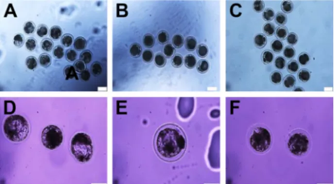
Fig. 1. In vitro development and morphology of embryos col- lected at the compacting 2-cell, morula and blastocyst stages. In the upper panels, 2-cell stages are shown fol- lowing 2 days cultures of uninjected embryos (A), buf- fer-injected embryos (B), and mitochondria-injected em- bryos (C). In the lower panels, morula and blastocyst stages are shown following 7 days cultures of uninjected embryos (D), a buffer-injected embryo (E) and mitochon- dria-injected embryos (F). Scale bar=200μm.
토콘드리아를 microinjection을 통하여 주입하였다. 직경이 1 mm인 capillary tube를 이용하여, 보정용 피펫(holding pip- ette)은 외경이 160~180 μm 그리고 주입용 피펫(injection pip- ette)은 외경이 20~30 μm가 되도록 제작하여 사용하였다. 미토 콘드리아가 주입된 단위발생 수정란은 mSOF 배양액에 45 μ/l (v/v) BME amino acids, 0.5 μ/l(v/v) MEM amino acids, 3 mg/ml BSA (fatty acid free) 또는 5% FBS를 첨가하여 5%
CO2, 5% O2, 90% N2, 39℃ 세포배양기에서 배반포 단계까지 7일간 체외배양하였다.
단백질 정량, 현광현미경 관찰 및 통계분석
분리된 미토콘드리아의 양은 통상적인 단백질 정량법인 Bradford assay로 결정하였다. 소 난자에 미세주입된 GFP- tagged 미토콘드리아의 전달 및 초기 발생 단계에서의 분포를 확인하기 위하여, 각 초기 발생 단계의 세포를 paraformalde- hyde로 고정한 후에 현광현미경(Leica DM2500, Wetzlar, Ger- many)으로 분석하였다. 또한, 세포의 핵은 Hoechst 33342 형 광염색으로 관찰하였다. 미토콘드리아 미세주입이 초기 발생 과정의 분할과 상실배/배반포 형성에 미치는 영향에 대한 통 계분석은 Prism 7 software (GraphPad, La Jolla, USA)를 이용 한 One-way ANOVA로 검정하였으며, 사후검증으로는 Tukey test를 실시하였다. p<0.05인 경우에 통계학적으로 유의하다고 판단하였다.
결과 및 고찰
미토콘드리아 분리, 소 난자로의 미세주입 및 초기 배아 발 생에 대한 영향
Mitochondrial targeting sequence가 연결된 GFP를 미토콘 드리아에서 과발현 시키고 있는 HeLa/mito-GFP 세포주를 배 양하고, 앞서 기술된 differential centrifugation에 기반한 미토 콘드리아의 생화학적 분리가 수행되었다. 분리된 미토콘드리 아에서 GFP의 존재를 현광현미경으로 확인하였다. 또한 소 난소에서 난자를 분리하여 체외배양하고 단위발생을 유도하 였다. 분리된 GFP가 표지된 미토콘드리아를 동물복제에 쓰이 는 미세주입 기법을 사용하여 단위발생이 유도된 난자에 전달 하였다. 대략 1~2 μg 정도의 GFP-표지 미토콘드리아가 하나의 난자에 미세주입되었으며, 미세주입된 난자는 배반포 단계까 지 체외배양하였다.
먼저, 미세주입된 GFP-표지 미토콘드리아가 초기 발생에 미치는 영향을 실체현미경 관찰을 통해 분석하였다. 2세포, 상실배 그리고 배반포 형성과 이러한 단계의 세포 형태를 실 체현미경으로 관찰하였을 때, 미토콘드리아 미세주입은 완충 액을 미세주입 한 경우와 미세주입이 없었던 경우와 비교하여 큰 차이가 없었다(Fig. 1). 이러한 결과는 소 난자에서 유도된 초기 발생을 크게 억제하지 않는 수준에서 미세주입 기법으로
미토콘드리아가 체외에서(in vitro) 전달될 수 있음을 제시한 다.
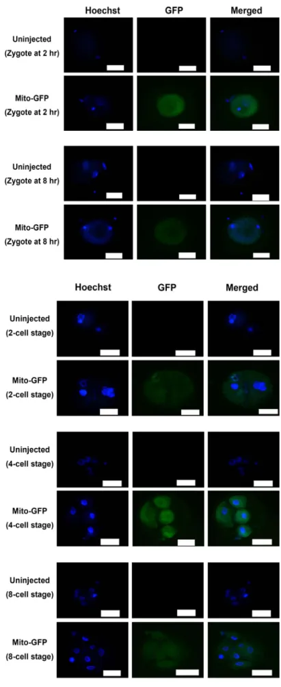
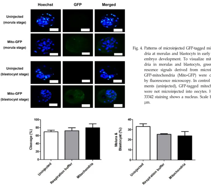
다음으로, 단위발생이 유도된 난자에 미세주입으로 전달된 GFP-표지 미토콘드리아의 전달과 분포를 초기발생의 여러 단 계에 있는 세포에서 형광현미경을 사용하여 관찰하였다. 우선 접합체의 경우(Fig. 2), GFP-표지 미토콘드리아는 미세주입 2 시간 경과 후에 세포질 전체에 균등히 퍼져 분포됨이 확인되 었다. 이러한 분포는 GFP-표지 미토콘드리아가 미세주입되어 8시간 경과된 접합체에서도 유사하게 확인되었다. 핵 DNA를 보여주는 Hoechst 33342 형광염색으로 접합체를 확인할 수 있었으며, 미세주입을 실시하지 않은 경우에서는 녹색형광이 관찰되지 않음이 GFP-표지 미토콘드리아 미세주입 경우와 뚜 렷이 대조되었다. 이와 유사하게, 2세포, 4세포, 8세포, 상실배 그리고 배반포 단계의 여러 세포에서도 GFP-표지 미토콘드리 아가 세포질에서 분포됨이 현광현미경으로 관찰되었다. 마찬 가지로 Hoechst 33342 형광염색으로 2세포에서 배반포까지 초기 발생의 여러 단계를 뚜렷이 구분할 수 있었고, 이러한 단계에서 생성되는 여러 세포가 최초 미세주입된 GFP-표지 미토콘드리아에서 생성하는 녹색형광을 세포질 내에 분포하 는 양상을 확인하였다(Fig. 3, Fig. 4). 이러한 형광현미경 관찰 결과는 미세주입으로 미토콘드리아가 체외에서(in vitro) 전달 될 수 있음을 분명히 보여주며, 또한 이러한 최초 미세주입된 미토콘드리아가 배반포 형성 단계까지의 초기 발생과정에서 생성되는 딸세포에게 전달될 수 있음을 제시한다.
마지막으로, 미토콘드리아 미세주입은 단위발생이 유도된 난자의 분할과정이나 상실배/배반포 형성과정을 크게 방해하 지 않았다(Fig. 5). 미세주입이 없었던 경우 그리고 완충액을 미세주입한 경우를 미토콘드리아를 미세주입한 경우와 비
Fig. 2. Patterns of microinjected GFP-tagged mitochon- dria at zygote stages (at 2 hr and 8 hr) in early bovine embryo development. To visualize mi- tochondria in zygotes, green fluorescence sig- nals derived from microinjected GFP-mitocho- ndria (Mito-GFP) were checked by fluorescence microscopy. In control experiments (uninjected), GFP-tagged mitochondria were not micro- injected into oocytes. Hoechst 33342 staining shows a nucleus. Scale bar=200 μm.
Fig. 3. Patterns of microinjected GFP-tagged mitochon- dria at 2-cell, 4-cell, and 8-cell stages in early bovine embryo development. To visualize mi- tochondria in 2-cell, 4-cell, and 8-cell stages, green fluorescence signals derived from micro- injected GFP-mitochondria (Mito-GFP) were checked by fluorescence microscopy. In control experiments (uninjected), GFP-tagged mitocho- ndria were not microinjected into oocytes.
Hoechst 33342 staining shows a nucleus. Scale bar=200 μm.
Fig. 4. Patterns of microinjected GFP-tagged mitochon- dria at morulas and blastocyts in early bovine embryo development. To visualize mitochon- dria in morulas and blastocyts, green fluo- rescence signals derived from microinjected GFP-mitochondria (Mito-GFP) were checked by fluorescence microscopy. In control experi- ments (uninjected), GFP-tagged mitochondria were not microinjected into oocytes. Hoechst 33342 staining shows a nucleus. Scale bar=200 μm.
Fig. 5. Effects of mitochondrial microinjection on the early development of bovine oocytes. Bovine oocytes are maturated in a matura- tion medium and culmulus cells were removed. Oocytes were then activated and cultured. The resulting embryos were microinjected with a respiration buffer only and GFP-tagged mitochondria suspended in a respiration buffer. Microinjected embryos were further cultured for evaluating cleavage rate (panel A) and morula/ blastocyst formation rate (panel B) relative to uninjected embryos. The number of uninjected oocytes is 52, the number of respiration buffer injected oocytes is 70, and the number of GFP-tagged mitochondria injected oocytes is 63.
교하였을 때, 이들 사이에서 분할율과 상실배/배반포 형성율 에 대한 통계적인 차이는 없었다. 이는 미세주입 기법 자체나 미토콘드리아 미세주입이 초기 발생에 큰 영향을 주지 않음을 반영하며, 미세주입이 미토콘드리아를 체외에서 전달하는 하 나의 방법이 될 수 있음을 제시한다.
미토콘드리아 사이의 fusion 및 fission으로 세포질 내의 미 토콘드리아는 서로 섞이게 된다. 이러한 과정으로 세포는 결 함 미토콘드리아를 정상 미토콘드리아로 보상하거나, 결함 미 토콘드리아를 격리시켜 autophagy로 제거할 수 있음이 잘 알 려져 있다[18, 20]. 따라서, 미세주입된 GFP-표지 미토콘드리 아가 초기 발생과정에서 나타나는 여러 세포의 세포질에 고루 분포하는 양상은 아마도 미토콘드리아의 fusion 및 fission에
기인할 것으로 추측된다. 또한 이러한 관점에서 정상 미토콘 드리아를 결함 미토콘드리아를 가진 세포에게 전달하는 시스 템을 개발하는 것은 매우 의미 있는 일이라 판단된다. 또한, 최근 연구에서 성체줄기세포 등이 주변의 세포에게 자신의 정상적인 미토콘드리아를 직접 전달하여 주변 세포의 미토콘 드리아 기능을 향상시킬 수 있다고 보고되고 있다[1, 8, 9]. 이 러한 연구결과는 미토콘드리아 전달이 실제로 체내에서 이루 어지며, 최소한 세포간의 에너지대사 복구에 필요함을 시사한 다. 따라서, 본 연구에서 제시된, 분리된 미토콘드리아를 미세 주입을 통하여 난자로 전달하는 시스템은 향후 유전성 mtDNA 질환의 제어나 동물복제에 기여될 것으로 사료된다.
감사의 글
본 연구는 교육부와 미래창조과학부의 재원으로 한국연구 재단의 지원(과제번호: NRF-2014R1A1A2A16055714, NRF- 2016R1D1A1B03931722, NRF-2017R1A2B1011438, 및 경상대 학교 BK21 프로그램)을 받아 이루어졌으며 이에 대해 감사드 립니다.
References
1. Ahmad, T., Mukherjee, S., Pattnaik, B., Kumar, M., Singh, S., Kumar, M., Rehman, R., Tiwari, B. K., Jha, K. A., Barhanpurkar, A. P., Wani, M. R., Roy, S. S., Mabalirajan, U., Ghosh, B. and Agrawal, A. 2014. Miro1 regulates inter- cellular mitochondrial transport & enhances mesenchymal stem cell rescue efficacy. EMBO J. 33, 994-1010.
2. Bratic, A. and Larsson, N. G. 2013. The role of mitochondria in aging. J. Clin. Invest. 123, 951-957.
3. Clayton, D. A. and Shadel, G. S. 2014 Isolation of mitochon- dria from tissue culture cells. Cold Spring Harb. Protoc. 10, 1109-1111.
4. Craven, L., Tuppen, H. A., Greggains, G. D., Harbottle, S.
J., Murphy, J. L., Cree, L. M., Murdoch, A. P., Chinnery, P. F., Taylor, R. W., Lightowlers, R. N., Herbert, M. and Turnbull, D. M. 2010. Pronuclear transfer in human embryos to prevents transmission of mitochondrial DNA disease.
Nature 465, 82-85.
5. Gogvadze, V., Orrenius, S. and Zhivotovsky, B. 2008. Mito- chondria in cancer cells: what is so special about them?
Trends Cell. Biol. 18, 165-173.
6. Green, D. R. and Kroemer, G. 2004. The pathophysiology of mitochondrial cell death. Science 305, 626-629.
7. Guzy, R. D., Hoyos, B., Robin, E., Chen, H., Liu, L., Mans- field, K. D., Simon, M. C., Hammerling, U. and Schumacker, P. T. 2005. Mitochondrial complex III is required for hypo- xia-induced ROS production and cellular oxygen sensing.
Cell Metab. 1, 401-408.
8. Hayakawa, K., Esposito, E., Wang, X., Terasaki, Y., Liu, Y., Xing, C., Ji, X. and Lo, E. H. 2016. Transfer of mitochondria from astrocytes to neurons after stroke. Nature 535, 551-555.
9. Islam, M. N., Das, S. R., Emin, M. T., Wei, M., Sun, L.,
Westphalen, K., Rowlands, D. J., Quadri, S. K., Bhattacharya, S. and Bhattacharya, J. 2012. Mitochondrial transfer from bone-marrow–derived stromal cells to pulmonary alveoli protects against acute lung injury. Nat. Med. 18, 759-765.
10. Jacobs, H. T. 2001. Making mitochondrial mutants. Trends Genet. 17, 653-660.
11. Klopstock, T., Klopstock, B. and Prokisch, H. 2016. Mito- chondrial replacement approaches: challenges for clinical implementation. Genome Med. 8, 126.
12. Kroemer, G. and Pouyssegur, J. 2008. Tumor cell metabo- lism: Cancer’s Achilles’ heel. Cancer Cell 13, 472-482.
13. Mishra, P. and Chan, D. C. 2014. Mitochondrial dynamics and inheritance during cell division, development and disease. Nat. Rev. Mol. Cell Biol. 15, 634-646.
14. Orrenius, S., Gogvadze, V. and Zhivotovsky, B. 2007. Mito- chondrial oxidative stress: Implications for cell death. Annu.
Rev. Pharmacol. Toxicol. 47, 143-183.
15. Park, C. B. and Larsson, N. G. 2011. Mitochondrial DNA mutations in disease and aging. J. Cell Biol. 193, 809-818.
16. Park, S. J., Shin, J. H., Kim, E. S., Jo, Y. K., Kim, J. H., Hwang, J. J., Kim, J. C. and Cho, D. H. 2012. Mitochondrial fragmen- tation caused by phenanthroline promotes mitophagy. FEBS Lett. 586, 4303-4310.
17. Reznichenko, A. S., Huyser, C. and Pepper, M. S. 2016.
Mitochondrial transfer: Implications for assisted repro- ductive technologies. Appl. Transl. Genom. 11, 40-47.
18. Sebastian, D., Palacin, M. and Zorzano, A. 2017 Mitochon- drial Dynamics: Coupling Mitochondrial Fitness with Heal- thy Aging. Trends Mol. Med. 23, 201-215.
19. Shadel, G. S. and Horvath, T. L. 2015. Mitochondrial ROS signaling in organismal homeostasis. Cell 163, 560-569.
20. Stewart, J. B. and Chinnery, P. F. 2015. The dynamics of mitochondrial DNA heteroplasmy: Implications for human health and disease. Nat. Rev. Genet. 16, 530-542.
21. Wallace, D. C. 2005. A mitochondrial paradigm of metabolic and degenerative diseases, aging, and cancer: A dawn for evolutionary medicine. Annu. Rev. Genet. 39, 359-407.
22. Wallace, D. C. 2013. A mitochondrial bioenergetic etiology of disease. J. Clin. Invest. 123, 1405-1412.
23. West, A. P. and Shadel, G. S. 2017. Mitochondrial DNA in innate immune responses and inflammatory pathology. Nat.
Rev. Immunol. 17, 363-375.
초록:미세주입을 이용한 난자로의 분리된 미토콘드리아 전달
백상기1†․변준호2†․김보규3․이아람3․조영수1․김익성1․서강미1․정세교4․이준희1*․우동균3*
(1경상대학교 축산생명학과, 2경상대학교병원, 3경상대학교 약학과, 4경상대학교 제어계측공학과)
미토콘드리아는 산화적 인산화와 연결된 전자전달을 통하여 에너지 생산에 중추적인 역할을 갖는다. 이 외에도 미토콘드리아는 신진대사, 세포자멸, 신호전달 그리고 활성산소 생성 등의 다양한 기능을 수행한다. 따라서, 미토 콘드리아의 기능장애는 여러 인체질환에 영향을 준다는 것이 명백하다. 또한, 미토콘드리아 DNA의 돌연변이는 에너지 신진대사에 결함이 있는 여러 유전성 질환의 원인을 제공한다. 불행하게도 아직 이러한 유전성 미토콘드 리아 DNA 질환의 치료법은 전무한 상태이다. 이러한 관점에서, 결함 미토콘드리아를 정상 미토콘드리아로 치환 하는 최근의 시도는 큰 주목을 받고 있다. 본 연구에서는 녹색형광단백질로 표지된 미토콘드리아를 원심분리에 기반하여 생화학적으로 분리하고, 분리된 미토콘드리아를 동물복제에 쓰이는 미세주입 기법으로 소 난자에 전달 하였다. 이러한 미토콘드리아가 미세주입된 난자에서 단위발생을 유도하여 배반포 단계까지의 초기 발생과정에 서 미토콘드리아 미세주입의 영향을 분석하였다. 미토콘드리아에 표지된 녹색형광단백질을 형광현미경으로 분석 함으로써 미세주입으로 난자에 전달된 미토콘드리아는 빠르게 세포질에서 분산되고, 이 후 발생되는 딸세포에게 전달됨이 확인되었다. 따라서, 본 연구에서 수행된, 미세주입을 이용한 미토콘드리아의 전달은 최근 활발히 연구 되는 미토콘드리아 치환 기법, 유전성 미토콘드리아 DNA 질환 치료법 및 동물복제 등에 유용한 모델로의 기여가 기대된다.